PLANT MORPHOLOGY - T. A. Sautkina - 2012
CHAPTER 7. THE FLOWER AS A SPECIAL REPRODUCTIVE ORGAN OF ANGIOSPERMS
7.7. Ontogenetic Development of the Pistil. Characteristics of Processes Occurring in the Ovule
During ontogeny, the future pistil either develops as a meristematic tissue protuberance with subsequent shape changes and fusion of the carpel margins, or originates as a ring-shaped primordium. As development proceeds, the pistil gradually takes shape, consisting of an Ovary, a stylodium, and a stigma. There may be several stylodia, or they may fuse to form a style. Sometimes the style is absent (opium poppy — Papaver), leaving a sessile stigma. In its Anatomical Structure, the pistil ovary bears a clear resemblance to a leaf. Externally, it is covered by an epidermis, beneath which lies the mesophyll with a well-developed Vascular System; the ovary cavity (seed chamber) is likewise lined with an epidermis.
7.7.1. Initiation and Development of the Ovule
At a certain stage of development, ovules (megasporangia) begin to form on the inner wall of the ovary (see Fig. 156).
Each ovule originates subepidermally (beneath the epidermis) as a protuberance of meristematic tissue. As this meristematic protuberance enlarges, the central part of the ovule—the nucellus, which Functions as the macrosporangium (megasporangium)—begins to take shape. The nucellus may be massive or weakly developed. At Cytology/cytology/16.html">Early stages of ovule development, its polarization becomes apparent: the chalazal and micropylar poles are clearly defined. Each pole is named after the respective ovule structures—the chalaza and the micropyle—which occupy diametrically opposite positions.
The chalaza of the ovule is of great importance as a trophic structure. In the region of the chalaza, the ovule attaches to the ovary wall by means of a stalk, or funiculus. A vascular bundle approaches the chalaza, passing through the funiculus, branching at the Base of the nucellus, and supplying the Cells of the developing ovule with Water and dissolved minerals.
From the chalaza, the integuments—the protective coats of the ovule—begin to form as lateral outgrowths (Fig. 178). The number of integuments varies among representatives of different angiosperm families. Polypetalous plants develop two integuments: an outer and an inner one. Sympetalous plants form a single integument, while some parasitic plants lack integuments altogether. The integuments perform a protective function. As the nucellus enlarges, the integuments grow around it on all sides, but they do not fuse at the apex of the ovule, thus forming the micropyle (pollen tube entry). Both integuments most commonly participate in forming the micropyle, with the inner integument forming the endostome and the outer one forming the exostome. Sometimes the micropyle is formed by a single integument. In rare cases, some apomictic plants lack a micropyle altogether.
Class="center">Fig. 178. Macrosporocyte (megasporocyte) of broad bean (Vicia faba) in the nucellus of the ovule: 1—nucellus; 2—macrosporocyte; 3—developing inner integument; 4—developing outer integument

7.7.2. Initiation of the Female Archesporium. The process of Macrosporogenesis (Megasporogenesis)
At early Stages of Ontogeny, when the integuments reach approximately half the height of the nucellus, one (Crassulaceae, Araceae) or, less frequently, several cells (Lemnaceae) in its micropylar part begin to differentiate subepidermally, becoming primary archesporial cells (cells of the female archesporium). The primary archesporial cells are larger than the remaining nucellus cells, possessing non-vacuolated Cytoplasm and a large Nucleus. In some plants, they divide mitotically to form parietal (shielding) cells and secondary archesporial cells (sporogenic cells, macrosporocyte mother cells) (Rosaceae), whereas in others (Hydrocharitaceae, Balsaminaceae, Fabaceae) they become macrosporocytes (megasporocytes) directly without division (see Fig. 178). Differences in The structure of the female archesporium are closely related to the STRUCTURE OF THE nucellus. Crassinucellate ovules usually have parietal cells, while tenuinucellate ovules lack them, although there are A number of exceptions. The parietal cells push the megasporocytes deeper into the nucellus. It is not feasible to establish the Different types of female archesporium as a phylogenetic marker, since the same archesporial types occur in representatives of both primitive and highly evolved families. For example, the multicellular archesporium, considered a primitive type of female archesporium, is found in such primitive families as Ranunculaceae and Rosaceae, as well as in evolutionarily advanced families such as Asteraceae and Apiaceae.
At a certain stage of development, the process of macrosporogenesis (megasporogenesis) takes place within the ovule nucellus. During this process, the macrosporocyte nucleus undergoes reduction division (Meiosis) followed by The formation of Cell walls, resulting in a tetrad of macrospores (megaspores). The Cell walls of the macrospores are laid down simultaneously. Macrospores in the tetrad are usually arranged linearly (Fig. 179). Other arrangements of macrospores in the tetrad are rare. Representatives of the family Onagraceae quite frequently form T-shaped tetrads, whereas members of the family Piperaceae form tetrahedral ones.
Fig. 179. Tetrad of macrospores (megaspores): 1—three degenerating macrospores; 2—macrospore that will develop into the female gametophyte (embryo sac); 3—nucellus cells

Each macrospore is enclosed in a callose wall and represents a typical haploid cell.
Macrosporogenesis begins later than microsporogenesis, but proceeds significantly faster, making its study difficult. Data concerning the cytochemical and submicroscopic features of archesporial cells and macrospores are fragmentary.
7.7.3. Development of the Female Gametophyte (Embryo Sac). Structure of a Typical Embryo Sac
Following macrosporogenesis, the macrospores enter a brief period of rest, after which a new process begins in the ovule nucellus—the Formation of the female gametophyte (embryo sac). In the majority of angiosperms, out of the four resulting macrospores, only one—most frequently the chalazal one (located closest to the chalazal pole)—remains functionally competent and capable of forming the female gametophyte. An embryo sac developing from a single megaspore is termed a monosporic or typical embryo sac, because it has been identified in the majority of studied angiosperms. Since it was first described in Polygonum divaricatum, a representative of the buckwheat family (Polygonaceae), it is also referred to as the Polygonum-type embryo sac (Fig. 180).
Fig. 180. Formation of macrospores and development of the typical Polygonum-type embryo sac: 1—ovule nucellus (megasporangium); 2—macrosporocyte; 3—ovule integuments; 4—macrospore tetrad; 5—enlarging chalazal macrospore (uninucleate embryo sac); 6—degenerating micropylar macrospores; 7—binucleate embryo sac; 8—tetranucleate embryo sac; 9—young 8-nucleate embryo sac; 10—mature embryo sac; 11—egg cell; 12—synergids; 13—polar nuclei; 14—antipodal cells

The Development of the embryo sac occurs through a series of mitotic Divisions of the megaspore nucleus and its derivatives. The embryo sac develops as a coenocytic structure, meaning that the formation of daughter nuclei during embryo sac development is not accompanied by the formation of cell walls between them.
The formation of a typical embryo sac begins with the enlargement of the chalazal macrospore, while the other three macrospores begin to compress and subsequently degenerate. Due to the enlargement of this macrospore, the adjacent cells of the nucellus are also destroyed, and their contents are utilized to nourish the developing female gametophyte.
The embryo sac goes through a series of stages in its development, each bearing a name that reflects the number of nuclei in the developing embryo sac and the degree of their differentiation.
The onset of development, when the developing embryo sac (effectively the enlarging macrospore) contains only a single nucleus, is referred to as the uninucleate embryo sac stage (Fig. 181). At this stage, The Nucleus occupies a central position and is surrounded by a layer of cytoplasm that connects with the peripheral cytoplasm.
Fig. 181. Uninucleate embryo sac of the broad bean (Vicia faba): 1—chalazal macrospore from which the embryo sac develops; 2—Nucleus of the enlarging macrospore; 3—degenerating micropylar macrospores; 4—nucellus cells

The first mitotic division of the macrospore nucleus culminates in the formation of two nuclei located in the center of the developing macrospore. This stage is known as the young binucleate embryo sac stage. Due to the formation of a large vacuole between the newly formed nuclei, they migrate toward opposite poles, rendering the embryo sac binucleate (Fig. 182). Once a single nucleus reaches both the chalazal and micropylar poles, subsequent nuclear divisions within the developing embryo sac proceed in strict synchrony.
Fig. 182. Stages of embryo sac development in Polygonum-type: A—"young" binucleate embryo sac; B—binucleate embryo sac: 1—nuclei in the equatorial zone of the developing embryo sac; 2—nucellus cells; 3—degenerating nucellus cells adjacent to the developing embryo sac; 4—nucleus at the micropylar pole; 5—nucleus at the chalazal pole

The subsequent, second mitotic division results in the formation of two nuclei at each pole, making the embryo sac tetranucleate. Finally, the third and final mitosis takes place, after which each pole of the embryo sac contains four nuclei. Cytoplasm concentrates around each group of four nuclei, and the two groups are interconnected by peripheral cytoplasmic strands. An embryo sac containing eight undifferentiated nuclei is termed a young 8-nucleate embryo sac. It forms a rather large cavity within the nucellus Tissues, bounded by its degenerating cells. Whether the embryo sac possesses its own distinct wall remains unclear: some researchers point to the presence of a cytoplasmic envelope surrounding the embryo sac, whereas others argue that it is absent and that the mature embryo sac is bounded solely by the adjacent nucellus cells.
Although the nuclei within the embryo sac arise via mitosis and might theoretically be expected to share identical properties, they actually prove to be heterogeneous, as evidenced not only by their Morphology but also by their functional characteristics. It is hypothesized that this heterogeneity is promoted by The Nature of the metabolic exchanges occurring between the nuclei of the micropylar and chalazal poles and the Tissues of the maternal Organism.
Upon completion of the mitotic divisions within the cavity of the young embryo sac, the Differentiation of the nuclei begins (see Fig. 180). One nucleus from each pole migrates into the central (equatorial) zone of the embryo sac, where they come to lie within a shared cytoplasm. These are known as polar nuclei. While the term "polar nuclei" carries no functional connotation, it indicates THE ORIGIN OF these nuclei and their migration to the center, or equatorial zone.
Cytoplasmic domains begin to delimit around the three nuclei of the micropylar pole, resulting in the formation of three cells that constitute the egg apparatus of the embryo sac. The egg apparatus comprises an egg cell and two synergids. These cells are readily distinguishable by morphological features: the egg cell is pyriform, with its nucleus occupying the apical region and a large vacuole located in the basal part. The synergids flank the egg cell and possess an elongated shape. Unlike the egg cell, the nucleus of a synergist resides in the basal portion of the cell, while a small vacuole is situated apically. The question regarding the presence of cell walls around the cells of the egg apparatus remains unresolved to this day.
Cytoplasm also segregates around the three nuclei remaining at the chalazal pole, forming three cells. These are termed antipodal cells, reflecting their characteristic position opposite the egg apparatus.
The cells of the embryo sac exhibit distinct functional differentiation. The egg cell functions as the sole female gamete. The synergids play a chemotropic role, "attracting" the pollen tube and guiding its orientation upon entry into the embryo sac. Occasionally, synergids may form micropylar haustoria, in which case they assume a trophic function by facilitating the supply of nutrients to the egg cell. The antipodals are trophic cells that utilize haustoria to deliver nutrients from the chalazal region of the ovule into the embryo sac until the endosperm is formed. Following Fertilization, the polar nuclei give rise to a specialized nutritive tissue—the endosperm. In some plants, the polar nuclei remain free until fusion with a sperm cell, whereas in others they fuse prior to fertilization to form the central nucleus of the embryo sac. Some embryologists believe that cytoplasm also segregates around this nucleus to form the central cell of the embryo sac.
Thus, the female gametophyte of angiosperms, much like the male, is heavily reduced; it arises within the tissues of the maternal organism, never leaves it, and develops at the expense of the maternal sporophyte's nutrients. This pronounced reduction of the female gametophyte should be viewed as an adaptive process that shortens the developmental duration of the sexual generation and accelerates the maturation of sex cells.
An embryo sac in which cellular differentiation has taken place is considered mature and ready for fertilization.
7.7.4. Types of Embryo Sac Development
In approximately 80% of studied angiosperms, the embryo sac develops According to the normal type, i.e., represented by the Polygonum-type embryo sac (see Fig. 180). At the same time, so-called variant types of embryo sacs have been discovered in representatives of various angiosperm families.
METABOLISM/2.html">THE CONCEPT OF embryo sac developmental types was definitively formulated by Professor I. D. Romanov. Characterizing a developmental type involves considering not only the number of megaspores giving rise to the embryo sac, but also the number of divisions occurring during its formation, the number of poles, and the total number of nuclei within the embryo sac.
Thus, the normal type of embryo sac (Polygonum type) is monosporic, triple-mitotic, bipolar, and 8-nucleate.
Variant types of embryo sacs differ substantially from the normal type. Depending on the number of macrospores forming them, they can be monosporic (developing from a single megaspore), bisporic (forming from a megaspore dyad), and tetrasporic (forming from a 4-nucleate coenocyte, i.e., a specialized "megaspore tetrad" in which cell plate formation has not occurred). The development of variant embryo sacs proceeds through either one (monomitotic embryo sacs) or two (dimitotic embryo sacs) mitotic divisions. The nuclear number in variant embryo sacs ranges from 4 to 16. In certain embryo sacs, the number of nuclei may be reduced via polyploidization, resulting in a mixture of haploid and triploid nuclei.
Depending on how nuclei are distributed within the cavity of variant embryo sacs during ontogeny, the latter may be unipolar, bipolar, or tetrapolar.
A peculiar variant monosporic embryo sac is the Oenothera-type, which develops in a unique manner (Fig. 183). This monosporic, dimitotic, unipolar embryo sac was first discovered by W. Hofmeister in members of the evening primrose family (Onagraceae). Development proceeds from the micropylar macrospore. It contains a total of four nuclei: three form the egg apparatus, while a single nucleus resides in the center and functions as the polar nucleus. To date, this developmental type has not been identified in any other plant family, lending it significant systematic value. Based on studies of its embryo sac developmental type, the genus Trapa L. (water chestnut), previously included in Onagraceae, was excluded and reassigned to its own family, Trapaceae. In water chestnut, the embryo sac develops according to the Polygonum type.
Fig. 183. Macrospore formation and developmental Features of the Oenothera-type embryo sac: 1—ovule nucellus (macrosporangium); 2—macrosporocyte; 3—ovule integuments; 4—macrospore tetrad; 5—enlarging chalazal macrospore (uninucleate embryo sac); 6—degenerating chalazal macrospores; 7—binucleate embryo sac; 8—tetranucleate embryo sac; 9—mature tetranucleate embryo sac; 10—egg cell; 11—synergids; 12—polar nucleus

Bisporic embryo sacs originate from a macrospore dyad and can be monomitotic, unipolar, and tetranucleate (Podostemum-type) or dimitotic, bipolar, and 8-nucleate (Allium-type) (Fig. 184).
Fig. 184. Macrospore formation and developmental features of the Allium-type embryo sac: 1—ovule nucellus (macrosporangium); 2—macrosporocyte; 3—ovule integuments; 4—micropylar and chalazal macrospore dyads; 5—enlarging chalazal macrospore dyad (young binucleate embryo sac); 6—degenerating micropylar macrospore dyad; 7—binucleate embryo sac; 8—tetranucleate embryo sac; 9—8-nucleate embryo sac; 10—mature embryo sac; 11—egg cell; 12—synergids; 13—polar nuclei; 14—antipodal cells

No less than 10 types of tetrasporic embryo sacs have been described. They exhibit remarkable structural diversity and occur in representatives of various families.
A distinct tetrasporic, two-mitosis, bipolar, eight-nucleate embryo sac is found in Fritillaria-type (Fig. 185). During the initial stage of development in the Fritillaria-type embryo sac, unequal polarization takes place: three nuclei of the four-nucleate coenocyte migrate to the chalazal pole, while one moves to the micropylar pole. Subsequently, the three chalazal nuclei fuse through polyploidization, forming a single triploid nucleus. Thus, As a result of two successive mitotic divisions, eight nuclei are formed within the embryo sac: four haploid and four triploid. At the micropylar pole, three haploid nuclei become cell-walled to form the egg cell and two synergids, whereas at the chalazal pole, three antipodal cells with triploid nuclei are formed. The two polar nuclei differ in their ploidy level—one is haploid, and the other is triploid.
Fig. 185. Megaspore formation and developmental features of the Fritillaria-type embryo sac: 1—ovule nucellus (megasporangium); 2—megasporocyte; 3—ovule integuments; 4—four-nucleate coenocyte; 5—unequal polarization of nuclei in the developing embryo sac; 6—binucleate embryo sac with a haploid nucleus at the micropylar pole and a triploid nucleus at the chalazal pole; 7—four-nucleate embryo sac; 8—eight-nucleate embryo sac; 9—egg cell; 10—synergids; 11—antipodals; 12—polar nuclei; 13—mature embryo sac

When the nuclei of the four-nucleate coenocyte migrate toward the micropylar, chalazal, and two lateral poles, four-pole tetrasporic embryo sacs are formed. These can be single-mitosis, eight-nucleate (Plumbago-type) (Fig. 186) or two-mitosis, sixteen-nucleate (Penaea-type) (Fig. 187).
Fig. 186. Megaspore formation and developmental features of the Plumbago-type embryo sac: 1—ovule nucellus (megasporangium); 2—megasporocyte; 3—ovule integuments; 4—four-nucleate coenocyte (four-nucleate megaspore); 5—developing four-pole embryo sac; 6—eight-nucleate four-pole embryo sac; 7—mature embryo sac; 8—egg cell; 9—polar nuclei

Fig. 187. Megaspore formation and developmental features of the Penaea-type embryo sac: 1—ovule nucellus (megasporangium); 2—megasporocyte; 3—ovule integuments; 4—four-nucleate coenocyte (four-nucleate megaspore); 5—developing four-pole embryo sac; 6—eight-nucleate four-pole embryo sac; 7—sixteen-nucleate four-pole embryo sac; 8—mature sixteen-nucleate embryo sac; 9—egg cell; 10—synergids; 11—antipodals; 12—polar nuclei; 13—cells at the lateral poles 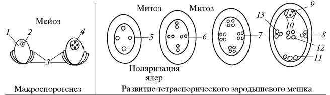
Most researchers consider the Polygonum-type to be the most primitive and ancestral type of embryo sac development. The reduction in the number of mitotic divisions during embryo sac formation represents one of the major evolutionary trends in the female gametophyte of angiosperms. This promotes a more rapid development of the female gametophyte and likely holds adaptive significance. However, according to plant embryologists, "rather numerous deviations from the normal type of Development and Structure of embryo sacs in flowering plants apparently call for broader and more detailed studies to elucidate their specific adaptive, ontogenetic, or ecological meaning."
Last update: 07/08/2026
Editorial and Educational Adaptation: This material has been compiled based on the primary/original source text. The project team performed an editorial review, corrected technical inaccuracies, structured sections, and adapted the content for an educational format.
What was processed:
- elimination of formatting defects (OCR errors, structural breaks, corrupted characters);
- editorial organization of content;
- standardization of terminology in accordance with academic sources;
- verification of factual statements against the original source text.
All mentions of the author, publication year, and origin of the primary text have been preserved in accordance with the source.