HETEROPHYLLY IN PLANTS - O.M. NEDUKHA - 2011
CHAPTER TWO. MECHANISMS OF HETEROPHYLLY EXPRESSION IN PLANTS
Class="center">
2.2. ENDOGENOUS MECHANISMS OF HETEROPHYLLY EXPRESSION IN PLANTS
2.2.3. Ca2+ as a secondary messenger in regulatory processes
In higher aquatic plants characterized by heterophylly, the transition from one leaf form to another typically occurs when the stem emerges from the Water onto the water surface. This process is marked by phenotypic plasticity of anatomical and ultrastructural leaf traits, as well as an increase in the intensity of CO2 fixation and a higher synthesis of chlorophylls and ABA (Nekrasova et al., 2003; Mommer, Visser, 2005). In submerged leaves, a decrease in The activity of carboxylating Enzymes and the content of photosynthetic products is accompanied by structural Changes in the cuticle of epidermal Cell walls, which facilitates The transport of CO2 and HCO3- via the apoplast (Rascio, 2002). Most of the aforementioned processes are calcium-dependent.
The control of Plant GROWTH AND DEVELOPMENT, as well as plant responses to environmental factors, is mediated by secondary messengers, including cyclic GMP, Lipids, Ca2+, and others. However, Calcium Ions alone exhibit an almost instantaneous response to virtually all stimuli. Ca2+ participates in The regulation of numerous processes across all plant Tissues, where Ca2+-dependent Proteins (calcium sensors) are activated or inhibited depending on the cytoplasmic calcium content [Ca2+]c (Sanders et al., 1999; Bush, 1995). This occurs due to the ability of these ions to coordinate multiple bonds (from 2 to 8) with Protein domains and alter their conformation (see review: Belyavskaya, 1994). In photosynthetic Cells, Ca2+ accumulates in the stroma and thylakoids of METABOLISM/14.html">Chloroplasts: in the stroma, it activates NAD kinase, which catalyzes The conversion of NAD to NADP; in the thylakoids, it activates Calvin cycle enzymes and participates in the repair of PSII reaction centers (Charles, Halliwell, 1980; Moore, Akerman, 1984; Matoo et al., 1989; Grove, Brudvig, 1998; Sai, Johnson, 2002). It has been established that chloroplast thylakoids can accumulate up to 30 nM Ca2+ per 1 mg of chlorophyll (Ettinger et al., 1999).
The potential role of Ca2+ fluxes in regulatory processes within the chloroplast and their connection to the regulation of cytosolic Ca2+ transport is of utmost importance. It has been demonstrated that isolated chloroplasts absorb calcium upon illumination, which is evidently transported across the chloroplast envelope. Light-induced calcium absorption has also been observed in in vitro experiments using Cells of the charophyte alga Nitellopsis sp. It was established that the cytosolic calcium content in cells decreased upon illumination; this phenomenon depended on photosynthetic electron transport (Sai, Johnson, 2002).
In cell walls, calcium ions bind to galacturonic acid residues of Pectins, inducing Conformational Changes in pectins and forming dimeric or multimeric pectin structures (Jarvis, 1982). Furthermore, calcium ions in cell walls can bind to Xylan polymers, inducing their crystallization and binding to Lignin molecules (Jarvis, 1982), which results in Cell wall reinforcement. It has been established that stomatal movement is also regulated by calcium, alongside other endogenous and exogenous factors such as potassium ions, CO2 and ABA content, as well as soil and air humidity (Kearns, Assmann, 1999; Xi-Qing et al., 1998). Investigating The Role of calcium in the functioning of Vicia faba L. Stomata, the authors found that millimolar concentrations of calcium ions (from 0.1 to 1 mM) inhibited stomatal opening by 60% on the lower leaf surface and by 45% on the upper surface (Xi-Qing et al., 1998).
Various stimuli, such as changes in illumination, osmotic and oxidative stress, Temperature increases, salinity, and others, trigger an increase in cytoplasmic calcium content (Bush, 1995; Knight, 2000; Knight et al., 1997). Changes in the calcium balance, which ensure plant adaptation to the environment, evidently involve both epidermal and mesophyll cells. Considering the above, it can be hypothesized that photosynthetic cells of plants growing submerged in water will differ in ionized calcium content from those growing in the air (above water).
In photosynthetic cells, ionized calcium, acting as a secondary messenger, participates in the regulation of Photorespiration and Photosynthesis—specifically, in the repair of PSII reaction centers, the activation of D-ribulose-1,5-bisphosphate carboxylase, and the regulation of chlorophyll synthesis (Hiedema, Prins, 1992; Sai, Johnson, 2002). In The Cell walls of mesophyll leaves, calcium ions bind to pectins and structural proteins, reducing wall plasticity and inhibiting apoplastic water transport (Mommer, Visser, 2005; Virk, Cleland, 1988). The involvement of calcium ions in these processes within the leaves of hydrophytes growing under optimal conditions or moderate water deficit remains scarcely studied. Previously, certain differences were established in the mesophyll Cell Structure of submerged and floating leaves of S. latifolium, as well as in the epidermal ultrastructure of Alisma plantago-aquatica leaves grown under different water supply conditions (Kordyum et al., 2003; Nedukha, 2005). We hypothesized that the ultrastructural differences between mesophyll cells of submerged and floating leaves of S. latifolium are mediated by the redistribution of calcium ions within the cells and changes in calcium-dependent chlorophyll synthesis (Nedukha, 2010b). Further investigation of calcium ion distribution in the mesophyll and epidermal cells of different S. latifolium leaves during the vegetative growth phase (in May), depending on growth conditions, revealed the following.
Sium latifolium, a helophytic ecoform in the virginal stage of ontogeny (in May), was characterized by The Development of morphologically distinct submerged and floating leaves. We investigated the distribution of Ca2+ ions in Organelles, cell walls, and the Cytoplasm of mesophyll and epidermal cells of submerged and floating leaves (Nedukha, 2010b). Analysis of calcium ion localization using a specific fluorescent indicator (Fluo-4) in mesophyll cells of submerged and floating leaves revealed a uniform green luminescence of the studied ions in chloroplasts, nuclei, cell walls, and the cytoplasm of the cells examined (Fig. 2.2.3.1, a–d; see insert XV). The Use of the "Pascal" software to construct corresponding histograms (Fig. 2.2.3.1, e, e', f, f') made it possible to determine the relative content of Ca2+ ions in the organelles and cell walls of leaves treated with the fluorescent indicator.
A comparison of calcium ion fluorescence intensity in the mesophyll cells of the two leaf types of S. latifolium showed that in submerged leaves, the fluorescence intensity of calcium ions in chloroplasts, nuclei, and mesophyll cell walls was 1.7-, 1.5-, and 1.4-fold lower, respectively, while in the cytoplasm it was 1.4-fold higher compared to the corresponding organelles and cell walls of floating leaves (Table 2.2.3.1). The Study of the luminescence of the fluo-4+calcium complex in the cell walls of the upper and lower epidermis of submerged and floating leaves showed that in the outer walls of the main epidermal cells, calcium fluoresced as individual green grains.
Calcium ion fluorescence in the anticlinal walls of the same cells and the walls of stomatal guard cells was uniform (Fig. 2.2.3.1, g, h).
In stomatal guard cells, we examined the cell walls forming the stomatal pore. In submerged leaves, the distribution of calcium ion fluorescence intensity in the walls of the upper and lower epidermis differed. In the upper epidermis, its intensity was significantly higher: 3.3-fold in the outer walls, 1.7-fold in the anticlinal walls, and 2.2-fold in the walls of stomatal cells compared to the corresponding cell walls of the lower epidermis (Table 2.2.3.2). In floating leaves, a similar trend was also observed regarding the distribution of calcium ions in the cells of the upper epidermis: a 1.3-fold increase in the anticlinal walls and a 1.12-fold increase in the walls of stomatal guard cells compared to the cells of the lower epidermis (Table 2.2.3.2).
TABLE 2.2.3.1. Distribution of Ca2+ ions in mesophyll cells of Sium latifolium leaves collected in May
Relative calcium ion content, arbitrary units, x ± Sx- |
||||
Structure |
Leaves of helophytic plants |
Leaves of terrestrial plants |
||
cells |
submerged |
floating |
with entire |
pinnatifid |
blade |
||||
Chloroplast |
125 ± 9.0 |
211 ± 12** |
95 ± 5.2 |
109 ± 7.1 |
83 ± 2.7 |
124 ± 5.7** |
87 ± 2.9 |
151 ± 9.1** |
|
Cell wall |
35 ± 2 |
50 ± 2.4* |
33 ± 1.9 |
41 ± 1.5* |
Cytoplasm |
60 ± 3.7 |
48 ± 2.4* |
53 ± 2.3 |
114 ± 1.2** |
Note: * - P ≤ 0.01; ** - P ≤ 0.001 (When comparing data of the two leaf types within each ecoform of S. latifolium)
TABLE 2.2.3.2. Distribution of Ca2+ ions in epidermal cells of Sium latifolium leaves collected in May
Relative calcium ion content, arbitrary units, x ± Sx- |
||||
Epidermis / wall type |
Leaves of helophytic plants |
Leaves of terrestrial plants |
||
submerged |
floating |
with entire blade |
pinnately dissected |
|
Upper epidermis: outer wall |
87 ± 4.3 |
49 ± 2.1*** |
91 ± 3.2 |
57 ± 4.4*** |
anticlinal wall |
157 ± 10.2 |
133 ± 11 |
101 ± 9.3 |
105 ± 10.1 |
stomatal walls |
174 ± 12.9 |
120 ± 8.9** |
110 ± 8.9 |
190 ± 13.2*** |
Lower epidermis: outer wall |
26 ± 1.7 |
43 ± 7*** |
90 ± 7.0 |
47 ± 2.9*** |
anticlinal wall |
90 ± 4.9 |
99 ± 4.4 |
89 ± 7.8 |
100 ± 9.9 |
stomatal walls |
79 ± 6.7 |
107 ± 9.7* |
97 ± 8.0 |
140 ± 12.7** |
Note: * - P ≤ 0.05; ** - P ≤ 0.01; *** - P ≤ 0.001 (When comparing data of the two leaf types within each ecoform of S. latifolium)
TABLE 2.2.3.3. Photosynthetic pigment content in Sium latifolium leaves collected in May
Pigment, mg/g fresh weight |
Pigment content, x ± Sx |
|||
Leaves of helophytic plants |
Leaves of terrestrial plants |
|||
submerged |
floating |
with entire blade |
pinnately dissected |
|
Chlorophyll a |
1.43 ± 0.08 |
2.16 ± 0.08** |
1.76 ± 0.2 |
1.59 ± 0.10 |
Chlorophyll b |
0.95 ± 0.02 |
1.18 ± 0.04** |
1.14 ± 0.11 |
1.21 ± 0.08 |
Total chlorophylls a+b |
2.38 ± 0.08 |
3.34 ± 0.09** |
2.89 ± 0.12 |
2.79 ± 0.12 |
Chlorophyll a/b ratio |
1.5 |
1.83 |
1.46 |
1.3 |
Carotenoids |
0.55 ± 0.02 |
1.01 ± 0.05* |
0.78 ± 0.05 |
0.88 ± 0.06 |
Note: * - P ≤ 0.05; ** - P ≤ 0.001 (When comparing data of the two leaf types within each ecoform of S. latifolium)
A comparison of the calcium ion content in the walls of the upper and lower epidermis of submerged and floating leaves of S. latifolium showed that leaf growth above water was accompanied by a decrease in the relative ion content in the outer walls of the main cells and stomatal guard cell walls of the upper epidermis, as well as an increase in the calcium ion content in the corresponding cell walls of the lower epidermis.
Parallel determination of photosynthetic pigment content in submerged and emergent leaves, which were examined for the localization and content of calcium ions, showed that the levels of chlorophyll a, b and carotenoids in submerged leaves were significantly lower than in emergent ones (Table 2.2.3.3).
S. latifolium is a terrestrial ecoform. The leaves of broadleaf water-parsnip plants growing in terrestrial conditions in May (during the vegetative growth phase) also exhibited varying blade shapes: entire (first to third leaves) and pinnatifid (fourth and fifth leaves) (Fig. 2.2.2.1, b; see insert XIII). Calcium ion fluorescence in the mesophyll cells of S. latifolium leaves with different blade shapes was identical to that in the leaf blades of the amphibious form of S. latifolium (Fig. 2.2.3.1, d, f; see insert XV). A comparison of the fluorescence intensity distribution of calcium ions across mesophyll cells of leaves with different shapes revealed the following: in The Nucleus, cell walls, and cytoplasm of cells in leaves with an entire blade, the fluorescence intensity of Ca2+ was 1.7, 1.3, and 2.1 times lower, respectively, than in identical structures of mesophyll cells in pinnatifid leaves (Table 2.2.3.1). At the same time, the relative content of calcium ions in mesophyll chloroplasts did not significantly differ between leaves with different blade shapes. Furthermore, the distribution of calcium ions in the cell walls of the upper and lower epidermis of the first three leaves with an entire blade of the terrestrial ecoform of S. latifolium also showed no differences (Table 2.2.3.2).
In pinnatifid leaves, the relative content of calcium ions varied between epidermal cell walls: in the outer walls of the main epidermal cells of the upper epidermis, it was 1.2 times higher, and in the walls of stomatal guard cells, 1.4 times higher than in the walls of the lower epidermis (Table 2.2.3.2). A comparison of the calcium ion content in the epidermal cell walls of the two leaf types of the terrestrial ecoform of broadleaf water-parsnip revealed a significant difference between the outer walls and the stomatal cell walls (Table 2.2.3.2).
Determination of the photosynthetic pigment content in the first leaves with an entire blade and the subsequent pinnatifid leaves of the terrestrial ecoform of the species showed no significant differences in the content of chlorophylls a, b, their sum (a + b), and carotenoids (Table 2.2.3.3). The obtained data regarding the absence of substantial changes in the distribution of calcium ion in mesophyll chloroplasts and the photosynthetic pigment content between the two leaf types of the terrestrial ecoform of S. latifolium apparently indicate that these traits remain relatively stable in the leaves of the terrestrial form during the vegetative growth phase.
Thus, cytochemical Methods and laser scanning confocal Microscopy have demonstrated that the leaves of the two ecological forms of S. latifolium during the vegetative growth phase are characterized by a specific distribution of calcium ions depending on the plant's growing conditions. We established that the relative content of Ca2+ in the mesophyll chloroplasts of emergent leaves of amphibious plants is higher than in the chloroplasts of submerged leaves. As is well known, the photosynthetic rate, the activity of carboxylating enzymes, and the content of photosynthetic products in submerged leaves of many wild and cultivated plant species are considerably lower compared to those in emergent leaves of the same species (Bowes, Salvucci, 1989; Ronzhina, Pyankov, 2001; Nekrasova et al., 2003; Ronzhina et al., 2004). In addition, it has been established that Rubisco activity and the synthesis of Photosynthetic Pigments depend on the calcium ion content within the chloroplast (Sai, Johnson, 2002; Lechowski, Bialczyk, 1993). Taking into account the aforementioned literature data and our experimental results, it can be hypothesized that in submerged leaves of S. latifolium, alterations in chlorophyll synthesis are mediated by a lower calcium ion content in the chloroplasts.
Comparative Analysis of the chlorophyll and carotenoid content in submerged and emergent leaves of the amphibious form of S. latifolium showed that their levels were significantly lower in submerged leaves. Similar data regarding low chlorophyll content in submerged leaves have been reported by other authors when comparing submerged and floating leaves of Ranunculus vulgaris L., Marsilea quadrifolia, and other plant species characterized by heterophylly (Bowes, Salvucci, 1989; Nekrasova et al., 2003; Ronzhina et al., 2004; Mommer, Visser, 2005). The inhibition of photosynthetic pigment synthesis underwater occurs due to low light intensity, an increase in far-red light, and slow diffusion of CO2 in water (Nekrasova et al., 2003; Mommer, Visser, 2005).
A comparison of the relative calcium ion content in the chloroplasts of emergent leaves of the amphibious ecoform of Sium latifolium with that in the mesophyll chloroplasts of the two leaf types of the terrestrial form of this species showed that the mesophyll chloroplasts of emergent leaves contained twice as many calcium ions as the mesophyll chloroplasts of the terrestrial form of broadleaf water-parsnip. Moreover, emergent leaves of amphibious plants contained more photosynthetic pigments than leaves of the terrestrial form. Previously, a similar correlation between pigment and calcium ion content was found in cucumber cotyledons when the calcium ion concentration in the nutrient medium did not exceed 10 mM (Tanaka, Tsuji, 1980).
It is known that chlorophyll synthesis is a calcium-dependent process (Sai, Johnson, 2002; Charles, Halliwell, 1980). In chloroplasts, Ca2+ activates NAD kinase and Calvin cycle enzymes (Charles, Halliwell, 1980; Grove, Brudvig, 1998). An analysis of literature data on the mechanisms of calcium ion Transport from the Cytosol into the chloroplast indicates that such transport depends on light intensity and the binding affinity of Ca2+ ions to PSII proteins (Ettinger et al., 1999; Johnson et al., 1995; Roh et al., 1998).
As a result of experimental work carried out with transgenic tobacco chloroplasts expressing cytosolic aequorin—a Ca2+-binding protein whose luminescence determines the concentration of ionized calcium—it was established that upon transferring tobacco seedlings from darkness to light, rapid transport of Ca2+ ions from the cytoplasm into the chloroplasts began (Fig. 2.2.3.2, a). It was noted that with increased duration of exposure to light, the Ca2+ ion content in the chloroplast increased. When seedlings were transferred back to darkness, reverse transport occurred within just 5 minutes: calcium ions were pumped out from the thylakoids into the chloroplast stroma, and from the stroma into the cytoplasm (Fig. 2.2.3.2, b) (Sai, Johnson, 2002).

Figure 2.2.3.2. Diagram of the influx (a) of Ca2+ ions from the cytoplasm into the chloroplast (upon illumination) and the efflux of Ca2+ ions (b) from the thylakoids and chloroplast stroma into the cytoplasm (in darkness). Designations: thy - thylakoid lumen, LR - light regulation, pet - photosynthetic electron transport, ap - Ca2+/H+ antiporter (Sai, Johnson, 2002).
Ca2+ ions located in the thylakoids play a crucial role in PSII function, specifically in molecular oxygen evolution during photosynthetic water oxidation. Using a model system—spinach leaf chloroplasts—it has been demonstrated (Shi Hua et al., 1998) that the regulation of this process occurs through the saturation or depletion of Ca2+ ions in PSII: calcium ions, by binding to D1 and D2 proteins (with molecular weights of 17 and 23 kDa), activate them. The core of these proteins forms a heterodimer comprising a tetranuclear cluster of four manganese atoms (Mn4) and two Cofactors: Ca2+ and Cl-.
J. Vrettos and G. Brudvig proposed a scheme (Vrettos, Brudvig, 2002) according to which Electron transport along the chain in PSII (from the S0 state to the S4 state) occurs only in the presence of Ca2+ ions (Fig. 2.2.3.3). The researchers suggest that Ca2+ ions can bind to the Glu and Asp residues of the main peptide chain and to water molecules of the tetranuclear Mn cluster (Shi Hua et al., 1998; Taka-aki Ono et al., 2001; Vrettos, Brudvig, 2002). X-Ray Diffraction Analysis has shown that the crystal structure of PSII molecules involved in photochemical oxygen evolution changes in the absence of Ca2+ ions, and the oxidation process of the Mn cluster from the S2 or S3 state is disrupted, halting photooxidation (Taka-aki Ono et al., 2001). Furthermore, it has been shown that secondary structural changes in PSII proteins (from α-helical to sheet) occur during photoinhibition, which is accompanied by a decrease in the content of Ca2+ ions associated with the PSII light-harvesting complex in the thylakoids (Shi Hua et al., 1998).
It was found that the relative content of Ca2+ in the mesophyll chloroplasts of terrestrial S. latifolium plants varies depending on the ontogenetic phase and the growing site of the species (Nedukha, 2010, c): during the budding phase, its content increases by 1.5 times, while during the fruiting phase, it decreases by 1.4 times compared to the chloroplasts of leaves from amphibious individuals of water-parsnip.
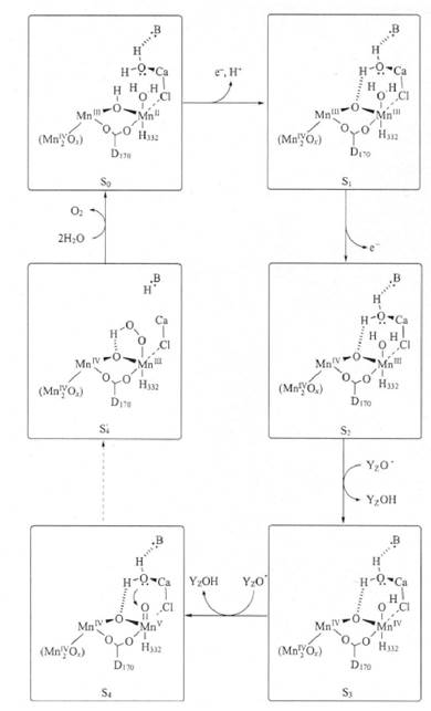
Figure 2.2.3.3. Scheme of Ca2+ ion binding in the Mn-retaining cluster of PSII during photooxidation according to J. Vrettos and G. Brudvig (Vrettos, Brudvig, 2002), according to which electron transfer along the chain in PSII (from the S0 state to the S4 state) occurs only in the presence of Ca2+ ions. Inhibitory analysis (chelation of Ca2+ ions) showed that oxygen evolution during photosynthetic water oxidation did not occur.
Based on our results and literature data, we hypothesize that during the budding phase of S. latifolium, the higher relative content of Ca2+ ions in the mesophyll chloroplasts of terrestrial plants compared to amphibious plants promotes enhanced chlorophyll synthesis and PSII activation in leaf cells under terrestrial growing conditions. Obviously, the activity of transport systems in the chloroplast envelope membranes for Ca2+ ions (Ca2+/H+ antiporter and calcium channels) changes in the mesophyll cells of broadleaf water-parsnip depending on the plant's growth stage and environmental conditions. Considering the aforementioned literature data and the significant changes we observed in the relative calcium content in the mesophyll chloroplasts of terrestrial broadleaf water-parsnip plants during the fruiting phase, the following can be inferred. Evidently, during the ontogenesis (specifically the fruiting phase) of terrestrial broadleaf water-parsnip, alterations in the calcium balance, resulting from the inhibition of Ca2+ ion transport from the cytoplasm into the chloroplasts, lead to a substantial decrease in Ca2+ ion content within the chloroplasts and a reduction in pigment levels.
In addition, an increase in the relative calcium ion content was found in the cell walls of stomata in pinnatifid leaves of the terrestrial ecoform of S. latifolium when compared to the emergent leaves of amphibious individuals of this species. According to researchers (Kordyum et al., 2003), calcium ions play an active role in regulating the turgor of stomatal guard cells and the opening/closing of the stomatal pore, with their levels regulated by both apoplastic calcium reserves and calcium bound to endomembranes. Taking into account our results and the literature data cited above, one can postulate an intensification of stomatal functioning in the upper and lower epidermis of the upper leaves of the terrestrial form of S. latifolium compared to the emergent leaves of the amphibious ecoform.
We also revealed a difference in the distribution of calcium ions between the cell walls of the upper and lower epidermis in submerged leaves of S. latifolium: a higher Ca2+ content was observed in the upper epidermal cell walls compared to the lower epidermis. It was previously established that submerged leaves of Potamogeton lucens and Elodea sp. are polarized: HCO3- uptake occurs on the lower side of the leaf, while hydroxyl and hydrogen ions are released on the upper side (Bowes, Salvucci, 1989). In the lower epidermis of submerged leaves of the aforementioned plants, a Ca2+-dependent Carbonic anhydrase is activated, which participates in generating CO2 from HCO3- (Bowes, Salvucci, 1989; Rascio, 2002). Furthermore, it is known that Ca2+ ions, by binding to the oxygen atoms of free carboxyl groups in pectins and to hydroxyl groups of proteins, reduce elasticity, mechanical strength of the wall, and its adsorption capacity (Preston, 1979; Virk, Cleland, 1988). Considering our experimental data on the increased calcium ion content in the upper epidermal walls of submerged water-parsnip leaves and the literature data mentioned above, it can
be hypothesized that the cell walls of the upper and lower epidermis of submerged S. latifolium leaves also exhibit functional polarity, in which unbound calcium ions are involved.
The observed differences in the relative calcium ion content in the epidermal cells of emergent leaves of the amphibious form of S. latifolium and the dissected leaves of the terrestrial form of this species have been noted (Nedukha, 2010, b). It is known that the outer cell walls of the upper and lower leaf epidermis participate in cuticular Transpiration, The rate of which depends on wall structure, pectin-bound calcium content, and the presence of cuticular pores (Schreiber, 2005; Virk, Cleland, 1988). The question regarding the relationship and effect of ionized calcium on The formation of cuticular pores and, consequently, on cuticular transpiration through the outer walls of epidermal cells remains open for now.
Thus, the obtained data on the distribution of calcium ions in various leaf types of amphibious and terrestrial plants of S. latifolium indicate that calcium ion content plays a crucial role in the phenotypic plasticity of broadleaf water-parsnip to environmental changes. Future research should focus on elucidating the mechanisms of calcium ion redistribution within leaf mesophyll and epidermal cells in response to alterations in water regime.
Last update: 07/08/2026
Editorial and Educational Adaptation: This material has been compiled based on the primary/original source text. The project team performed an editorial review, corrected technical inaccuracies, structured sections, and adapted the content for an educational format.
What was processed:
- elimination of formatting defects (OCR errors, structural breaks, corrupted characters);
- editorial organization of content;
- standardization of terminology in accordance with academic sources;
- verification of factual statements against the original source text.
All mentions of the author, publication year, and origin of the primary text have been preserved in accordance with the source.