PLANT HETEROPHYLLY - O.M. NEDUKHA - 2011
CHAPTER ONE. MORPHOLOGICAL AND STRUCTURAL-FUNCTIONAL CHARACTERISTICS OF VEGETATIVE ORGANS IN HETEROPHYLLOUS PLANTS
1.8. ULTRASTRUCTURAL FEATURES OF LEAF AND ROOT CELLS
1.8.1. Leaves of Sium latifolium
S. latifolium growing in Water exhibits heterophily: leaves of different shapes develop on a single SHOOT, including submerged leaves (bi- or tripinnatisect with thread-like segments), emergent leaves (pinnatisect with lanceolate segments), and "transitional" leaves, in which the blade is positioned above water and the petiole remains submerged (with blade shapes similar to those of submerged leaves) (see subsection 1.3.3 and Fig. 1.3.1 in Insert II). An ultrastructural Study of the three leaf types in water parsnip revealed both common features and specific differences in the ultrastructure of mesophyll Cells (Nedukha, 2004).
Submerged leaves. An Analysis of the mesophyll Cell ultrastructure in submerged leaves demonstrated that cells of both palisade and spongy mesophyll possessed a large central vacuole, with the Cytoplasm and Organelles located along The Cell walls. METABOLISM/14.html">Chloroplasts of the palisade and spongy mesophyll, lens-shaped or elongated (depending on the plane of section), exhibited a similar ultrastructure. The number of chloroplasts per cell section was 9.0 ± 2.4 for the palisade parenchyma and 6.7 ± 0.3 for the spongy parenchyma (Table 1.8.1.1). The average chloroplast size was 4.3 × 1.2 µm, with a well-developed grana-type membrane system (Fig. 1.8.1.1, a, b); starch grains were rarely observed. A characteristic feature of the Plastids was the presence of a high number of thylakoids per granum (Table 1.8.1.2), ranging in diameter from 0.2 to 0.4 µm, with the number of grana per chloroplast section varying from 6–8 to 12–14. In some chloroplasts, both grana and stroma thylakoids showed increased electron density. Plastoglobules up to 50 nm in diameter were occasionally observed in the dense stroma. Mitochondria featured a well-developed cristae system (Fig. 1.8.1.1, c). Numerous Ribosomes and rough Endoplasmic reticulum were detected in the cytoplasm. Cell wall thickness was 0.2–0.4 µm. The Nucleus was round or slightly elongated, with dense nucleoplasm, and both granular and fibrillar components were identified within the nucleolus (Fig. 1.8.1.1, d).
"Transitional" leaves. Mesophyll cells of transitional leaves (with petioles submerged in water and blades positioned above water) differed in their ultrastructural characteristics. They were conventionally divided into two types: in some cells, the ultrastructure of endomembranes and organelles remained intact (Fig. 1.8.1.2, a, b), whereas in others, degradation of organelles and endomembranes was observed (Fig. 1.8.1.2, c, d).
In the first type of cells, mitochondria were of the condensed type, the Golgi apparatus was represented by groups of short dictyosomes, and The endoplasmic reticulum was rough. Ribosomes and Polysomes were visible in the hyaloplasm.
The cell wall thickness was 0.3–0.4 µm. Chloroplasts differed from those in the previous variant in their linear dimensions, the number of starch grains (Table 1.8.1.1), and thylakoids per granum (Table 1.8.1.2); grana with 20 or more thylakoids were almost 2.5 times less frequent than in chloroplasts of submerged leaves.
Class="center">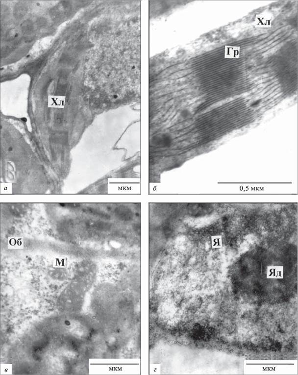
Fig. 1.8.1.1. Fragments of mesophyll cells from submerged Leaves of Sium latifolium:
M - mitochondrion, Chl - chloroplast, Gr - granum, CW - cell wall, N - nucleus, Nu - nucleolus;
a, b - palisade parenchyma, c, d - spongy parenchyma
A specific feature of these cells was the presence of nearly round, electron-dense structures 10–15 nm in size that contacted the organelle membranes and The Plasma Membrane (Fig. 1.8.1.2, a, b). In Cells of the second type (Fig. 1.8.1.2, c, d), clear signs of degradation typical of senescing cells were detected: disruption of the integrity of the plasma membrane and tonoplast, degradation of the nucleoplasm, Condensation of the nucleolus, and increased electron density of organelles in the cytoplasm.
TABLE 1.8.1.1. Quantitative characteristics of chloroplasts in mesophyll cells of Sium latifolium
Leaf type |
Number of chloroplasts per cell section in different mesophyll Tissues |
Chloroplast size, µm |
Starch grain size, µm |
Number of starch grains per chloroplast section |
|||
Palisade |
Spongy |
Long axis |
Short axis |
Long axis |
Short axis |
||
Submerged |
9.0 ± 2.4 |
6.7 ± 0.3 |
4.3 ± 0.3 |
1.2 ± 0.1 |
1.4 ± 0.1 |
0.6 ± 0.19 |
0.7 ± 0.1 |
"Transitional" |
10 ± 0.1 |
6.5 ± 0.3 |
6.0 ± 0.2 |
1.9 ± 0.1 |
1.3 ± 0.1 |
0.5 ± 0.1 |
2.3 ± 0.2 |
Emergent |
7.5 ± 0.3 |
6.6 ± 0.4 |
4.4 ± 0.1 |
1.3 ± 0.1 |
1.2 ± 0.1 |
0.3 ± 0.02 |
1.1 ± 0.1 |
TABLE 1.8.1.2. Quantitative characteristics of thylakoids in chloroplasts of Sium latifolium mesophyll
Leaf type |
Distribution of grana by number of thylakoids, % |
||||
2 - 3 |
4 - 6 |
7 - 10 |
10 - 20 |
over 20 |
|
Submerged |
- |
8.6 |
26.6 |
30.0 |
34.8 |
"Transitional" |
11.1 |
18.5 |
14.8 |
40.8 |
14.7 |
Emergent |
5.3 |
66.2 |
28.5 |
- |
- |
TABLE 1.8.1.3. Pigment content in leaf blades of Sium latifolium
Pigments |
Leaf |
||
submerged |
"transitional" |
emergent |
|
Chlorophyll a |
|||
mg/g fresh weight |
0.654 |
0.411 |
1.053 |
mg/g dry weight |
1.712 |
2.020 |
1.316 |
Chlorophyll b |
|||
mg/g fresh weight |
0.443 |
0.315 |
0.391 |
mg/g dry weight |
1.180 |
1.298 |
0.510 |
Total chlorophylls (a+b) |
|||
mg/g fresh weight |
1.097 |
0.726 |
0.391 |
mg/g dry weight |
2.892 |
3.318 |
1.826 |
Carotenoids |
|||
mg/g fresh weight |
1.179 |
0.114 |
0.598 |
mg/g dry weight |
3.148 |
0.310 |
0.780 |
Chlorophyll a/b ratio |
|||
per fresh weight |
1.48 |
1.31 |
2.69 |
per dry weight |
1.45 |
1.55 |
2.58 |
Chloroplasts were characterized by either a dense stroma or complete stroma translucency, containing starch grains and plastoglobules; degraded grana and stroma thylakoids exhibited high electron density. Numerous plastoglobules were present in the chloroplast stroma, whereas ribosomes were absent. Rough endoplasmic reticulum, ribosomes, and polysomes were not detected. Such cells contained round or oval electron-dense formations (10–100 nm in size) contacting endomembranes, including organelle membranes, the plasma membrane, and the endoplasmic reticulum (Fig. 1.8.1.2, c, d). In terms of localization and size, these structures resemble calcium sites found in ROOT and leaf cells of Alisma plantago-aquatica and pea roots (Belyavskaya, 1994; 2003; Kordium et al., 2003).
Emergent leaves. Organelles in the mesophyll cells of S. latifolium were distributed along the cell walls (Fig. 1.8.1.3, a–g). The shape of chloroplasts in palisade and spongy parenchyma cells was lens-shaped or slightly elongated, depending on the plane of section (Fig. 1.8.1.3, b, d). Chloroplasts contained starch grains (Table 1.8.1.1); numerous grana consisted of a small number of thylakoids (Table 1.8.1.2) with a diameter of 0.2–0.5 µm. Ribosomes were visible in the dense stroma of the plastids.

Fig. 1.8.1.2. Fragments of mesophyll cells from "transitional" leaves of Sium latifolium: M - mitochondrion, Chl - chloroplast, CW - cell wall, N - nucleus; a, b - spongy parenchyma, c, d - palisade parenchyma
Mitochondria were of the condensed type, round or slightly elongated, with a long-axis length of 0.5–0.8 µm. Contacts between Mitochondria and chloroplasts were frequently observed. The cytoplasm was rich in ribosomes; short channels of rough endoplasmic reticulum were located along the membranes. The Golgi apparatus was represented by groups of dictyosomes consisting of 3–4 cisternae and several vesicles. The nucleus was round or oval with translucent nucleoplasm (Fig. 1.8.1.3, a, d).

Fig. 1.8.1.3. Mesophyll cell fragments from emergent leaves of Sium latifolium: Chl - chloroplast, CW - cell wall, N - nucleus; a, c - palisade parenchyma, b, d - spongy parenchyma
The content of Photosynthetic Pigments, including chlorophylls a and b as well as carotenoids, differed between submerged and emergent leaves (Table 1.8.1.3). When evaluated on a fresh weight basis, the chlorophyll a content in emergent leaves was almost twice as high as that in submerged leaves, whereas on a dry weight basis, it was higher in submerged and "transitional" leaves. The chlorophyll b content (per dry weight) was higher in submerged and transitional leaves. The total amount of chlorophylls a + b (calculated per both dry and fresh weight) was also significantly higher in submerged and transitional leaves compared to emergent ones. The chlorophyll a to chlorophyll b ratio was 1.7–1.9 times higher (on both fresh and dry weight bases) in emergent leaves.
Thus, ultrastructural analysis of leaf cells in the heterophyllous plant water parsnip (Sium latifolium) demonstrated that the mesophyll Cell Structure of both submerged and emergent leaves is typical of mesophytic terrestrial plants (Gamalei & Kulikov, 1978). However, we identified specific structural features characteristic exclusively of submerged leaves. The first distinct feature is the presence of grana with a high number of thylakoids. As is well known, this trait is typical of chloroplasts in shade-tolerant plant leaves and is driven by enhanced synthesis of chlorophyll b (Gorishina, 1989; Nikolayeva & Vlasova, 1990). Given that the water surface partially reflects and absorbs light (Landsberg, 1986), it can be stated that the submerged leaves of this water ecotype received considerably less light than the emergent ones, which directly affected their cellular structure.
The second feature distinguishing the chloroplasts of submerged water parsnip leaves from those of emergent leaves is the lower Abundance of starch grains within the plastid. The number of starch grains in the chloroplasts of submerged leaves was 1.7 times lower than in emergent leaves and threefold lower compared to "transitional" leaves. We hypothesize that submerged leaves are characterized by reduced starch synthesis. This is supported by the findings of G.F. Nekrasova et al., who used radioisotope Methods to study CO2 fixation rates and photosynthetic product content in the submerged and emergent leaves of 42 hydrophyte species; they established that plants with submerged leaves exhibited a substantially lower incorporation of 14C into sucrose and starch (averaging 30% and 9%, respectively) compared to emergent and floating leaves (45% and 15%), while simultaneously showing 1.5 times more 14C in C4 acids (Nekrasova et al., 2003).
Another characteristic feature of submerged leaves identified in our study is a high content of chlorophyll b and a nearly twofold decrease in the chlorophyll a/b ratio compared to the same parameters in emergent leaves. These traits are well known to be characteristic of plants growing under shade and low-light conditions (Silaeva, 1978; Gorishina, 1989). Taking these findings together with literature reports indicating that chlorophyll b is predominantly localized in grana thylakoids (Goodwin & Mercer, 1983), we can conclude that There is a strong correlation between the chloroplast ultrastructure of submerged leaves and their pigment composition.
The partial membrane destruction (of the tonoplast and Plasmalemma) and the reduction or complete absence of ribosomes in the cytoplasm of certain mesophyll cells in "transitional" leaves are typical features of senescing leaves (Gamalei & Kulikov, 1978) as well as leaf mesophyll subjected to stress factors (Silaeva, 1978). The electron-dense structures we observed in such cells are similar in shape, size, and localization to globular formations known in the literature as calcium sites of PLANT AND ANIMAL cells (Belyavskaya, 1994, 2003). The latter are formed when cytoplasmic calcium levels exceed physiological thresholds, either due to the inhibition of plasmalemma Ca2+-ATPase activity or via the activation of cytoplasmic membrane Lipid Peroxidation, which ultimately disrupts cellular Calcium Homeostasis and induces senescence (Roux & Slocum, 1982; Belyavskaya, 1994, 2003).
The appearance of numerous well-developed mitochondria in the mesophyll cells of submerged and transitional (floating) leaves during the vegetative growth phase indicates that intensive Respiration occurs concurrently with Photosynthesis, supplying the cells with ATP necessary for sugar and Polysaccharide synthesis during plant growth (Krömer, 1995). Furthermore, the proliferation of mitochondria in the mesophyll cells of transitional leaves—which also display signs of other organelle degradation—likely points to the induction of programmed cell death triggered by elevated levels of reactive oxygen species within the cells. A similar phenomenon has been previously described in cells of Arabidopsis thaliana (L.) Heynh., leading to ATP depletion, an oxidative burst, and subsequent cell death (Keiko Yoshinaga et al., 2002; Maike et al., 2004). The appearance of numerous Peroxisomes in close contact with chloroplasts within the mesophyll cells of transitional Sium latifolium leaves apparently reflects enhanced Photorespiration accompanying the signs of cellular senescence.
Last update: 07/08/2026
Editorial and Educational Adaptation: This material has been compiled based on the primary/original source text. The project team performed an editorial review, corrected technical inaccuracies, structured sections, and adapted the content for an educational format.
What was processed:
- elimination of formatting defects (OCR errors, structural breaks, corrupted characters);
- editorial organization of content;
- standardization of terminology in accordance with academic sources;
- verification of factual statements against the original source text.
All mentions of the author, publication year, and origin of the primary text have been preserved in accordance with the source.