PLANT BIOPHYSICS - Y. I. Posudin - 2004
II. TRANSPORT PROCESSES IN THE SOIL-PLANT-ATMOSPHERE SYSTEM
6. MASS TRANSPORT
6.9. MEASUREMENT OF WATER AND SOLUTE FLUXES
Class="center">6.9.1. Observation of ROOT Systems
Rhizotrons. One of the Methods for observing plant root systems relies on The Use of specialized chambers known as rhizotrons (from the Greek words rhizos, meaning root, and tron, meaning an instrument for study), which are equipped with Glass walls. The details of The Root System are investigated using a specially designed Microscope and camera. In addition, this setup allows for the measurement of Water potential, Temperature, gas concentration, and humidity. Overall, rhizotrons are characterized by high cost and complex installation, which is why minirhizotrons are more frequently employed. These are root periscopes made of plastic tubes approximately 70 mm in diameter and 110 cm in length, inserted into the soil [Bohm, 1979]. The Main Components of a minirhizotron include an above-ground darkened tube section (1), a transparent underground tube section (2), a mirror (3), a light source (4), a lens system (5), or a miniature video camera (Fig. 6.19). The device provides information on root system density, root growth, and the effects of water and nutrient supply on the roots. A Modification of the minirhizotron is a fiberscope, which consists of two light guides—one to transmit light to the roots and the other to capture the image. All rhizotron systems make it possible to observe plant root systems in situ without causing damage.
Fig. 6.19. Main components of a minirhizotron (explanations in the text).
Perforated soil system. A soil monolith placed in a box with transparent front and rear walls contains periodically arranged numerous channels through which roots can be observed [Tweel and Schalk, 1981]. It is assumed that root growth in the soil and in the channels does not differ significantly. This system is used under laboratory conditions; in field studies, a similar perforated system can be created by digging two trenches at a distance of 20–30 cm with holes connecting them.
NMR imaging method. The principles of NMR techniques were discussed in the section "Measurement of Water Potential and Its Components." Soil is a medium transparent to both static and radiofrequency magnetic fields. The application of proton (1H) nuclear magnetic Resonance makes it possible to assess water distribution in the soil and obtain images of wet roots in situ [Rogers and Bottomley, 1987].
6.9.2. Monitoring Mineral Nutrient uptake by Plants
As early as the mid-19th century, an experiment was conducted (Sachs, 1860, cited in [Hopkins, 1999]) whose main objective was to determine the minimum set of nutrients required for a plant. The plant was grown with its root system immersed in a nutrient solution. The composition of this culture medium is given in Table 6.4.
Table 6.4. Nutrient composition required for plant cultivation [Sachs, 1860]
Salt |
Formula |
Concentration, mmol |
Potassium nitrate |
KNO3 |
9.9 |
Calcium phosphate |
Ca3(PO4)2 |
1.6 |
Magnesium sulfate |
MgSO4·7H2O |
2.0 |
Calcium sulfate |
CaSO4 |
3.7 |
Sodium chloride |
NaCl |
4.3 |
Iron sulfate |
FeSO4 |
Trace |
Notably, the nutrient solution was subjected to aeration. This experiment laid the foundation for a technology called hydroponics. Today, the latest advances in analytical instrumentation are used for precise (10-6 g mL-1) determination of nutrient content.
Atomic absorption spectrometry. This method involves the vaporization of the test substance (in a graphite tube or high-frequency plasma discharge flame), after which optical radiation is passed through the resulting vapor. Recording the degree of attenuation of the emission lines of the target element makes it possible to estimate its concentration in the sample. Currently, this method allows for the determination of up to 70 different chemical elements. Limitations of the method include the short residence time of atoms in the flame, METABOLISM/18.html">The Influence of viscosity, surface tension, and chemical processes on the measurement results, and the lengthy Sample preparation required to extract the elements.
Atomic emission spectrometry. The Essence of the method lies in studying the emission spectra of atoms, molecules, and ions excited by various sources of electromagnetic radiation for the qualitative and Quantitative determination of substance composition. In this process, an aerosol of the solution containing an oxidizer and fuel gas is introduced into the test substance and excited by a burner flame, arc, electric spark, or plasma. Under the influence of high temperature, ionization of the substance begins, and its atoms emit light of a specific wavelength. Modern flame spectrophotometers are capable of determining 30–35 elements in a single sample within one minute. Limitations of the method include the need to maintain a constant temperature, the low concentration of excited atoms compared to the total atom concentration, and the loss of some energy by excited atoms via inelastic collisions with surrounding particles, which disrupts the linear relationship between emission intensity and atom concentration. Furthermore, emission spectra are multiline, posing a risk of spectral line overlap from different atoms.
NMR spectrometry. The nuclear magnetic resonance method (the principles of which were reviewed in Section 6.7, "Measurement of Water Potential and Its Components") makes it possible to study in vivo the fluxes of ions such as P, Na, K, H, etc., from the culture medium to the root surface, as well as ion transport processes across Cell membranes and between cell Organelles; to determine Changes in the chemical forms of essential mineral elements within and between Cells in situ; and to investigate the Functions of micronutrients. NMR spectrometry provides insight into the roles of various nutrient elements in plant metabolism, facilitates The Study of ion transport processes in plant cells, and helps evaluate THE CONTRIBUTION OF specific nutrients (such as calcium) to maintaining cell membrane integrity.
Electron paramagnetic resonance. The EPR spectroscopy method is based on the resonant absorption of electromagnetic energy by substances containing paramagnetic particles (paramagnets are substances capable of magnetization in an external magnetic field along the field direction). In a constant magnetic field, the Energy Levels of a paramagnetic particle split, enabling the observation of an EPR spectrum. Objects of Study include atoms and molecules with an odd number of electrons (N, H, NO), free radicals of chemical compounds with unpaired electrons (e.g., CH), and ions with partially filled inner shells, among others. EPR spectroscopy allows researchers to investigate The properties of protein structures, cytoplasmic viscosity, and Membrane Functions and structures. An important application of EPR techniques is the study of how deficiencies in various nutrients affect metabolic processes and membrane functions in plant cells.
Use of radioisotopes. A major breakthrough in the study of Plant Mineral Nutrition has been the application of radioactive isotopes to trace the pathways of nutrient transport along the soil-plant-consumer chain. However, implementing this technology requires long-lived isotopes; moreover, the use of radioactive isotopes in field conditions is quite problematic due to potential soil contamination and the high cost of the technology.
Mass spectrometry. This method is based on the Separation of ionized molecules and atoms according to their masses under the influence of electric and magnetic fields on ion beams traveling in a vacuum. One of the latest advancements is the coupling of argon plasma technology (used as a source for atomic emission spectrometry) with mass spectrometry. The high ionization efficiency of argon plasma, combined with the sensitivity of mass spectrometry, makes it possible to analyze about 90% of the elements of the periodic table at relative concentrations of 10-10–10-8 with high speed—approximately 30 elements per minute. Consequently, this technique demonstrates powerful potential for the quantitative measurement of elemental composition across various types of mineral nutrition.
Column Chromatography. Chromatographic methods are based on the separation, detection, and identification of substances due to their differential behavior in a system of two immiscible phases: mobile and stationary. The Mobile phase can be a liquid (a solution of the analyzed substance mixture) or a gas (a gas mixture), while the stationary phase can be a solid or a liquid adsorbed onto a solid support, known as the carrier. As the mobile phase moves along the stationary phase, each component of the mixture is deposited (sorbed) onto the stationary phase (sorbent) in accordance with the sorbent material, becoming retained and slowing down its movement. Because different components possess varying affinities, spatial separation of these components occurs—certain components are retained near the beginning of the path, others advance further forward, and so on. Column chromatography involves separation within a column, where individual components are retained at different sections of the column and can be isolated for subsequent chemical analysis using a computer system. This method is used for the rapid isolation of trace amounts of metal contaminants from mineral nutrition solutions, for the preparation of ultrapure solutions, and in research on the potential Applications of trace metals in supporting plant growth.
6.9.3. Analysis of Phloem Sap
To prove that photoassimilates are transported specifically via the phloem, Analysis of the phloem sap is required. Here, researchers encounter A number of difficulties arising from the fact that the Functions of the phloem translocation system are carried out by living, functioning cells that contain Cytoplasm and do not release their contents as readily as xylem vessels. Furthermore, cutting a SHOOT or leaf can lead to The oxidation of the phloem exudate followed by gel formation. To overcome these challenges, original methodological approaches are employed.
A sophisticated method for studying the contents of phloem sieve elements is the use of aphid stylets while the insect feeds on phloem sap. After anesthetizing the aphid, its stylet is severed, allowing the sap contents—which may continue to exude for several days—to be analyzed. This technique revealed that CARBOHYDRATES account for more than 90% of the total dissolved solutes, with sucrose predominating at concentrations ranging from 0.2 to 0.5 M. The advantage of this method is the absence of contamination in the phloem sap. At the same time, the limitation of the method lies in the fact that it is restricted to analyzing only those plants that are attractive to aphids.
Another promising method involves the use of radioactive isotopes, such as 14C. An example of this technique is the study of photoassimilate translocation in the petioles of sugar beet Beta vulgaris. A detached leaf is placed for 10 min. in a chamber with a source of radioactive carbon dioxide 14CO, after which the labeled photoassimilates are immobilized using liquid nitrogen. Next, a thin section of the frozen petiole is prepared and placed on an X-ray film. The film is exposed in the areas where the labeled photoassimilates accumulate. This method has proven that photoassimilates and other organic substances are transported specifically through the phloem tissue (Fig. 6.20).
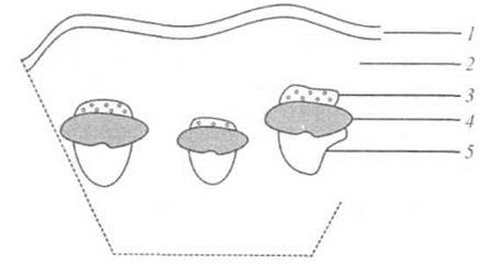
Fig. 6.20. The use of radioactive isotopes (14C) to study photoassimilate translocation in sugar beet Beta vulgaris petioles: 1 — epidermis; 2 — parenchyma; 3 — fiber; 4 — phloem; 5 — xylem.
1. What are the methods for measuring water potential and its components?
2. What drives the flow of water and solutes within a cell?
3. What is surface tension?
4. What is meant by excess pressure?
5. Write out and explain the Laplace formula.
6. What is the wilting point?
7. Characterize the pathways of water transport through the plant root system.
8. Explain the mechanisms of xylem transport and phloem translocation.
9. Explain the main methods for monitoring mineral nutrient uptake by plants.
Last update: 07/08/2026
Editorial and Educational Adaptation: This material has been compiled based on the primary/original source text. The project team performed an editorial review, corrected technical inaccuracies, structured sections, and adapted the content for an educational format.
What was processed:
- elimination of formatting defects (OCR errors, structural breaks, corrupted characters);
- editorial organization of content;
- standardization of terminology in accordance with academic sources;
- verification of factual statements against the original source text.
All mentions of the author, publication year, and origin of the primary text have been preserved in accordance with the source.