General Microbiology - Schlegel, H. 1987
Fundamental mechanisms of metabolism and energy conversion
Nutrient uptake by cells
For an exogenous substrate to be utilized by a Cell, it must cross its boundary layers. While The Cell wall does not present a significant barrier to small molecules and ions, it retains macromolecules with a molecular weight exceeding 600 Da. The boundary layer responsible for The transport of nutrients into The Cell is The Plasma Membrane.
The transport of nutrients across the plasma membrane is generally specific: only those substances for which a corresponding transport system exists can be taken up. With few exceptions, transport depends on the presence of specific permeases or translocases. These are Membrane Proteins whose very name indicates that they possess enzymatic properties—that is, they can be induced by the substrate, are substrate-specific, and are synthesized only under conditions where Protein Synthesis is possible.
Regarding The Mechanism of substance transport, a distinction is made among several different processes. Two of these are capable of mediating only transport rather than the intracellular accumulation of substances; in contrast, Active Transport processes lead to the accumulation of substances inside the cell (Figs. 7.18 and 7.19).
Simple diffusion. The nonspecific entry of substances into the cell occurs via passive diffusion. Molecular size and the degree of lipophilicity are crucial for diffusion. The rate of transport via diffusion is low. Such processes have not been detected for sugars and are highly unlikely. Poisons, inhibitors, and other foreign substances apparently enter the cell through simple diffusion.
Facilitated Diffusion. In facilitated diffusion, a nutrient present in the medium is transported into the cell "down" its concentration gradient. This process is driven by a substrate-specific permease and requires no metabolic Energy Expenditure. Over a wide range, the transport rate depends on the Substrate Concentration in the medium (Fig. 7.19). Nutrients cannot accumulate within the cell against a concentration gradient.
Class="center">
Fig. 7.18. Schematic representation of four Mechanisms of Substance transport into the cell. Pink circle: transported substrate; c: permease (carrier protein); c with a gray rectangle: energized carrier; PEP: phosphoenolpyruvate; HPr: heat-stable protein. Explanations in the text.
Active transport. Active transport and group translocation share with facilitated diffusion the characteristic that these processes involve substrate-specific transport proteins. However, unlike facilitated diffusion, this type of transport requires energy expenditure. Utilizing metabolic energy, a substance can accumulate inside the cell against a concentration gradient. The primary difference between active transport and group translocation lies in The Nature of the molecule entering the cell.

Fig. 7.19. Saturation curves for the uptake of two substrates by intact bacterial Cells (plotted from O2 consumption data [Respiration rate]). Active and passive substrate uptake can be distinguished by the shape of the curve. Because substrate A is taken up via active transport and accumulates in the cell, respiration reaches its maximum level even at very low substrate concentrations. Substrate B is taken up passively, and the respiration rate reaches a maximum only at a relatively high substrate concentration (on the order of 10-20 mM/L).
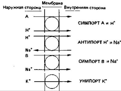
Fig. 7.20. Various types of active transport driven by the proton motive force Δp.
During active transport, the exact same molecule that was taken up from the nutrient medium enters the Cytoplasm. During group translocation, the transported molecule is chemically altered during transport, for example, by being phosphorylated.
All theories explaining active transport incorporate METABOLISM/2.html">THE CONCEPT OF specific transport proteins residing in the membrane. These proteins have been given names that reflect their function: permeases, translocases, transporter proteins, and carriers. Transport processes differ from one another primarily in their energy source—the proton motive force Δp (Fig. 7.20), ATP, or phosphoenolpyruvate (Fig. 7.18).
The energy of the proton motive force is utilized for the transport of numerous substances, including inorganic and organic ions as well as sugars (see pp. 243-244). Bacterial cells maintain the proton motive force by continuously pumping protons and other ions (such as Na+) out of the cell. Specific transport proteins in the membrane serve this purpose.
Each of these proteins has a strictly defined function. For example, There is a protein that catalyzes the simultaneous, unidirectional transport of a single proton and a single sugar molecule (lactose, melibiose, glucose). Such cases are referred to as the symport of two (or more) substances. Other transport proteins catalyze the simultaneous counter-transport of two particles, such as a proton and another ion (Na+ or an organic acid anion); these cases are known as antiport. Sugar transport coupled with ion transport presumably always utilizes H+ or Na+ ions. In prokaryotes, symport with H+ ions predominates, whereas in eukaryotes, symport with Na+ is more common (Fig. 7.20).
The actual existence of transport proteins of this type in Bacteria has been confirmed (a) by purifying and subsequently reconstituting the carrier protein into protoplasts or so-called Liposomes, and (b) by isolating mutants lacking the respective protein and its specific function. Regarding transport driven by the proton motive force, this is likely the most widespread mechanism of active substrate uptake.
The concept that specific carrier proteins participate in ion transport is supported by data on the action of various Antibiotics and synthetic compounds known as ionophores. These are relatively low-molecular-weight compounds (500–2000) whose molecules are hydrophobic on the outside and hydrophilic on the inside. Due to their hydrophobic properties, they diffuse into the lipid membrane. Among the antibiotic ionophores, valinomycin is the best known; it diffuses into the membrane and catalyzes the transport (uniport) of K+, Cs+, Rb+, or NH4+ ions. Consequently, the presence of such cations in the suspension medium leads to charge equalization across both sides of the membrane (effectively causing a short circuit) and, consequently, a collapse of the proton motive force. Other ionophores form channels through which ions can pass. There are also synthetic compounds that increase the proton conductance of membranes; the most prominent proton carrier is carbonyl cyanide m-chlorophenyl hydrazone (or carbonylcyanide-p-trifluoromethoxyphenylhydrazone). It acts as an uncoupler—disrupting the coupling of ATP synthesis with electron transport by translocating protons into the cell bypassing ATP synthase. Studies of membrane transport have yielded significant results that are consistent with and reinforce the chemiosmotic theory of energy conversion.
Alongside transport systems that utilize the proton motive force, ATP-dependent systems also exist. Periplasmic binding proteins play a specific role here (Fig. 2.28). The plasma membrane of animal cells neither transports protons nor generates a proton gradient. The Membrane Potential is presumably maintained exclusively by ATP-driven pump mechanisms, such as the sodium-potassium pump, while the sodium gradient, in turn, provides the energy for the symport of nutrients alongside Na+ ions.
Group translocation. In this type of transport, the molecule is chemically modified; for instance, while a sugar is taken up as such, it enters the cell in a phosphorylated form. Fructose, glucose, mannitol, and related substances are taken up via the phosphoenolpyruvate-dependent phosphotransferase system. This system consists of non-specific and specific components. The non-specific component is a heat-stable protein that is phosphorylated by phosphoenolpyruvate with the participation of enzyme I located in the cytoplasm. The second component is a membrane-bound, inducible enzyme II specific to a given sugar; it catalyzes the Transfer of phosphate from the heat-stable protein (HPr) to the sugar during the latter's Transport Across the membrane:

Enzyme II likely Functions simultaneously as both a permease and a phosphotransferase (see Fig. 7.18).
In other respects, the cellular uptake of substances is a highly complex process that remains poorly understood. Many metabolic inhibition effects and competition phenomena observed among simultaneously available substrates are presumably related to the Specific features of regulatory mechanisms that already manifest during transport processes.
Efflux of substances from the cell. Considerably less is known about the release of metabolites into the environment than about the mechanisms of cellular uptake. Apparently, their export from the cell also occurs both via transport systems and through uncontrolled diffusion. Substances leave the cell when, As a result of overproduction, they accumulate within it and reach concentrations exceeding normal levels. Such accumulation may result from incomplete oxidation, regulatory disruption, or Fermentation processes.
Iron transport. Microbial cells possess a specialized mechanism for the transport of this macronutrient. Under anaerobic conditions, iron exists as the divalent ion (Fe2+) and can reach concentrations of up to 10-1 M/L, thus not limiting Microbial growth. However, under aerobic conditions at pH 7.0, iron occurs as an almost insoluble Fe3+ hydroxide complex, resulting in a ferric ion concentration of merely 10-18 M/L. It is hardly surprising, therefore, that microorganisms excrete substances that convert iron into a soluble form. These substances—known as siderophores—bind Fe3+ ions into a complex and transport them in this form; they are primarily low-molecular-weight, Water-soluble compounds (with a molecular weight under 1500) that bind iron via coordination bonds with high Specificity and a high affinity (stability constants on the order of 1030). Chemically, these can be phenolates or hydroxamates. Enterochelin belongs to the former group; it possesses six phenolic hydroxyl groups and is secreted by certain enterobacteria. Upon release into the environment, it binds iron, and the resulting ferric enterochelin is taken up by the cell. Inside the cell, the iron is released via the Enzymatic Hydrolysis of ferric enterochelin (Fig. 7.21).
For the same purpose, many Fungi produce ferrichromes, which are classified as hydroxamate siderophores. These are cyclic hexapeptides that bind ferric iron using three hydroxamate groups. Like enterobactin, they are secreted from The Cell as iron-free compounds, bind iron in the nutrient medium, and are reabsorbed as ferrichromes. Inside the cell, iron is reduced to Fe2+, for which ferrichromes have very low affinity, thereby releasing it. Similar functions are performed by ferrioxamines (in actinomycetes), mycobactins (in mycobacteria), and exochelins (also in mycobacteria).

Fig. 7.21. Examples of iron transport mechanisms into microbial cells involving siderophores. Top: the enterobactin transport system typical of many bacteria; bottom: the ferrichrome system found in many fungi.
Microorganisms typically secrete siderophores into the nutrient medium only when iron is growth-limiting. Siderophore secretion is a consequence of the derepression of their synthesis. In the presence of dissolved, complexed iron, siderophores are synthesized in only small amounts and remain retained within the cell wall. Under these conditions, they function solely to transport iron into the cell.
Interestingly, in this context, one of the natural defense mechanisms of higher organisms is the "depletion" of iron from their internal environment. Specialized proteins bind available iron so tightly that it becomes inaccessible to microorganisms. For instance, chicken egg white contains conalbumin, milk, tear fluid, and saliva contain lactotransferrin, and Blood serum contains serotransferrin. When bacteria are inoculated onto egg white, they grow only if iron ions (in the form of citrate) are added simultaneously with the inoculum. Thus, iron plays a crucial role in the antagonistic relationships between higher organisms and bacteria. The winner of this competition is the partner that produces the substance with a higher affinity for iron.
Last update: 13/08/2026
Editorial and Educational Adaptation: This material has been compiled based on the primary/original source text. The project team performed an editorial review, corrected technical inaccuracies, structured sections, and adapted the content for an educational format.
What was processed:
- elimination of formatting defects (OCR errors, structural breaks, corrupted characters);
- editorial organization of content;
- standardization of terminology in accordance with academic sources;
- verification of factual statements against the original source text.
All mentions of the author, publication year, and origin of the primary text have been preserved in accordance with the source.