BIOLOGY Volume 1 - A Guide to General Biology - 2004
8. HETEROTROPHIC NUTRITION
8.3. The Human Alimentary Canal
8.3.7. The Small Intestine
The first section of the Small Intestine is called the duodenum, which is about 25 cm long. It receives ducts from the Pancreas and the gall bladder. The duodenum leads into the ileum, which is approximately 3 m long in a living person (it relaxes and elongates after death) (Figs. 8.11 and 8.21). The submucosa and mucosa are folded (Fig. 8.21, A). In addition, the mucous membrane features numerous finger-like projections known as villi. The walls of the villi are richly supplied with Blood and Lymphatic capillaries and also contain smooth Muscle fibers (Fig. 8.21, D, E). The villi constantly contract and relax, thereby maintaining close contact with the food inside the small intestine. The free surfaces of the epithelial Cells on the villi are covered with microscopic microvilli (Fig. 8.21, F; 6.16, and section 5.10.8). These microvilli significantly increase the surface area of the small intestine (Table 8.2).
Class="center">Table 8.2. Structural features that increase the surface area of the small intestine
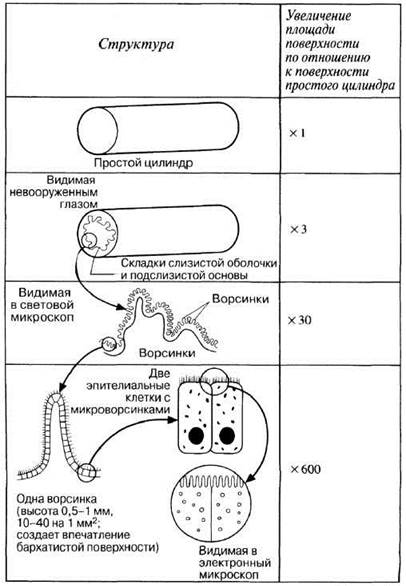

Fig. 8.21. A. Vertical section through the ileum. B. Crypts of Lieberkühn of the ileum. C. Diagrammatic cross-section of the small intestine. D. Diagram of a cross-section of the ileum showing The Structure of a villus. E. Photomicrograph of villi On the surface of the small intestine (x200). F. Electron micrograph of epithelial cells showing microvilli (x18,000).
8.4. a) Describe the Structural Features of the small intestine that increase its surface area.
b) What are the advantages of such a structure?
Between the villi are long, tubular depressions called crypts of Lieberkühn (Fig. 8.21, D). It is here that new epithelial cells are produced to replace the constantly sloughed-off Cells of the villi (the average lifespan of these cells is about five days). In addition, the cells of the crypts secrete succus entericus (intestinal juice), a mildly alkaline fluid containing Water and mucus that helps increase the volume of the intestinal contents. Paneth cells, located at the Base of the crypts, secrete Lysozyme—an antibacterial enzyme previously mentioned in the Discussion of saliva.
Throughout the length of the small intestine are specialized epithelial cells known as goblet cells; these cells secrete mucus, the Functions of which were discussed in section 8.3.1 (see the Description of the mucous membrane). The duodenum also secretes an alkaline fluid that neutralizes the acid from the gastric juice and maintains a pH of 7–8, which is optimal for the functioning of small intestinal Enzymes.
8.5. What would happen to The activity of intestinal enzymes if the pH in the small intestine remained at 2?
Last update: 06/08/2026
Editorial and Educational Adaptation: This material has been compiled based on the primary/original source text. The project team performed an editorial review, corrected technical inaccuracies, structured sections, and adapted the content for an educational format.
What was processed:
- elimination of formatting defects (OCR errors, structural breaks, corrupted characters);
- editorial organization of content;
- standardization of terminology in accordance with academic sources;
- verification of factual statements against the original source text.
All mentions of the author, publication year, and origin of the primary text have been preserved in accordance with the source.