Protein Structure and Function: Applications of Bioinformatics Methods - John Rigden 2014
Protein Dynamics: From Structure to Function
Molecular Dynamics Calculations
Appendices
Structure/8.html">Molecular Dynamics simulations have become a standard methodology in protein research and are routinely applied to a wide range of problems. However, protein conformational dynamics remains a challenging task for MD simulations because functionally significant conformational transitions often occur on timescales ranging from microseconds to seconds, which exceeds the limits of publicly available computational algorithms and power.
Class="center">9.1.2.1. Nuclear Transport Receptors
Despite their high demand for computational resources, MD simulations have been successfully employed to investigate protein function. To illustrate this, we will discuss in some detail a recent study by Zachariae and Grubmüller (2006), which revealed a remarkably fast conformational transition of the exportin CAS (Cselp in Yeast) between its open and closed states. The CAS/Cselp protein is a 960-amino-acid nuclear transport receptor that binds importin-a and the RanGTP protein in The Nucleus. The heterotrimeric complex (Fig. 9.1) can pass through nuclear pores and dissociates via catalytic GTP Hydrolysis in the Cytoplasm, thereby representing a vital part of the nucleocytoplasmic transport cycle in Cells.
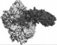
Fig. 9.1. (For the color version of the figure, see the insert.) The heterotrimeric complex of Cselp (shown in cyan), RanGTP (shown in yellow), and importin-a (shown in red). Cselp adopts a superhelical conformation and binds RanGTP and importin-a. The complex can penetrate through nuclear pores and dissociates via catalytic GTP hydrolysis in the cytoplasm
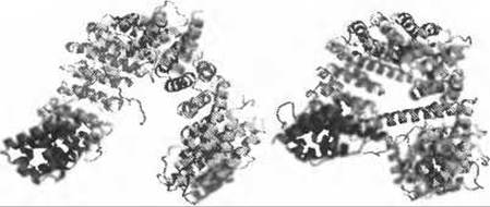
Fig. 9.2. (For the color version of the figure, see the insert.) Nucleoplasmic (left) and cytoplasmic (right) forms of Cselp. In the nucleoplasmic form, Cselp is bound to RanGTP and importin-a (both not shown). Upon dissociation in the cytoplasm, Cselp undergoes major conformational changes and forms a ring-like conformation that closes the RanGTP-binding site and prevents complex re-formation. Structures are colored According to the spectrum from cyan (N-terminus) to red (C-terminus)
For the importin-a/CAS system to function properly, it is required that upon complex dissociation in the cytoplasm, the CAS/Cselp protein undergoes a large conformational change that would prevent the re-Formation of the complex. The X-ray STRUCTURE OF THE Cselp protein demonstrates that its loaded conformation possesses a superhelical structure, encompassing the bound RanGTP (Fig. 9.2, left), whereas the cytoplasmic form of this protein adopts a closed-ring conformation, resulting in the closure of the RanGTP-binding site (Fig. 9.2, right). To understand The Mechanism of this conformational switch, the authors performed an MD simulation of the Cselp protein starting from its loaded conformation. They found that, primarily driven by Electrostatic Interactions, the Cell/13.html">Protein Structure abruptly folds and relatively quickly, within 10 ns, adopts a conformation close to the experimentally observed cytoplasmic form. MD simulations of mutant protein forms with different surface electrostatic potential values revealed no significant Conformational Changes in them. Conversely, these forms retained an open conformation, which is in good agreement with experimental observations (Cook et al. 2005). This example shows that functionally significant conformational changes occurring on small timescales can be studied using MD. However, in this particular case, the simulation did not start from an equilibrium conformation because importin-a and RanGTP were removed, and therefore, overcoming a significant energy barrier to reach the closed conformation was presumably unnecessary. When a simulation starts from a Free energy minimum, as is usually the case, the attainable simulation times are frequently too short to overcome high energy barriers and, consequently, to observe functionally relevant conformational transitions. This is known as the sampling problem and represents a general challenge for all MD simulations.
9.1.2.2. Lysozyme
MD modeling of T4 bacteriophage lysozyme (T4L), an enzyme six times smaller than Cselp, clearly illustrates the sampling problem for relatively long MD trajectories. T4L has been extensively studied using X-ray crystallography (Faber and Matthews 1990; Kuroki et al. 1993) and, having been crystallized in various Conformations, represents one of the rare cases where information on functionally important states can be obtained at atomic resolution directly from experimental data (Zhang et al. 1995; de Groot et al. 1998). The domain architecture of this enzyme is pronounced (Matthews and Remington 1974), and based on differences between the crystallographic structures of various T4L mutant forms, it was suggested that the hinge-bending motion of T4L (Fig. 9.3) is an intrinsic feature of this molecule (Dixon et al. 1992).
Furthermore, domain fluctuations are predicted to be essential for enzyme function, allowing the substrate to enter the Active Site and products to leave it in the open conformation, whereas catalytic transformations proceed in the closed conformation.
The wealth of experimental data also provides an opportunity to evaluate the reliability of computational Methods and sampling efficiency. Two MD simulations were carried out using the closed (Simulation 1) and open (Simulation 2) conformations as starting points. To assess sampling efficiency, Principal Component Analysis (PCA, see Section 9.2 below) of the ensemble of experimentally determined structures was performed, and this ensemble, along with the two MD trajectories, was projected onto the first two eigenvectors.
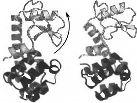
Fig. 9.3. Hinge-bending motion of T4 phage lysozyme. Domain fluctuations (domains are shown in different colors) are essential for the enzyme function, allowing the substrate to enter the active site in the open conformation and the products to leave it
The first eigenvector corresponds to the hinge-bending motion, whereas the second eigenvector corresponds to the twisting of the T4L domains. The projections are shown in Fig. 9.4. The ensemble of X-ray structures is represented by points, each corresponding to a single conformation. Motion along the first eigenvector (abscissa axis) describes the collective transition from the closed to the open state. It is easily noticed that none of the MD trajectories represented by lines spans the entire conformational space containing the X-ray structure ensemble by itself, although the simulation times (184 ns for run 1 and 117 ns for run 2) are an order of magnitude longer than the previously discussed MD simulation time of the Cselp protein. The phase space density suggests the existence of an energy barrier between the closed and open states, and neither simulation captures a complete transition from the closed state to the open state or vice versa.
9.1.2.3. Aquaporins
Aquaporins are a prime example of the contribution that MD simulations have made to understanding protein function from a dynamic and energetic perspective. Aquaporins facilitate the efficient and selective permeation of Water across Introduction/36.html">Biological Membranes. Related aquaglyceroporins additionally allow the passage of small neutral solute molecules such as glycerol. Available high-resolution structures have provided invaluable insight into the molecular mechanisms operating in aquaporins (Fu et al. 2000; Murata et al. 2000; de Groot et al. 2001; Sui et al. 2001). However, such structures provide predominantly static information, and thus we cannot directly observe aquaporins "at work." Moreover, there is still no experimental method providing sufficient spatial and temporal resolution to monitor permeation through aquaporins at THE MOLECULAR LEVEL. Thus, MD simulations Complement experimental results by depicting the motion of the biomolecular system with atomic resolution. Since permeation is known to occur on a nanosecond timescale, one can expect spontaneous permeation to take place during multi-nanosecond simulations, allowing direct observation of functional dynamics. For this reason, such simulations have been termed "real-time simulations" (de Groot and Grubmüller 2001).
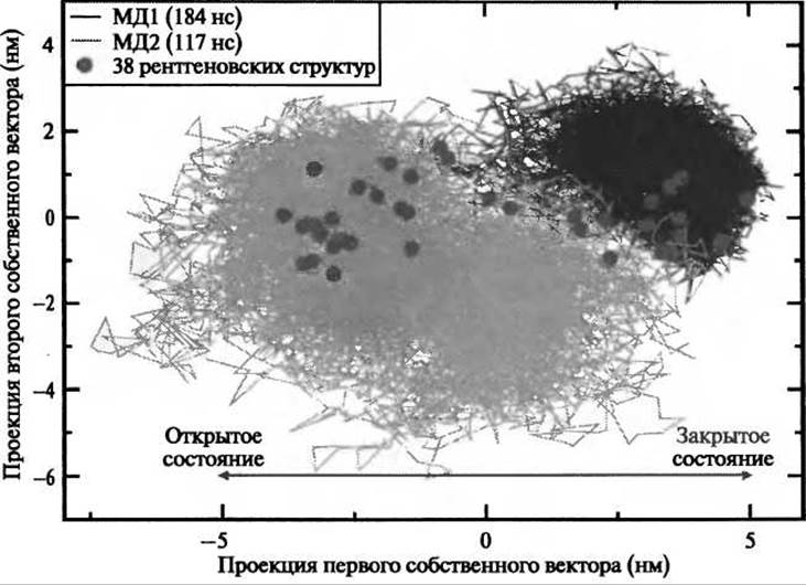
Fig. 9.4. Principal component analysis for T4 phage lysozyme. The ensemble of X-ray structures is shown by points, and MD trajectories by lines. Motion along the first eigenvector (abscissa axis) corresponds to the hinge-bending motion. Neither simulation 1, starting from the closed conformation, nor simulation 2, starting from the open conformation, demonstrates a complete transition due to the energy barrier separating the conformational states
Indeed, events of spontaneous permeation have been observed in MD simulations of aquaporin-1 and the aquaglyceroporin GlpF. These simulations revealed that the high water permeation efficiency is explained by the hydrogen-bonding complementarity inside the channel—comparable to that in bulk water—which results in a low energy barrier for this process (de Groot and Grubmüller 2001; Tajkhorshid et al. 2002). The simulations also clarified that the selectivity of these channels is governed by a two-stage filter. The First stage of the filter is located in the central part of the channel within the conserved asparagine-Proline-Alanine (NPA) region; the Second Stage is located at the extracellular surface of the channel within the aromatic-Arginine (ar/R) constriction region (Fig. 9.5). Since water permeation occurs on a nanosecond scale, permeation coefficients can be calculated directly from MD simulations and compared with experimental values. The quantitative agreement obtained from this comparison confirms the reliability of the simulations.
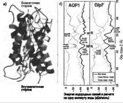
Fig. 9.5. (For a color version of this figure, see the insert.) a) Water molecules are highly ordered within the aquaporin-1 channel, with their dipole moments pointing away from the central NPA region (de Groot and Grubmüller 2001). Water dipoles (shown by yellow arrows) flip by approximately 180 degrees as they traverse the AQP1 channel. Red and blue indicate local electrostatic potentials, negative and positive, respectively, b) Hydrogen-bonding energy per water molecule (shown by black lines) in AQP1 (left) and GlpF (right). Protein-water Hydrogen Bonds (shown in green) compensate for the loss of water-water hydrogen bonds (shown in cyan). The main Protein-Water Interaction centers are the ar/R region and the NPA region
For a long time, the mechanism by which protons are excluded from the water pore remained an unresolved question in The Study of aquaporins. MD simulations focusing on water permeation revealed a distinct pattern of water dipole orientation within the channel, with a center of Symmetry at the NPA region (de Groot and Grubmüller 2001). It was found that during the dynamics, water molecules flip by 180 degrees along their pathway through the channel (Fig. 9.5a). In a series of simulations aimed at elucidating the proton exclusion mechanism, it was found that this orientation of water molecules is driven by an electric field within the channel centered at the NPA region (de Groot et al. 2003; Chakrabarti et al. 2004; Ilan et al. 2004). Thus, electrostatic effects provide the basis for proton exclusion. THE ORIGIN OF this electrostatic barrier remains a subject of debate, with both direct electrostatic effects caused by helix dipole moments (de Groot et al. 2003; Chakrabarti et al. 2004) and specific deshielding/desolvation effects having been proposed (Burykin and Warshel 2003). Recent results suggest that both effects make approximately equal contributions (Chen et al. 2006).
Furthermore, MD simulations have helped to clarify the mechanism of neutral solute selectivity in aquaporins and aquaglyceroporins. It was found that aquaporins are permeable only to small polar molecules like water, with the rare exception of the ammonium ion, whereas aquaglyceroporins are additionally permeable to nonpolar molecules like CO2 and larger molecules like glycerol, but not to urea (Hub and de Groot 2008). For aquaporins, an inverse relationship was found between solute permeation ability and its Hydrophobicity—molecules that compete with permeating water molecules for hydrogen bonding with the channel impose a permeation barrier. Thus, size and hydrophobicity Selection forms The basis of selectivity in aquaporins and aquaglyceroporins.
Last update: 06/08/2026
Editorial and Educational Adaptation: This material has been compiled based on the primary/original source text. The project team performed an editorial review, corrected technical inaccuracies, structured sections, and adapted the content for an educational format.
What was processed:
- elimination of formatting defects (OCR errors, structural breaks, corrupted characters);
- editorial organization of content;
- standardization of terminology in accordance with academic sources;
- verification of factual statements against the original source text.
All mentions of the author, publication year, and origin of the primary text have been preserved in accordance with the source.