Protein Structure and Function: Application of Bioinformatics Methods - John Rigden 2014
Protein Dynamics: From Structure to Function
Principal Component Analysis
Principal component analysis (PCA) is a well-established technique for obtaining low-dimensional descriptions of high-dimensional data. Its Applications include data compression, Image Processing, data visualization, scientific Data analysis, pattern recognition, and time series prediction (Duda et al. 2001). In the context of biomolecular calculations, PCA has become an essential tool for extracting and classifying meaningful information regarding large Conformational Changes in protein structural ensembles obtained experimentally or theoretically (Garda 1992; Go- et al. 1983; Amadei et al. 1993). In addition to PCA, A number of other Structure/131.html">Similar Methods are currently in use, among which Normal Mode Analysis (NMA) (Brooks and Karplus 1983; Go et al. 1983; Levitt et al. 1983), quasi-harmonic analysis (Karplus and Kushick 1981; Levy et al. 1984a, b; Teeter and Case 1990), and singular value decomposition (Romo et al. 1995; Bahar et al. 1997) are particularly noteworthy.
Principal component analysis is based on the observation that the vast majority of spatial fluctuations in Proteins occur along a small number of axes associated with collective degrees of freedom. This was first recognized during the analysis of normal vibrations in small proteins (Brooks and Karplus 1983; Go- et al. 1983; Levitt et al. 1983). In such an analysis (see Section 9.4.1), the potential energy surface is assumed to be harmonic, and the collective variables are determined by diagonalizing the Hessian matrix1 at a local energy minimum. Quasi-harmonic analysis, principal component analysis, and singular value decomposition of Molecular Dynamics trajectories, which do not assume harmonicity of vibrations, have demonstrated that the potential energy fluctuations during dynamics are indeed dominated by a limited number of collective coordinates, with the principal modes frequently exhibiting strong anharmonicity. These methods have made it possible to identify the collective degrees of freedom that best approximate all observed vibrations.
The variables that undergo The most significant changes form a set of generalized internal coordinates that can be employed to efficiently describe protein dynamics. Often, utilizing 5–10% of the total degrees of freedom yields a surprisingly accurate approximation. Unlike internal coordinates represented in the form of torsional angles, these collective internal coordinates are not known a priori; rather, they must be determined using either experimental structures or an ensemble of model structures. Once these collective degrees of freedom are found, this information can be utilized to analyze calculations and to design improved dynamics protocols aimed at enhancing conformational sampling (Grubmüller 1995; Zhang et al. 2003; He et al. 2003; Amadei et al. 1996).
1 The matrix of second derivatives of the potential energy d2V/dxidxj. Author's note.
Class="center">
Fig. 9.7. Illustration of the principal component analysis (PCA) method for a two-dimensional case. Two coordinates (x, y) are required to determine THE POSITION OF a point from the ensemble (b), whereas a single coordinate x' is sufficient for an approximate position determination (a)
Essentially, principal component analysis is a multi-dimensional least-squares method in configuration space. An ensemble of structures of a molecule with N atoms can be represented in a 3N-dimensional configuration space as a cloud of points, where each configuration is represented by a single point. For such a cloud, an axis along which the maximum dispersion of points is observed can always be defined. As shown for the two-dimensional case (Fig. 9.7), if such a line fits the data well, the position of each point can be approximated solely by its projection onto this axis, yielding a reasonable approximation even when projections onto the remaining directions perpendicular to this axis are discarded. If this axis is chosen as a coordinate axis, the position of the point can be described by a single coordinate. In the general 314-dimensional case, the Procedure is carried out analogously.
Knowing the axis that best describes the data as a first approximation, one can choose orthogonal directions for a second approximation, a third approximation, and so on (the principal components). Together, these directions span the 314-dimensional space. Mathematically, these directions are defined by the eigenvectors μi of the atomic fluctuation covariance matrix

where the angle brackets <•> denote ensemble averaging. The eigenvalues λi correspond to the mean-square spatial fluctuations along the respective eigenvectors and, thus, indicate THE CONTRIBUTION OF each principal component to the overall fluctuation (Fig. 9.8)
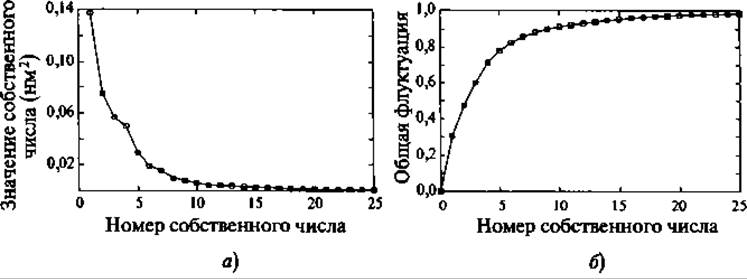
Fig. 9.8. Typical eigenvalue spectrum in the principal component analysis method (MD ensemble of backbone structures of guanylin). The first five eigenvectors (panel a) cover 80% of all observed fluctuations (panel b)
The application of such multi-dimensional fitting Procedures to protein configurations from MD calculations has demonstrated in several Examples that typically the first 10–20 principal components account for 90% of the protein fluctuations (Kitao et al. 1991; Garda 1992; Amadei et al. 1993). These principal components correspond to collective coordinates receiving contributions from every atom of the protein. In a number of cases, it has been shown that these principal modes form part of the functional dynamics of the studied proteins (Amadei et al. 1993; Van Aalten et al. 1995a, b; de Groot et al. 1998). For this reason, the subspace responsible for the majority of fluctuations has been termed the essential subspace (Amadei et al. 1993).
The fact that a small fraction of the total degrees of freedom (the essential subspace) dominates protein molecular dynamics arises from the large number of internal constraints imposed by atomic interactions within the biomolecule. These interactions include both strong covalent bonds and weak non-covalent interactions, while the constraints are dictated by the dense packing of atoms in the native structure.
In general, protein dynamics at physiological temperatures has been described as diffusion among a multitude of minima (Kitao et al. 1998; Amadei et al. 1999; Kitao and Go- 1999). Short-time dynamics are dominated by vibrations near the local minimum, corresponding to eigenvectors with low eigenvalues. Long-time large-scale fluctuations are dominated by anharmonic diffusion between numerous potential wells. Such slow dynamic transitions are typically represented in principal component analysis by modes with large amplitudes. Unlike normal mode analysis, the application of principal component analysis to an MD trajectory does not rely on the assumption of a harmonic potential. In fact, principal component analysis can be used to investigate the degree of anharmonicity in The Molecular Dynamics of the simulated system. It has been demonstrated that for proteins at physiological Temperature, the principal modes of collective fluctuations, which are frequently functionally significant, are dominated by anharmonic fluctuations (Amadei et al. 1993; Hayward et al. 1995).
Last update: 06/08/2026
Editorial and Educational Adaptation: This material has been compiled based on the primary/original source text. The project team performed an editorial review, corrected technical inaccuracies, structured sections, and adapted the content for an educational format.
What was processed:
- elimination of formatting defects (OCR errors, structural breaks, corrupted characters);
- editorial organization of content;
- standardization of terminology in accordance with academic sources;
- verification of factual statements against the original source text.
All mentions of the author, publication year, and origin of the primary text have been preserved in accordance with the source.