Biochemistry - The Chemical Reactions of Living Cells Volume 3 - D. Metzler 1980
Cell Growth, Differentiation, and Chemical Communication
Neurochemistry
Neural Pathways and Systems
Imagine that a nerve receptor in the Skin or another sensory organ perceives a signal. This signal travels along a sensory neuron (afferent fiber) up to the Brain. After passing through two or more synapses (typically one in the Spinal Cord and one in the thalamus), the signal eventually reaches a specific sensory area of the Cerebral Cortex. From here, in a modified form, it spreads via interneurons across virtually the entire cortex. Both in the synapses and in the cortex, the Propagation of the signal activates inhibitory Neurons that dampen impulses in adjacent fibers. Moreover, if the impulse triggered by the signal is not sufficiently strong, it fades out before ever reaching the cerebral cortex. Among the most crucial sensory neurons are the fibers originating from the light-sensitive Cells of the eye (7 million cones and 100 million rods). Nerve signals exit the retina via millions of ganglion Cell axons and project, alongside other brain regions, to the visual cortex (Fig. 16-5).
Class="center">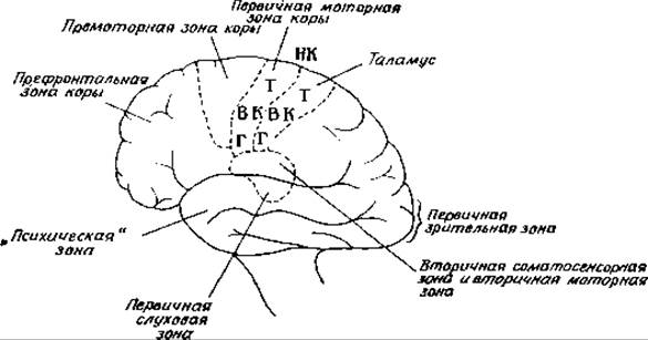
FIG. 16-5. Arrangement of A number of functional areas of the cerebral cortex. The primary motor and somatic sensory areas of the cortex show which body parts they control. Abbreviations: H – HEAD, BK – upper limbs, NK – lower limbs, T – trunk ([26a], p. 193).
The processes occurring in the cerebral cortex are extremely complex and remain insufficiently understood. We still do not know precisely how the brain initiates voluntary Muscle movements. It has been established, however, that signals emerging from the brain toward the Muscles via efferent fibers are generated in large motor neurons of the motor cortex; this area is organized as a band spanning the entire brain and adjacent to the sensory area (Fig. 16-5). The axons of motor neurons form the pyramidal tract, which conducts impulses downward to synapses in the spinal cord and from there to neuromuscular junctions. The latter are specialized synapses where Acetylcholine is released, transmitting the signal directly to muscle fibers. A depolarization wave sweeping across The Cell surface and T-tubules (Ch. 4, Sec. E, 1; Fig. 4-22, A) triggers calcium release and Muscle contraction.
While motor neurons send the primary signal to the muscles, excitation also spreads to other PARTS OF THE brain, including the olive, which sends a signal to the Cerebellum. The cerebellum Functions much like a computer, fine-tuning the impulses delivered to the muscles. Damage to the cerebellum disrupts fine motor coordination. The Influence of the cerebellum, transmitted via Purkinje cells, is always inhibitory. Within the cerebellar nuclei, Purkinje cells form synapses with neurons that Relay impulses back to the cerebral cortex, the thalamus, and downward into the spinal cord. This pathway to the cortex completes a feedback inhibition loop—a phenomenon widely prevalent throughout The Nervous system.
In addition to the somatic motor system, which regulates the movements of voluntary (skeletal) muscles via the pyramidal tract, there is also the Autonomic nervous system, which controls the function of involuntary (smooth) muscles, glands, as well as Heart rate, Blood pressure, and body Temperature. The highest centers of the autonomic nervous system are located in the CEREBRAL CORTEX AND the Hypothalamus. The autonomic nervous system is subdivided into the sympathetic and parasympathetic divisions. Fight-or-flight responses are mediated by the sympathetic system. Its postganglionic fibers (originating from sympathetic ganglia) release noradrenaline (norepinephrine); The adrenal medulla, composed of specialized neurons known as chromaffin cells, also belongs to the sympathetic system. The parasympathetic system is more closely associated with maintaining Homeostasis and regulating the functions of various body systems. Biochemically, this system is characterized by the release of acetylcholine as its neurotransmitter.
The hypothalamus—a Structure weighing merely 4 g—attracts significant attention from biochemists because, as the highest center of the autonomic nervous system, it plays a major role in maintaining homeostasis and regulating the secretory activity of Endocrine glands. We have already mentioned that the hypothalamus produces neurohormones that stimulate pituitary function (Sec. A). In addition, the hypothalamus is involved in regulating body temperature, Water balance, and presumably blood glucose concentration.
There are two other important systems in the brain: the Reticular Formation and the limbic system. The former mediates the Sleep-wake cycle and also determines the appearance of characteristic waves on the Electroencephalogram. The limbic system mediates emotional states as well as instincts; its Anatomical Structure is complex, with centers located in the amygdala and other subcortical nuclei, as well as in the limbic gyrus of the cortex. The latter formation is arranged in a ring, mainly within the longitudinal fissure between the two cerebral hemispheres, and includes the olfactory area, the hippocampus, and other evolutionarily ancient Regions of the cerebral cortex. Pleasure centers are located within the limbic gyrus. An animal with electrodes implanted in these centers will continuously press levers to trigger electrical stimulation of these areas. There are also punishment centers, the repeated stimulation of which the animal strives to avoid.
Last update: 06/08/2026
Editorial and Educational Adaptation: This material has been compiled based on the primary/original source text. The project team performed an editorial review, corrected technical inaccuracies, structured sections, and adapted the content for an educational format.
What was processed:
- elimination of formatting defects (OCR errors, structural breaks, corrupted characters);
- editorial organization of content;
- standardization of terminology in accordance with academic sources;
- verification of factual statements against the original source text.
All mentions of the author, publication year, and origin of the primary text have been preserved in accordance with the source.