BIOCHEMISTRY - Laboratory Manual - NAU 2015
MODULE II
ENZYMES AND METABOLIC PATHWAYS.
Laboratory Work 9
EXPERIMENTAL VERIFICATION OF RESPIRATORY CHAIN FUNCTIONING
Objective: to isolate Cell/35.html">Mitochondria and the submitochondrial fraction containing Respiratory Chain Enzymes, identify individual Components of the respiratory chain, and investigate their functioning.
Basic Theoretical Background
The Enzymes of the respiratory chain (RC) are localized on The inner mitochondrial membrane. Therefore, to study RC functioning in laboratory experiments, it is first necessary to obtain mitochondria. This is achieved by tissue mincing, disruption, and differential centrifugation. The isolation of individual enzyme Proteins and complexes is performed by extraction with Buffer solutions. The respiratory chain ensures The transfer of reducing equivalents (hydrogen atoms and electrons) to oxygen, with the final reaction resulting in The formation of Water. Thus, the RC provides tissue Respiration—the uptake of oxygen by Tissues. Driven by The energy released during The transport of reducing equivalents along the RC, Oxidative Phosphorylation takes place—the synthesis of ATP from ADP and inorganic phosphate.
The process of electron transfer along the RC is convenient to study using submitochondrial fragments. The Keilin-Hartrie preparation consists of fragments of the inner mitochondrial membrane almost devoid of Krebs cycle enzymes, yet retaining a full Complement of RC components. To obtain this preparation, the tissue is disrupted by grinding with abrasive Materials, followed by differential centrifugation and extraction with salt buffer solutions.
The ability of mitochondria to consume oxygen characterizes their metabolic intensity. The manometric method proposed by O. Warburg can be used to study processes that occur with oxygen uptake—Oxidative Deamination, aerobic Glycolysis, RC functioning—as well as processes accompanied by the release of gaseous products. By determining the pressure change in a closed system of known volume during specific reactions, the volume of the gaseous product formed or consumed is calculated.
The polarographic METHOD FOR DETERMINING oxygen concentration is based on the electrochemical reduction reaction of dissolved oxygen to hydrogen peroxide and water:
Class="center">О2 + 2 ĕ + 2 Н+ → Н2О2
О2 + 4 ĕ + 4 Н+ → 2 Н2О
Under certain conditions, the electric current is linearly dependent on oxygen concentration, making it possible to determine the respiration of biological objects—tissues, Suspensions of cellular Organelles, including mitochondria. Most commonly, a Clark electrode is used, which is made of platinum separated from the reaction mixture by a thin Teflon membrane. The electrode is filled with a saturated KCl solution. Oxygen from the reaction mixture diffuses into this solution, where the electrochemical reduction reaction takes place.
The acceptor of hydrogen atoms from NADH+H+ in the RC is flavin enzymes, which contain a prosthetic group derived from vitamin B2 (riboflavin)—flavin mononucleotide (FMN). The prosthetic group of the enzyme succinate dehydrogenase is flavin adenine dinucleotide (FAD). FMN and FAD as parts of enzymes participate in reversible oxidation-reduction reactions:
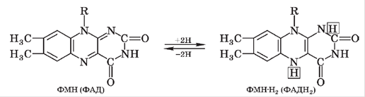
The oxidized forms of FMN and FAD have a red, brown, or green color, and therefore these RC components can be detected by Light absorption.
The cytochrome system participates in electron transfer along the RC. The prosthetic group of Cytochromes is heme. During RC functioning, the valence of the metal contained within the cytochrome heme changes (Fe2+ ↔ Fe3+, Сu+ ↔ Cu2+), with the cytochrome accepting or donating a single electron. At least two cytochrome chains operate simultaneously in The Cell. All cytochromes, especially in their reduced form, exhibit characteristic light absorption spectra. Cytochromes b, c1, and c function as intermediate electron carriers, whereas cytochromes a and a3 (cytochrome c oxidase) act as the respiratory enzyme that directly interacts with oxygen. The oxidized form of cytochrome c oxidase accepts electrons from the reduced cytochrome, transitioning to a reduced form, which is subsequently reoxidized by molecular oxygen. The resulting active oxygen О2- combines with two protons to form a water molecule.
Cytochrome c oxidase is highly sensitive to cyanides, sulfides, azides, carbon monoxide, and other substances that interact with the metal within the heme moiety. The inhibition of cytochrome c oxidase causes tissue oxygen starvation associated with the inability to utilize oxygen as an electron and proton acceptor in the mitochondrial RC.
Equipment: test tubes, a test tube with a stopper and delivery tube, gauze, filter paper, pipettes, scissors, Petri dishes, beakers, ice, a propeller stirrer, a Glass homogenizer, a high-speed refrigerated centrifuge, a meat grinder, a manual press, a porcelain mortar with pestle, a fluorimeter.
9.1. Isolation of Mitochondria from Animal Liver
Materials and Reagents: animal liver, 0.25 M sucrose solution, isolation medium containing 0.25 M sucrose and 0.001 M EDTA (ethylenediaminetetraacetate), pH 7.4, 1 M HCl solution; 0.1 M HCl solution; 1 M KOH solution; 0.1 M KOH solution.
Procedure
The liver tissue is washed 2–3 times with a small amount of isolation medium pre-cooled to 0 °C. 15–20 g of tissue is minced with scissors in a Petri dish placed on ice. The minced tissue is transferred to a beaker with fresh isolation medium, washed again 2–3 times, allowing the mixture to settle each time, and the supernatant is carefully decanted. The tissue is homogenized for 30–40 s with 40 ml of isolation medium, and centrifuged for 10 min at 600 g and 0–2 °C to remove nuclei and unfragmented tissue pieces. To calculate g (centrifugal field), the formula is used
g = 1118 · 10-8 R N2,
where R is the radius measured from the rotor center to the point where the centrifugal field is determined (to the bottom of the test tube), cm; N is the rotor speed, rpm.
The supernatant is decanted and kept on ice; the pellet is homogenized for 20 s with 20 ml of isolation medium and centrifuged at 600 g for 10 min. The supernatants are pooled.
To sediment the mitochondria, the supernatant is centrifuged at 14,000 g for 10 min. The pellet is suspended in 0.5 ml of isolation medium. With gentle shaking, 40 ml of isolation medium is added in small portions, and the mitochondria are sedimented again at 14,000 g for 10 min. The pellet is suspended in 0.25 M sucrose and recentrifuged at 14,000 g for 10 min. The supernatant is decanted, 0.2–0.3 ml of 0.25 M sucrose is carefully layered onto the mitochondrial pellet, the upper loose layer of the pellet is washed off with gentle shaking, and the procedure is repeated 2–3 times. The dense mitochondrial pellet is thoroughly suspended in 0.4–0.5 ml of 0.25 M sucrose. The resulting thick mitochondrial suspension is kept in a test tube on ice.
9.2. Preparation of Submitochondrial Fragments
(Keilin–Hartree Preparation)
Materials and Reagents: beef Heart, phosphate-borate buffer, pH 7.4 (mixing equal volumes of 0.15 M H3BO3 and 0.15 M Na2HPO4); 0.1 M phosphate buffer, pH 7.4; 0.02 M phosphate buffer, pH 7.4; 1 M HCl solution; 0.1 M HCl solution; 1 M KOH solution; 0.1 M KOH solution.
Procedure
Either fresh or frozen heart tissue can be used for this preparation. The activity of NADH-CoQ reductase in the preparation from frozen heart decreases, whereas succinate dehydrogenase activity remains virtually unchanged.
The Heart is trimmed of fat, Blood Vessels, and Connective Tissue; 100 g of the tissue is chopped and minced in a meat grinder. It is then mixed with 1 l of tap water and stirred with a propeller stirrer for 20 min. The water is drained, and the sediment is filtered through 4 layers of cheesecloth and squeezed thoroughly. This step is repeated several times. All subsequent operations are carried out in a cold room or on ice using solutions pre-cooled to 4 °C. The mass is poured into 1 l of 0.1 M phosphate buffer and left for 2 h with continuous stirring. The extract is decanted, and the remaining tissue mass is washed twice with 0.5 l of distilled water each time, squeezed thoroughly (preferably using a manual press), and placed in a porcelain mortar. Then, 10 ml of 0.02 M phosphate buffer and 10 g of quartz sand are added for grinding. The mixture is ground with a pestle for 1.5 h until a homogeneous, paste-like consistency is obtained. Next, 25 ml of 0.02 M phosphate buffer is added, and the mixture is thoroughly stirred.
Treatment with phosphate buffer washes out the Krebs cycle enzymes and cytochrome c; therefore, high enzymatic activity of the preparation can only be achieved by adding cytochrome c to the incubation medium.
The resulting preparation is centrifuged under refrigeration for 30 min at 2,500 rpm. The supernatant is carefully decanted and centrifuged at 40,000 g for 1.5 h at a Temperature of
1 — 2 °C. The nearly transparent supernatant is discarded. To the pellet, 1 ml of phosphate-borate buffer is added, and it is suspended in a glass homogenizer at the lowest pestle rotation speed.
The resulting Keilin–Hartree preparation is stored in a test tube at 0 °C for several days without any noticeable loss of succinate dehydrogenase activity.
9.3. Detection of Flavin Coenzymes
Materials and Reagents: 1% solutions of flavin adenine dinucleotide and flavin mononucleotide.
Procedure
The oxidized forms of FAD and FMN exhibit ultraviolet-induced fluorescence, the intensity of which depends on their concentration. Reduced forms of flavins do not fluoresce.
Pipette 10 drops of 0.002% FAD solution into one test tube, and 10 drops of 0.002% FMN solution (prepared by diluting the 1% solutions) into a second test tube. Add 5 ml of water to each tube and mix by shaking. Irradiation of the tube contents with ultraviolet light induces fluorescence. Compare the fluorescence intensity of the two samples using a fluorimeter.
Add a spatula-tip amount of sodium sulfite powder (a reducing agent) to each test tube and observe the quenching of fluorescence.
9.4. Determination of Cytochrome c
Materials and Reagents: 1% cytochrome c solution, concentrated Hydrochloric acid, metallic zinc, 0.5 M ascorbic acid solution.
Procedure
A cytochrome c solution is red in color, but the color is lost upon reduction. Hydrogen gas generated during the reaction between concentrated hydrochloric acid and metallic zinc can be used as an extracellular reducing agent. Ascorbic acid can reduce cytochrome c both in vitro and in vivo.
Place a small portion of the cytochrome c solution into the first test tube. Into a second test tube equipped with a gas outlet tube, pour a few milliliters of concentrated hydrochloric acid and add a small piece of metallic zinc. Stopper the second tube and submerge the outer end of the delivery tube into the first test tube so that hydrogen bubbles pass through the cytochrome c solution. After a short time, the solution becomes noticeably paler (compare the color with a control cytochrome c solution). Upon standing, the cytochrome c solution gradually regains its original color due to oxidation by atmospheric oxygen. Pipette 1–2 ml of cytochrome c solution into a third test tube and add 3 ml of 0.5 M ascorbic acid solution. Observe the disappearance of the color.
9.5. Determination of Cytochrome Oxidase
Materials and Reagents: fresh Muscle tissue; reducing reagent: a mixture of equal volumes of 1% dimethyl-p-phenylenediamine solution, 1.5% sodium carbonate solution, and 1% alcoholic α-naphthol solution. The reagent must be dark brown without a pink tint and should be prepared in advance (1 hour before the class);
1% sodium azide solution.
Procedure
Cytochrome c oxidase is capable of oxidizing not only cytochromes in the presence of oxygen, but also certain Other Compounds, such as α-naphthol and N,N′-dimethyl-p-phenylenediamine (a reducing reagent). Their oxidation yields a colored product, indophenol blue:

Chop 5 g of fresh muscle tissue with scissors and grind it thoroughly in a mortar, adding 20 ml of water in portions. Filter the muscle homogenate through a double layer of cheesecloth and wash it repeatedly with water until the washings are colorless. Squeeze the colorless mass—containing cytochrome c oxidase, a complex of cytochromes, and some dehydrogenases—between sheets of filter paper. Divide the resulting paste into three portions: transfer one into a test tube, and leave the other two on the filter paper. Add 2 ml of water to the test tube containing the paste and boil the mixture in a water bath for 1 min. Cool the test tube, carefully decant the liquid, and transfer the muscle paste using a glass rod onto another sheet of filter paper. Apply 2–3 drops of the reducing reagent to the first portion of muscle paste (boiled) and the second portion (unboiled); moisten the third portion (unboiled) first with a 1% sodium azide solution, and after 3 min, apply 2–3 drops of the NADI reagent. Compare the coloration of the three samples.
Processing of Experimental Data
Construct a diagram showing the successive stages of Isolation of the mitochondrial fraction and submitochondrial fragments from animal tissues. Write out the oxidation-reduction reactions involving respiratory chain cytochromes. Write the formula of the heme moiety in cytochrome b.
Selection/5.html">Control Questions and Tasks
1. Name the intramitochondrial localization of the TCA cycle and Electron Transport Chain enzymes.
2. Why is it necessary to add a cytochrome c solution to the reaction mixture to detect dehydrogenase activity in submitochondrial fragments (the Keilin–Hartree preparation)?
3. Write out the transformations of FAD, FMN, NAD, and NADP during the oxidation-reduction reaction. Indicate the Regions of the molecules that undergo change.
4. Explain the Principle of the method for detecting cytochrome c in the test solution.
5. What protein-lipid complexes make up The electron transport chain?
6. Name the coupling sites of respiration and phosphorylation.
7. What determines The sequence of enzymes and carriers in the electron transport chain?
8. Which substances are Inhibitors of the electron transport chain?
9. Which substances uncouple the processes of respiration and oxidative phosphorylation?
10. Which substances are inhibitors of oxidative phosphorylation? Which enzyme do they affect?
11. Explain Mitchell's chemiosmotic theory.
References: [1; 4—7].
Last update: 06/08/2026
Editorial and Educational Adaptation: This material has been compiled based on the primary/original source text. The project team performed an editorial review, corrected technical inaccuracies, structured sections, and adapted the content for an educational format.
What was processed:
- elimination of formatting defects (OCR errors, structural breaks, corrupted characters);
- editorial organization of content;
- standardization of terminology in accordance with academic sources;
- verification of factual statements against the original source text.
All mentions of the author, publication year, and origin of the primary text have been preserved in accordance with the source.