Chemistry and Biology of Proteins - F. Haurowitz 1953
Protein-water interactions
Proteins at interfaces
Proteins are surface-active agents, and their concentration at The surface of an aqueous solution is higher than in the bulk of the solution. The diffusion of protein molecules into the surface layer is a slow process that sometimes takes hours [45]; an elastic monomolecular film is formed On the surface of the protein solution. The most stable films are obtained when they are formed on the surface of solutions with a pH close to the isoelectric point [46, 47]. Protein films can be obtained by several Methods. One of them is based on the property of protein solutions to spread over the Water surface and consists in applying an aqueous protein solution to the water surface using a micropipette. Another method involves applying a small amount of dry protein to the water surface, which gradually dissolves and forms a protein film [48].
Highly soluble proteins, such as egg albumin or Hemoglobin, readily form surface films on water or dilute salt solutions. Less soluble proteins, such as Myosin, form films only after brief Treatment with Trypsin [46]. Gelatin does not form films at all on salt-free water, but is capable of forming films on the surface of an ammonium sulfate solution.
Studies of protein films have established a very interesting fact: all proteins form films of the same type. The area covered by 1 mg of protein is approximately 0.7–0.85 m3; since the specific volume of proteins is approximately 0.75, the volume occupied by 1 mg of protein is 0.75 mm3, and the film thickness is equal to The ratio of the protein volume to the area occupied by the film, i.e., approximately 9–10 Å [45]1. This value is significantly smaller than the diameter of most protein molecules and even less than the thickness of films formed by Fatty acids. It follows that the peptide chains of globular protein molecules unfold during film formation, and the film is formed by a monomolecular layer in which the main-valence chains are arranged parallel to the solution surface.
The lateral pressure exerted by protein films can be measured using a film balance. Expressing graphically the relationship between the area occupied by the film and the film pressure yields curves similar to the one shown in Fig. 25 [42]. The lower part of the curve characterizes The behavior of the film in the low-pressure region. In this region, protein molecules behave like a two-dimensional gas. They can move freely in two directions parallel to the surface, whereas movement in the third direction—up and down—is impossible. The lateral pressure developed by the film is analogous to osmotic pressure and depends on the number of protein molecules. Expressing the number of molecules forming the film as n, we obtain the following equation:
Class="center">FA = nRT,
where F is the force (in dynes), A is the area (in square centimeters), R is the gas constant (8.31 ∙ 107 erg/deg ∙ mol), and T is the absolute Temperature. If W denotes the weight of the entire protein film and M the Molecular Weight of the protein forming the film, then M = W/n [42]. This method has been used to calculate the molecular weights of several proteins. They were found to be 17,100 for ß-lactoglobulin, 20,100 for zein, and 34,400 for Pepsin [49]. This method was also used to determine the molecular weight of Peptides formed from egg albumin As a result of Hydrolysis by pepsin [50].
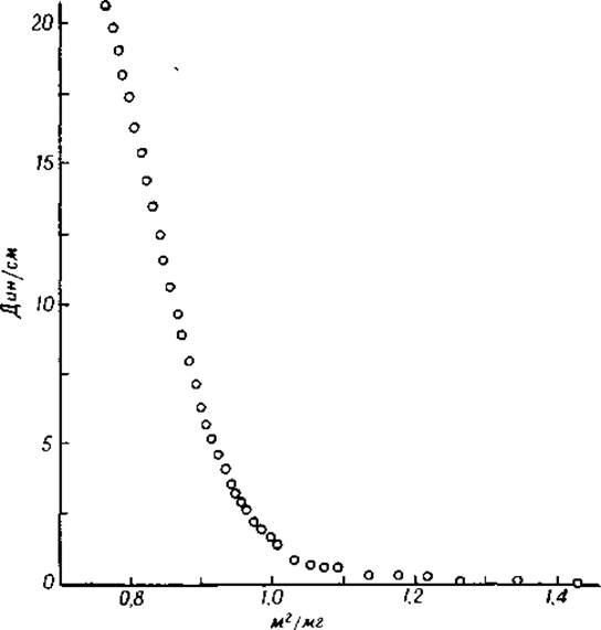
Fig. 25. Curve characterizing the force distribution in an egg albumin film formed on the surface of a 35% ammonium sulfate solution [42].
1 According to recently published data by M. M. Zaalishvili (Biokhimiya, 16, 321, 1951), myogenic A forms a film 7.5 Å thick on the water surface. The area covered by 1 mg of this protein is 1 m2. The main polypeptide chain lies on the surface, while the side groups form an angle approaching 90° with this surface. — Ed. note.
Table 9 shows that the area occupied by a protein film at low pressure is significantly larger than the area occupied by the same amount of protein at high pressure [51].
Table 9 Area of protein films formed by 1 mg of protein, m2 [51]
|
Protein |
Low-pressure region extrapolated to zero pressure |
High-pressure region at the point of film collapse |
|
Egg albumin |
1.3 |
0.77 |
|
1.65 |
0.77 |
|
|
Serum albumin |
1.45 |
0.79 |
|
Gliadin |
1.65 |
0.50 |
|
Cytochrome c |
1.55 |
0.70 |
The Mechanism of protein film formation has been very little studied. Presumably, in protein molecules located at the solution surface, hydrophilic ionic groups are immersed in water, while nonpolar groups, located above the layer of main-valence chains, project above the water surface [41, 52]. Protein films possess a definite Structure, which can be demonstrated using indicator oils. When spreading over the film surface, indicator oils form spots of various geometric shapes (e.g., star-shaped). These spots are visible due to the iridescent coloration arising from light Interference in thin films [53].
Calculations of the area occupied by films are based on the assumption that the films are homogeneous and continuous. If the films contain breaks or multimolecular regions, these can be detected by examining the film surface under a dark-field ultramicroscope [54]. Multimolecular egg albumin films are obtained when the protein solution concentration exceeds 0.1% [55]. Applying proteins to stearates yields inhomogeneous films [41].
When proteins are mixed with Lipids, mixed protein-lipid films are formed. The properties of such mixed films are of particular biological interest, since similar films may be present in living Cells. Protein-lipid films have been shown to be permeable to both water-soluble and fat-soluble substances. The penetration of water-soluble molecules presumably occurs through the protein Regions of the film, whereas the penetration of fat-soluble substances occurs through the regions formed by lipids. Molecules possessing both properties likely penetrate through the boundary regions of the water-lipid films in such a way that the polar part of the complex molecule is directed toward the water-protein phase, and the nonpolar part toward the lipid phase [56, 57].
Proteins spread not only across the surface of aqueous solutions, but also across the interface between water and organic Solvents. For example, gliadin and serum albumin have been found to form films at the water-benzene interface [58]. The area occupied by an egg albumin film at the water-bromobenzene interface is larger than the area of the film formed at the water-air interface [59].
The surface tension at the water–n-xylene interface is significantly lowered by serum albumin, egg albumin, or pepsin. This effect is particularly pronounced in globin solutions [60].
Globular proteins undergo partial or complete Denaturation in monolayers; therefore, The process of monolayer formation at a surface is irreversible. This topic will be discussed in Chapter VII.
Last update: 06/08/2026
Editorial and Educational Adaptation: This material has been compiled based on the primary/original source text. The project team performed an editorial review, corrected technical inaccuracies, structured sections, and adapted the content for an educational format.
What was processed:
- elimination of formatting defects (OCR errors, structural breaks, corrupted characters);
- editorial organization of content;
- standardization of terminology in accordance with academic sources;
- verification of factual statements against the original source text.
All mentions of the author, publication year, and origin of the primary text have been preserved in accordance with the source.