Biochemical Engineering Fundamentals, Part 1 - Bailey J., Ollis D. 1989
Chemical Foundations of Life
Biochemical Compounds of Mixed Structure
Cell walls; peptidoglycans and lipopolysaccharides
In the section on Lipids, we have already discussed how Cell membranes play a vital role in regulating the Transport of substances into and out of The Cell. In this regard, other Structure/83.html">Structural elements of the outer surfaces of microbial and tissue Cells are equally important. From the standpoint of environmental conditions, microorganisms, such as Bacteria, live in a much more variable and less controlled environment than animal tissue cells, such as Liver cells. Therefore, microorganisms must possess much greater rigidity, resistance to physical stress, and tolerance to abrupt changes in osmotic pressure. Consequently, The structure of bacterial outer envelopes must differ significantly from those of animal cells.
In biochemical engineering processes, cell envelopes are of interest in several aspects. First, The properties of outer cell surfaces determine their ability to adhere to each other, as well as to the walls of bioreactors, piping, and separators. As we will see in subsequent chapters, such phenomena must be taken into account when preparing immobilized cell catalysts. They also affect the operation of continuous microbial bioreactors and cell-liquid Separation processes. The chemical and mechanical CHARACTERISTICS OF THE envelopes also determine the cell's resistance to physical, enzymatic, and Chemical factors, which is essential in Cell Disruption and component recovery processes.
Class="center">
FIG. 2.24. Schematic representation of the wall structure of Gram-positive and Gram-negative bacteria. Abbreviations: CM — cytoplasmic membrane; PG — peptidoglycan; PP — periplasmic space; OM — outer membrane. (Reproduced from: Stanier R. Y., Adelberg E. A., Ingraham J. L., Wheelis M. L., Introduction to the Microbial World, p. 128, Prentice-Hall, Englewood Cliffs, N.Y. 1979.)
As mentioned above in the Discussion of the Gram stain, the envelope structures of Gram-positive and Gram-negative cells differ significantly from each other (Fig. 2.24). In both types, the envelopes consist of several layers, but their Location, thickness, and composition are far from identical. This figure does not show the various Proteins located within The cell membrane or anchored to its surface; we will consider them below, in Chapter 5, during our study of membrane transport.
Gram-positive bacteria have a single membrane, whereas Gram-negative cells have two similar membranes. In Gram-negative bacteria, the region between the outer and Cytoplasmic membranes is called the periplasmic space or periplasm. The periplasmic space, which accounts for 20 to 40 percent of the total cell mass, contains A number of Enzymes as well as proteins that bind sugars and Amino Acids.
In both Gram-positive and Gram-negative bacteria, the peptidoglycan layer lies directly adjacent to the outer surface of the cytoplasmic membrane. Peptidoglycans are composed of disaccharide residues [consisting of N-acetylmuramic acid (NAM) and N-acetylglucosamine (NAG) linked by a β-1,4-glycosidic bond], a pentapeptide bridge consisting solely of Glycine residues, and a tetrapeptide. The STRUCTURE OF THE latter depends on the microorganism species; in Staphylococcus aureus, for example, this tetrapeptide contains L-Alanine, D-glutamine, L-Lysine, and D-alanine residues (Fig. 2.25, a; note the presence of rare D-amino acids). All these structural elements of peptidoglycans are extensively cross-linked, forming, as it were, a single giant macromolecule that surrounds the entire cell (Fig. 2.25, b).
The enzyme Lysozyme, whose structure is shown in Fig. 2.17, is an effective antibacterial agent. Its bactericidal action is based on the Hydrolysis of glycosidic β-1,4-bonds between the NAM and NAG residues of peptidoglycan, which leads to the destruction and removal of the bacterial peptidoglycan envelope and thus to cell rupture (lysis) in native hypotonic solutions. Under laboratory conditions in an isotonic medium, it is possible to obtain viable cells lacking peptidoglycan envelopes. After Treatment with lysozyme under such conditions, cells are formed that lose their characteristic shape and become spherical; they are called spheroplasts. If it is known that The Cell wall has been completely removed, such cells are called protoplasts.
Lipopolysaccharides are important Components of the outer membrane of Gram-negative bacteria. Some lipopolysaccharides are called endotoxins because they are highly toxic to animals. The toxicity of lipopolysaccharides is one reason why E. coli bloodstream infections can be extremely dangerous. For the same reason, the purification of proteins synthesized by genetically engineered E. coli requires the meticulous removal of endotoxins.
Outer membrane lipopolysaccharide molecules consist of three regions: a) lipid A, consisting of six Unsaturated Fatty acids whose hydrocarbon chains extend into the membrane and are linked to a diglucosamine residue; b) a core oligosaccharide region composed of ten monosaccharide residues, some of which are rare sugars; c) an O-specific side chain consisting of many repeating tetrasaccharide units. The core region and the O-chain extend outward from the cell into the medium. Thus, it is the outer O-side chains that interact with The Immune System of the infected animal. Through Mutations, bacteria can rapidly alter the structure of their O-chains, which serves as part of their defense against the host immune system.

FIG. 2.25. a — structure of the main structural elements of peptidoglycans; b — schematic representation of the peptidoglycan in the cell wall of the Gram-positive bacterium Staphylococcus aureus, containing a high degree of cross-linking. (From: Lehninger A., Principles of Biochemistry, vols. 1–3. — Moscow: Mir, 1985, vol. 1, p. 317.)
However, this is not the end of the story regarding Bacterial cell wall structure. In many microbial species, the outer membrane is surrounded by a capsule or a slime layer, which is polysaccharide in nature. The capsule of one pneumococcal strain (the bacteria that cause Pneumonia) is composed of alternating glucose and glucuronic acid residues. Mutants lacking this polysaccharide capsule are non-pathogenic. The production of extracellular Polysaccharides is an important industrial process, which, however, is significantly complicated by the non-Newtonian flow behavior of the liquid medium (Chapter 8). Slime layers also play a role in bacterial flocculation, which is a key step in wastewater treatment processes using activated sludge.
The cytoplasmic membrane of Yeast cells consists of lipids, proteins, and mannose-containing polysaccharides. Moving further outward, the periplasmic space lies beyond the cytoplasmic membrane, which in turn is surrounded by the cell wall. In baker's yeast, Saccharomyces cerevisiae, the cell wall contains 6 to 8% protein (including several enzymes), and approximately 30% (by weight) each of glucan (a polysaccharide composed of D-glucose residues joined by β-1,6-linkages as well as β-1,3-crosslinks) and mannan (a polymannose with α-1,6-linkages and α-1,2-side chains). Treatment of S. cerevisiae with a glucan-hydrolyzing enzyme, such as 1,3-glucanase, yields the corresponding protoplasts. Such treatment is commonly performed when introducing recombinant DNA molecules into yeast cells.
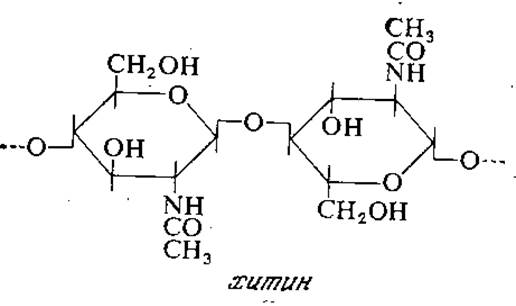
The cell walls of yeast and many Molds contain Chitin, a macromolecule composed of N-acetylglucosamine residues joined by β-1,4-glycosidic bonds (the structure of chitin is shown on the following page).
Animal cells, which typically exist in a strictly controlled isotonic environment, lack cell walls. In addition to Phospholipids and proteins, their Plasma Membranes contain 2 to 10% CARBOHYDRATES. The latter have been found on the outer surface of all mammalian cells studied to date; they are linked to lipids and proteins in the form of Glycolipids and Glycoproteins, respectively.
Last update: 06/08/2026
Editorial and Educational Adaptation: This material has been compiled based on the primary/original source text. The project team performed an editorial review, corrected technical inaccuracies, structured sections, and adapted the content for an educational format.
What was processed:
- elimination of formatting defects (OCR errors, structural breaks, corrupted characters);
- editorial organization of content;
- standardization of terminology in accordance with academic sources;
- verification of factual statements against the original source text.
All mentions of the author, publication year, and origin of the primary text have been preserved in accordance with the source.