ECOLOGICAL BIOCHEMISTRY - Textbook - V. M. Isaenko 2005
Chapter I. VIRAL INFECTIONS AND ORGANISMAL RESPONSES THERETO (General Concepts)
1.3. Plant Viruses
Plant Viruses, or more commonly referred to as phytoviruses, infect both lower and higher plants. Notably, the very discovery of viruses by D. I. Ivanovsky (1892) occurred during his research on the tobacco mosaic virus (TMV).
Viruses of higher plants, particularly angiosperms and especially agricultural crops, have been studied most extensively. In contrast to seed plants (gymnosperms and, above all, angiosperms), viruses infecting spore plants (bryophytes, rhyniophytes, psilophytes, lycophytes, and equisetophytes) remain considerably less explored. Nevertheless, these plants are also susceptible to phytoviral infections. At the same time, research into phytoviruses affecting lower plants has recently commenced, driven by the need to understand their role in maintaining biodiversity stability. For instance, relatively large, well-defined polyhedral viruses with a diameter of 150–250 nm have been identified in Algae in most cases. Specifically, they were found in Cells of green algae such as Oedogonium spp. and Uronema gigas, as well as the brown alga Chorda tomentosa. Nuclei of the green alga Coleochaete scutata may harbor clusters of an isometric virus measuring 40 nm in diameter (a DNA-containing virus). Similar viruses have been detected in the Cells of the red alga Sirodotia tenuissima. The virus infecting the green alga Chara corallina contains single-stranded RNA and resembles TMV in shape, albeit larger, with a length of 530 nm. Undoubtedly, the list of investigated viruses in lower plants is significantly broader.
Spread of Viruses Within the Plant. Experimental studies on higher plants demonstrate that viruses introduced via inoculation propagate relatively slowly through non-vascular tissue from primarily infected cells to adjacent ones. For example, the propagation rate of TMV is approximately 1 mm per day. Apparently, a virus entering a Cell initially replicates within it and subsequently penetrates neighboring cells via intercellular channels or plasmodesmata. Several hours elapse before the virus begins to move from the primarily infected cell to an adjacent one. Both intact virions and viral nucleic acid can migrate from one cell to another.
A virus entering the vascular tissue of a leaf from neighboring parenchymal cells, or introduced directly by vectors (particularly insects), moves first through the Veins, then the leaf petiole, and finally reaches the stem. In general, a plant can become systemically infected, with The rate of this process depending on The properties of both the virus and the host plant. Most plant viruses are classified as "unrestricted" viruses capable of penetrating all Tissues of an infected plant. Long-distance transport of viruses occurs primarily via the phloem, although they are also capable of migrating through the xylem. Seed transmission of viruses is relatively rare, and pollen transmission even rarer. The Morphological Characteristics of flower development apparently act to some extent as a barrier preventing viruses from penetrating Gametes.
Infection symptoms can vary and depend on the plant species, duration of infection, viral strain, and environmental conditions. For instance, disease symptoms are most pronounced in plants grown under bright illumination and moderate temperatures, whereas they may be less noticeable under low light and high temperatures. The incubation period—the time from virus entry into plant cells to the appearance of symptoms—likewise depends on the viral strain, host plant, and environmental conditions. For herbaceous plants, the incubation period typically spans several days or weeks, whereas in woody plants, it may exceed a year.
Disruption of Cellular METABOLISM by Viruses. The severity of plant viral infection is determined by cellular responses to the pathogen. The most pronounced manifestation of this response is necrotic lesion formation. In such cases, cells die so rapidly that they may fail to transmit the virus to neighboring cells. However, the most common response to viral infection involves nearly asymptomatic infections, characterized by very mild symptoms or identified solely because the virus induces disease in other plant species. In these instances, infected cells retain their capacity to divide. Another plant cellular response to infection involves intensive Cell Division and even tumor transformation.
Most metabolic disturbances in virus-infected plant cells presumably arise as a consequence of indirect viral impacts on metabolic processes.
Virus-induced metabolic alterations resemble those observed during plant senescence. For instance, infection of tobacco with cucumber mosaic virus leads to an increased content of alcohol-soluble nitrogen (amino, amide, and ammonium nitrogen), as well as RNA and DNA phosphorus, whereas the concentration of other organic phosphorus compounds decreases. Infection of tobacco leaves with TMV exerts little effect on total nitrogen content and only slightly increases protein nitrogen content, even though the virus may comprise up to 75% of leaf protein; this indicates that viral Protein Synthesis occurs at the expense of normal leaf Proteins.
Certain viruses are known to influence the functional activity of Chloroplasts in a highly specific manner. For example, during the peak Replication period of turnip yellow mosaic virus in leaves, transient changes occur, manifested by the utilization of photosynthetic products for the synthesis of Amino Acids and other organic acids rather than sugars, alongside an enhanced activity of phosphoenolpyruvate carboxylase and aspartate aminotransferase. Other chloroplast alterations are evidently less specific and typically manifest following the viral replication phase. They result in a reduced rate of Photosynthetic Carbon Fixation, decreased levels and activity of Enzymes (particularly ribulose bisphosphate carboxylase), lower chlorophyll content, and a diminished concentration of chloroplast Ribosomes. Conversely, cytoplasmic ribosome levels may remain unchanged.
Plant leaves affected by yellowing-inducing viruses, such as sugar beet yellows virus, contain an excess of glucose, fructose, and sucrose. The primary cause of this sugar accumulation is believed to be not a blockage of their transport, but rather an increased "resistance" along The pathway of sugar translocation in the petioles; consequently, a steeper concentration gradient is required to maintain the previous rate of sugar export from leaves to roots.
Another widespread effect of viral infection is the reduction in both daytime starch synthesis within leaf cells and the rate of nocturnal starch export from leaves.
Viral infection generally leads to an increased rate of Plant Respiration. More pronounced respiratory stimulation is typically caused by viruses or viral strains that induce the most conspicuous disease symptoms, whereas no respiratory changes occur when symptoms are mild. Enhanced respiration in Nicotiana leaves developing local lesions upon TMV infection is primarily associated with the Uncoupling of respiration and Oxidative Phosphorylation. Infection with certain other viruses may lead to the activation of Pentose Phosphate Pathway enzymes.
When upper, young, healthy leaves of a plant are infected via inoculated lower leaves, an acquired resistance to infection is observed, which correlates with peroxidase activation in the upper leaves. It is hypothesized that peroxidase activation in tissues promotes the rapid accumulation of Quinones, which trigger cell death during infection foci and thereby slow down viral spread.
Viruses that induce enations or other morphological growth abnormalities affect the balance of growth regulators in plants.
The aforementioned list of plant cellular metabolic alterations during infection is by no means exhaustive. Other common manifestations of infection include cell enlargement, tissue maceration, and Water balance disorders resulting from ROOT damage or increased Transpiration.
Vectors of Plant Viruses. Among the diverse modes of plant virus dissemination, vectors hold a position of particular importance. They acquire viruses, maintain them in a viable state, and introduce them into the host plant, ultimately resulting in infection. If any of these steps fails to occur, viral propagation is prevented.
Vectors of plant viruses include Arthropods, nematodes, and Fungi (Fig. 1.1).
Class="center">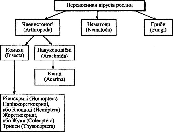
Fig. 1.1. Vectors of plant viruses (after Ryzhkova et al., 2002)
Let us examine in somewhat greater detail the involvement of specific plant virus vectors in the infection process.
Insects. Homopteran insects comprise approximately 25,000 species. Vectors of plant viruses include Representatives of the suborders Cicadinea (leafhoppers), Aleyrodinea (whiteflies), Coccinea (scale insects), and Aphidinea (aphids).
Female leafhoppers lay their eggs primarily in plant stems. Nymphs of most species migrate into the soil, where they feed on underground plant parts. Adults feed on aerial plant parts and frequently fly from one plant to another, which accounts for their ability to transmit phytoviruses.
Cicadellids most commonly transmit plant viruses of the families Caulimoviridae (genus Rice tungo bacilliform-like viruses), Rhabdoviridae (genera Cytorhabdovirus, Nucleorhabdovirus), Sequiviridae (genus Wairavirus), and Geminiviridae (genera Mastrevirus, Curtovirus).
Whitefly nymphs and adults feed on the undersides of leaves. They transmit plant viruses from the families Geminiviridae and Closteroviridae, as well as certain genera of Potyviridae (Ipomovirus) and others. Geminiviruses are among the plant viruses vectored by whiteflies, predominantly causing yellowing, leaf curl, and stunting. Typical representatives include Tomato leaf curl virus, Tomato golden mosaic virus, and Potato yellow mosaic virus, among others.
Closteroviruses transmitted by whiteflies primarily cause interveinal yellowing and reddening of the leaf blades. Examples of such viruses include Lettuce infectious yellows virus, which affects lettuce, sugar beets, and melons, as well as Tomato infectious chlorosis virus.
Numerous other whitefly-borne plant viruses have been identified, including sweet potato speckling virus and zucchini yellow leaf curl virus.
Coccoids (families Coccidae, Pseudococcidae) predominantly transmit viruses belonging to the genera Vitivirus, Badnavirus, and Closterovirus.
Aphids are among the most common vectors of plant viruses. In some species, a portion of the population continuously reproduces on the primary host plant, while another migrates to the secondary host. Such aphids settling on young woody plants may remain non-migratory throughout the entire season, whereas those on older trees may relocate to herbaceous plants. For certain aphid species, migration to other plants is essential to complete their life cycle starting from the second or third generation. Typical plant viruses transmitted by aphids include lettuce necrotic yellows virus (family Rhabdoviridae), which replicates within the insect, and barley yellow dwarf virus (family Luteoviridae), which does not replicate in its vector. A wide range of other viruses are also aphid-borne, including potato viruses (potato leafroll virus, potato virus Y, potato virus A, potato virus M, and potato virus S).
Insect vectors of plant viruses include beetles from three families: leaf beetles (Chrysomelidae), weevils (Curculionidae), and ladybird beetles (Coccinellidae). Viruses transmitted by beetles belong to the genera Carmovirus (Bean mild mosaic virus, Turnip crinkle virus, etc.), Comovirus (Cowpea mosaic virus, Squash mosaic virus, etc.), Bromovirus (Broad bean mottle virus, etc.), Sobemovirus (Southern bean mosaic virus, Rice yellow mottle virus, etc.), Tymovirus (Turnip yellow mosaic virus, Andean potato latent virus, etc.), and Machlomovirus (Maize chlorotic mottle virus, etc.).
Thrips inhabit grasses and sedges, leaves and stems of shrubs and trees, leaves and shoots of herbaceous plants, and the bark of dead trees. A significant number of species live in inflorescences, feeding on Ovary sap, nectar, and pollen. They transmit viruses of the genera Tospovirus (Bunyaviridae) and Machlomovirus (Tombusviridae). Typical plant viruses transmitted by thrips include Tomato chlorotic spot virus, Groundnut bud necrosis virus, Chrysanthemum stem necrosis virus, and Watermelon silver mottle virus, among others.
Mites. Among plant virus vectors, mites belonging to the class Arachnida, phylum Arthropoda, are of considerable importance (Fig. 1.1). These are primarily mites of the families Tenuipalpidae, Phytoptidae, and Eriophyidae within the order Acariformes. Eriophyid mites are the most prominent in this regard, with Aceria tulipae being a prime representative.
Mites transmit a variety of plant viruses, notably those of the genera Pymovirus (Agropyron mosaic virus, Hordeum mosaic virus, etc.), Tritilovirus (Wheat streak mosaic virus, etc.), Foveavirus (Blackcurrant reversion associated virus), Allexivirus (Garlic virus C, Onion mite-borne latent virus, etc.), as well as the family Rhabdoviridae (Coffee ringspot
virus, etc.) and Bromoviridae (Prunus necrotic ringspot virus, etc.). The transmission of Tobamovirus and Potexvirus genera by mites has also been demonstrated.
Nematodes. Ectoparasitic nematodes that attack plant root tips or adjacent cells are typical vectors. They cause plant growth retardation or gall formation independently of viral infection. At the same time, nematodes are capable of transmitting plant viruses. These include nematodes of the families Longidoridae (genera Xiphinema and Longidorus) and Trichodoridae (Trichodorus and Paratrichodorus).
Nematode-borne viruses are RNA-containing and belong to the genera Nepovirus (e.g., Tobacco ringspot virus, Grapevine fanleaf virus, Tomato black ring virus, Cherry leafroll virus, etc.) and Tobravirus (Tobacco rattle virus, Pea early-browning virus, etc.).
Nematodes of the genera Xiphinema and Longidorus are relatively large, with a length of 5–12 mm. In contrast, members of the family Trichodoridae measure 0.6–1.2 mm in length.
Both adult nematodes and juveniles at various developmental stages are capable of transmitting viruses. Viruses can persist in the nematode vector for a relatively long period, ranging from several weeks to months. Unlike Xiphinema, adult nematodes of the genus Longidorus require continuous access to virus-containing food to retain their infectivity over extended periods. Acquisition of the viral infection by nematodes occurs within 15 minutes to 1 hour of feeding. Trichodorus nematodes typically feed on epidermal cells, moving from one to another within a few minutes. Meanwhile, members of the genus Xiphinema feed at a single site for an hour or more. The acquisition access period required for a nematode to infect a plant with a virus ranges from 1 hour to several days.
Fungi. Plant virus-vectored fungi are unicellular organisms. They are intracellular parasites that go through two stages: a motile zoospore and a thick-walled resting spore. Approximately 30 plant viruses are known to be transmitted by fungi of the class Chytridiomycetes (genus Olpidium) and the class Plasmodiophoromycetes (genera Polymyxa and Spongospora). For instance, Olpidium bornovanus transmits Cucumber necrosis virus, Olpidium brassicae transmits Tobacco necrosis virus, Polymyxa betae transmits Beet necrotic yellow vein virus, Polymyxa graminis transmits Rice stripe necrosis virus, and Spongospora subterranea transmits Watercress yellow spot virus, among others.
The life cycle of fungi of the genus Olpidium is relatively simple. Asexual reproduction occurs via flagellated zoospores. Carried by soil water, a zoospore moves toward a plant root, attaches to it, retracts its flagellum, forms a cyst wall, and pours its contents into a plant epidermal cell. This results in The formation of a thallus that develops into a multinucleate zoosporangium. Zoospores escape through a punctured Cell wall. The entire cycle takes 5–10 days.
During Sexual reproduction in fungi of the genus Olpidium, thick-walled resting spores are formed. They are released into the soil after root decay and can remain viable for many years.
The Life Cycle of plasmodiophorid fungi of the genera Polymyxa and Spongospora is similar to that of Olpidium.
Fungi transmit viruses to plants in two ways. In the first mechanism, free-soil viruses become localized (adsorbed) on the outer membrane of zoospores, which move from plant to plant via soil water. The source of zoospores can be vegetative sporangia or spores that have passed through a resting stage, while the source of the virus is infected plant roots or plant debris. It is believed that viruses enter the zoospore protoplasm via the flagellum, which retracts during cyst formation and on whose surface the viruses are localized. Viruses thus accumulated in the zoospore protoplasm are transferred into the plant root cells during the pouring of cyst contents. Note that this infection pathway is characteristic exclusively of fungi of the genus Olpidium. Examples of plant infection via this method include the transmission of Cucumber necrosis virus by Olpidium bornovanus and Tobacco necrosis virus by Olpidium brassicae.
The second pathway of plant infection by fungi involves spores. The fungus acquires viruses from a plant cell as it develops within it. The viruses persist within thick-walled spores. Resting spores remain viable and retain viruses for several years. Virus transmission to plant roots in this case is carried out by zoospores, just as in the previous mechanism. This mode of fungal-mediated plant virus transmission is common to all three genera: Olpidium, Polymyxa, and Spongospora. Examples of this infection route include the transmission of Tobacco stunt virus by Olpidium brassicae, Barley yellow mosaic virus by Polymyxa graminis, and Potato mop-top virus by Spongospora subterranea.
Among numerous fungal viruses, only a small fraction are morphologically similar to plant viruses. Consequently, it has been hypothesized that
in most cases of plant infection, fungi merely serve as a storage site (reservoir) for plant viruses. Indeed, tobacco mosaic virus has been isolated from conidia of powdery mildew pathogens Sphaerotheca lanestris and Erysiphe graminis. Other examples of plant virus localization in fungi are also known.
Last update: 06/08/2026
Editorial and Educational Adaptation: This material has been compiled based on the primary/original source text. The project team performed an editorial review, corrected technical inaccuracies, structured sections, and adapted the content for an educational format.
What was processed:
- elimination of formatting defects (OCR errors, structural breaks, corrupted characters);
- editorial organization of content;
- standardization of terminology in accordance with academic sources;
- verification of factual statements against the original source text.
All mentions of the author, publication year, and origin of the primary text have been preserved in accordance with the source.