ECOLOGICAL BIOCHEMISTRY - Study Guide - V. M. Isaienko 2005
Chapter 8. PLANT AND ANIMAL RESISTANCE AND ADAPTATION
8.3. Animal Resistance and Adaptation to Environmental Conditions
8.3.6. Bioluminescence
The ability to emit light is widely distributed among Bacteria, Fungi, Algae, and animals. The vast majority of luminous animals are marine deep-sea, coastal, and planktonic organisms. However, fireflies, slugs, and certain dipteran larvae are also capable of luminescence.
Luminescence (from the Latin lumen — light, and the suffix -escent — weak action) in animals results from chemiluminescence, meaning it is caused by Chemical Reactions in which The oxidation of a substrate produces light emission. In intracellular luminescence, these reactions occur within granules located in the Cytosol of luminescent tissue Cells. Extracellular luminescence takes place in light-generating Organs consisting of unicellular or multicellular glands that secrete substances externally. Sometimes these glands contain two distinct Cell types that separately house the substrate (luciferin) and the enzyme (luciferase) involved in the light-generation process.
Organismal luminescence plays a specific role in survival and has evolved alongside The Development of specialized cells and organs. Furthermore, this phenomenon can be part of an Organism's general irritability and response to stimuli.
The phenomenon of luminescence primarily serves a defensive function—acting as a warning, deterrent, and mechanism to confuse predators. It helps animals conceal themselves within a luminous cloud, as seen in the deep-sea marine shrimp Acanthophyra and the squid Heteroteuthis. It is also possible that the triggered luminescence of one individual serves as a danger warning to others, allowing them to escape harm.
Another function of animal luminescence is signaling for mate attraction during the breeding season. A typical example is the attraction of males by female fireflies. In many species, females—sometimes even wingless—sit in the grass and flash light signals to flying males. The female responds to the male's luminescent signals only within her own species, using highly specific patterns: flashes of a particular frequency, color, intensity, duration, and count.
Animal luminescence can also drive aggressive mimicry, where females of certain species lure males of other species only to prey upon them. This occurs, for instance, in female Photuris fireflies that hunt males of other species within the same family.
In Protozoa, luminescent granules are scattered throughout the Cytoplasm, whereas in higher animals, photogenerating cells or organs are localized. In cephalopods, crustaceans, fish, and insects, light-emitting cells constitute only a part of an organ that also includes layers of cells acting as "light filters" (which absorb and reflect light), light-refracting bodies, and nerve endings.
Sometimes the luminescence of organisms is not intrinsic, but rather caused by luminescent bacteria residing within or On the surface of the body.
Prevalence of Bioluminescence among animals. Among protozoa, many marine species are luminescent, notably radiolarians and dinoflagellates, with Gonyaulax and Noctiluca being the best studied. Luminescent granules are localized in microsomes, with the highest concentration at The Cell periphery. They emit light in response to mechanical or electrical stimulation.
In Gonyaulax, Peridinium, and Pyrocystis, luminescence occurs both in response to excitation and spontaneously. The intensity of their luminescence can vary depending on the time of day.
Among Coelenterates, many species of polyps, jellyfish, siphonophores, and sea anemones are capable of luminescence. This phenomenon arises in response to stimulation. The area directly subjected to mechanical irritation responds with light emission first,
after which a wave of luminescence spreads outward from the point of origin. In some coelenterates, the luminescence may be extracellular in nature.
Evidently, all ctenophores (phylum Ctenophora) luminesce in response to stimulation. Light exposure inactivates this emission.
Among nemerteans, only Emplectonema randai are capable of luminescence. Photogenerating cells are distributed throughout the body, and local tactile stimulation elicits a localized response. Among Annelids, marine polychaetes and terrestrial oligochaetes possess luminescent abilities.
Certain earthworms, such as Eisenia submontana, when disturbed, expel glowing mucus through their Mouth, anus, or dorsal pores. In the polychaete Chaetopterus, stimulation causes the greater part of its body surface to glow. Photogenerating cells are located in the hypodermis alongside mucous cells, and their contents are jointly secreted outward. Luminescence in annelids is regulated by The Nervous system; stimulation of one part of the body triggers a wave of light traveling up and down from the excitation point.
Among Arthropods, crustaceans and insects, as well as some myriapods and arachnids, are capable of luminescence. Studies of the luminescence of crustaceans of the genus Cypridina and fireflies have provided fundamental insights into the chemical processes of bioluminescence. In Cypridina, the light-generating organ is a gland situated near the mouth, where some secretory cells contain the substrate and others contain the enzyme.
In euphausiids and shrimp, photogenerating organs are distributed across the entire body surface and regulated by the nervous system. The deep-sea shrimp Acanthophyra purpurea, In addition to typical photogenerating organs near the mouth, possesses glands that expel a luminous substance, allowing the shrimp to shroud itself in a glowing cloud to evade predators.
Among insects, luminous species are found in only a few orders. These include several species of springtails (Collembola), larvae of the fungus gnat Ceratoplatus, and larvae and adults of the fungus gnat Bolitophila, in which luminescence is generated within the Malpighian tubules.
The most extensively studied luminous insects are beetles of the order Coleoptera, specifically fireflies (Lampyridae) and click beetles (Elateridae). In these insects, the photogenerating organ is located in the posterior region of the abdomen and consists of a layer of photogenerating cells backed by a dorsal (or inner) layer of light-reflecting cells. Insect luminescence is regulated by the nervous system.
In the South American click beetle Pyrophorus, a pair of greenish luminescent organs is located on the anterior part of the prosternum, while an orange-yellow organ is situated on the ventral surface
of the first abdominal segment. The HEAD of the South African "railroad worm" (Phrixothrix) glows with red light, and green luminescent patches are distributed segmentally along its body. Luminescence in this insect occurs in response to irritation. In contrast, the North American insect genus Phengodes glows continuously and lacks a distinct head light organ.
Some species of centipedes secrete mucus that glows similarly to that of annelids. Luminescence has also been reported in sea spiders.
Among Mollusks, cephalopods exhibit the greatest capacity for luminescence. In many of them, such as the squid Loligo, the light-emitting organ is located externally and contains luminous bacteria. The Loligo squid itself does not possess intrinsic luminescent organs. Nevertheless, most mollusks do have organs capable of luminescence. For instance, in the squid Heteroteuthis, a single luminescent organ opens into the mantle cavity, from which a glowing cloud is expelled through the siphon when disturbed. In the squid Watsenia, photogenic organs are distributed across the entire body, with three located on each arm.
In Echinoderms, luminescent organs consist of unicellular photogenic structures scattered throughout the body. Their luminescence occurs in response to any form of mechanical or environmental stimulation.
Lower Chordates and Fishes are also capable of luminescence. In tunicates (e.g., Pyrosoma), a wave of light propagates across the entire colony upon stimulation. Some species of Balanoglossus secrete glowing mucus.
Numerous luminescent species are found among cartilaginous and bony fishes, particularly deep-sea dwellers. Well-developed photogenic organs on the head are characteristic of Astronesthes and Stomias.
In some fishes (Photoblepharon, Anomalops, Physiculus, Leiognathus, Monocentrus, etc.), the light is generated by symbiotic bacteria housed in specialized organs, which are expelled outward when the animal is disturbed.
Chemical mechanisms of animal luminescence. As previously noted, bioluminescence is a form of chemiluminescence. Research has shown that this phenomenon arises from the oxidation of a substrate called luciferin (from Latin lucis – light, and ferre – to bear) by molecular oxygen, catalyzed by the enzyme luciferase. This process can be represented by the equation (Fig. 8.7).
Different organisms contain specific substrates and Enzymes, but the underlying mechanism of luminescence is similar. Therefore, the terms "luciferin" and "luciferase" are used to denote the substrate and enzyme in all chemical reactions accompanied by light emission.
Class="center">
Fig. 8.7. Equation of the enzyme-substrate reaction accompanied by luminescence:
S — substrate (luciferin); E — enzyme (luciferase); E - S* — enzyme-substrate complex in an electronically excited state; hv — quantum of light (h — Planck's constant, v — frequency)
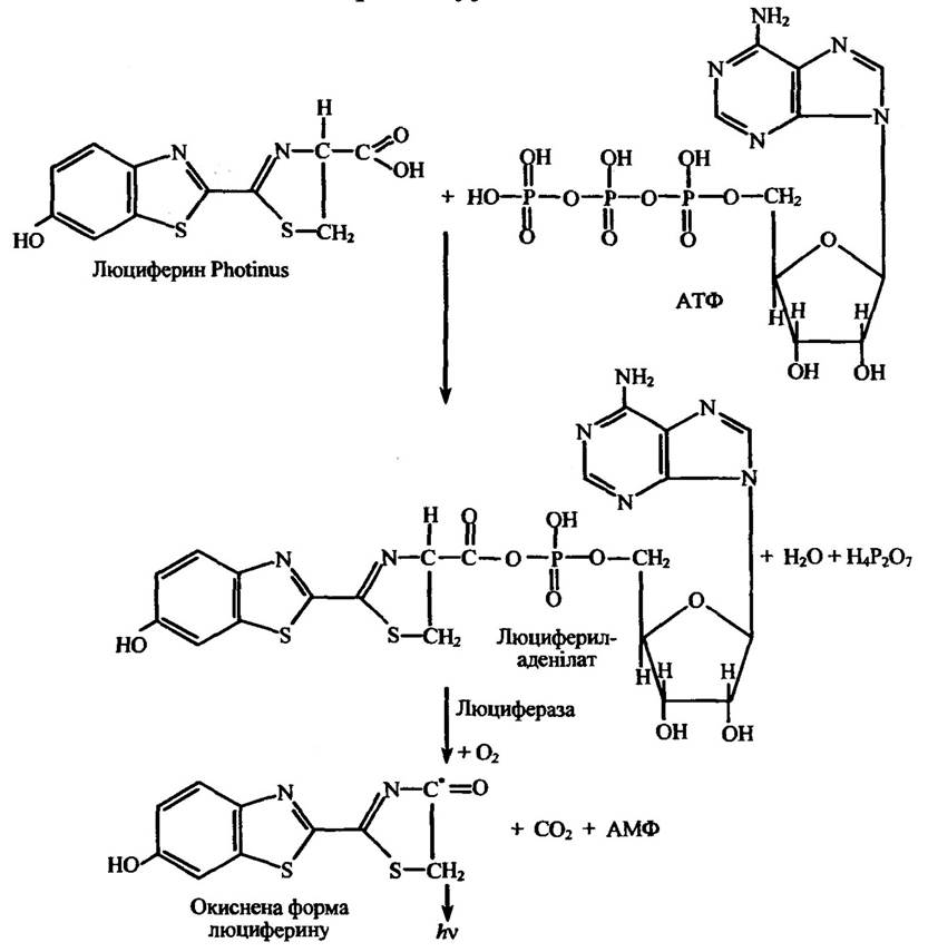
Fig. 8.8. Interaction of firefly (Photinus) luciferin with ATP in the presence of luciferase,
accompanied by luminescence
Note. The asterisk (*) indicates that the given atom is in an electronically excited state.
Let us examine the reactions of luciferin and luciferase in the firefly Photinus and the crustacean Cypridina.
In the firefly, the luminescence reaction occurs intracellularly. Its luciferin is activated through a reaction with ATP to form luciferyl adenylate in an electronically excited state; the return of this complex to the ground state is accompanied by light emission in the red region of the spectrum (X = 560 nm). This is schematically illustrated in Fig. 8.8.
Variations in the luminescence spectra among different firefly species are primarily due to differences in the Structure and conformation of the luciferase enzyme.
The luminescence-coupled interaction between the luciferin and luciferase of the crustacean Cypridina is shown in Fig. 8.9.

Fig. 8.9. Interaction of Cypridina luciferin in the presence of luciferase, accompanied by luminescence
Note. The asterisk (*) indicates that the given atom is in an electronically excited state.
The biochemical processes underlying luminescence have been studied in detail for several cnidarian species. For example, in Renilla corals, this process involves two stages (Fig. 8.10).
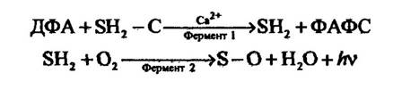
Fig. 8.10. Equations of substrate and enzyme reactions in Renilla accompanied by luminescence:
PAPSW — 3',5'-phosphoadenosine; SH2 — luciferin; SH2 — luciferin with a removed group; PAPS — phosphoadenosine phosphosulfate
At The First stage, luciferin activation occurs via the removal of a specific group identified as a sulfate. This reaction is stimulated by 3',5'-phosphoadenosine, resulting in The formation of activated luciferin and phosphoadenosine phosphosulfate. Structurally, activated luciferin resembles the luciferin of the crustacean Cypridina. Its oxidation, which proceeds via a mechanism similar to that of Cypridina luciferin, gives rise to luminescence.
In jellyfish of the genera Aequorea and Halistaurea, as well as certain other jellyfish species, a specialized protein (photoprotein) has been discovered that emits light upon interaction with Calcium Ions in the presence of oxygen. This protein is presumably an intermediate in a reaction cascade similar to that described for the soft coral Renilla. At The final stage of transformation, the photoprotein interacts with oxygen in the presence of calcium ions.
In some coelenterates, luminescence involves energy transfer: the reaction initially yields an electronically excited product, which subsequently transfers its energy to another substance. When this second substance returns from its excited state to the ground state, luminescence is emitted (Fig. 8.11).
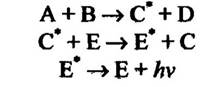
Fig. 8.11. Equations of luminescence reactions involving energy transfer in coelenterates
Note. The asterisk (*) indicates that a given atom is in an electronically excited state.
The luminescence System of the polychaete worm Chaetopterus comprises a photoprotein, two Cofactors (one resembling a nucleoprotein and the other a lipid), peroxides (H2O2 or organic peroxides), oxygen, and iron.
In fish capable of intrinsic luminescence rather than relying on symbiotic bacteria (such as Apogon and Parapriacanthus), The production of luciferin is potentially attributable to their diet of Cypridina crustaceans. However, the luciferase of these fish is distinct from that of Cypridina.
Bacteria exhibit continuous intracellular luminescence driven, for instance, by the Oxidation of reduced riboflavin-5'-phosphate (flavin mononucleotide) (Fig. 8.12).
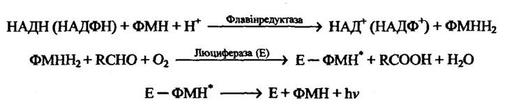
Fig. 8.12. Oxidation reaction of riboflavin-5'-phosphate accompanied by bacterial bioluminescence
Note. The asterisk (*) indicates that a given atom is in an electronically excited state.
Fungal luminescence involves at least two stages: 1) reduction of dehydro- or oxyluciferin mediated by NADH (NADPH) and an enzyme; 2) oxidation of the reduced product by oxygen in the presence of the enzyme luciferase, accompanied by light emission.
Thus, despite all variations in the pathways of luminescence-associated chemical reactions, the common denominator is the luciferase-mediated oxidation of luciferin, which generates an electronically excited product whose return to the ground state results in light emission.
Last update: 06/08/2026
Editorial and Educational Adaptation: This material has been compiled based on the primary/original source text. The project team performed an editorial review, corrected technical inaccuracies, structured sections, and adapted the content for an educational format.
What was processed:
- elimination of formatting defects (OCR errors, structural breaks, corrupted characters);
- editorial organization of content;
- standardization of terminology in accordance with academic sources;
- verification of factual statements against the original source text.
All mentions of the author, publication year, and origin of the primary text have been preserved in accordance with the source.