Biotechnology - Yu.O. Sazykin 2006
Applied Biotechnology
Challenges in the Discovery, Development, and Application of Antibiotics in Medical Practice
Antibiotic Resistance - Molecular Mechanisms
There are four main Mechanisms of Antibiotic resistance:
✵ Modification of the conformation of the intracellular target for a given antibiotic. The antimicrobial agent penetrates The Cell, but its target (peptidoglycan transpeptidase, ribosome, DNA gyrase, etc.) fails to "bind" it, and METABOLISM is not suppressed;
✵ reduction of the permeability of the microbial cell envelope to the antibiotic. Although the antibiotic penetrates the cell, it does so in insignificant amounts;
✵ emergence of an active "efflux" system within the cell envelope that pumps out the penetrating antibiotic, preventing its intracellular concentration from reaching high levels;
✵ enzymatic inactivation of the antibiotic by protective Enzymes. This latter type of microbial defense is the most effective and a very common cause of antibiotic therapy failure. All Major Groups of Antibiotics undergo enzymatic inactivation: Penicillins and Cephalosporins, Aminoglycosides, erythromycin, and several Other Antibiotics.
The Development of these defense mechanisms in bacterial Cells is driven by the appearance of "resistance genes" not only in the chromosome. Extrachromosomal (plasmid) genetic elements of the microbial cell—circular DNA molecules hundreds of times smaller than Chromosomes—also attract significant attention. Plasmids carrying Antibiotic Resistance genes are called R-plasmids (an older term is R-factors).
The main danger of plasmid-mediated resistance from a genetic perspective is that plasmids are transferred from Cell to Cell via conjugation (an analog of the sexual process)—without Cell Division, during which the plasmid nonetheless replicates. Thus, a single cell can rapidly transmit resistance to a large population of cells. The multicopy nature of certain types of plasmids further facilitates this process. This has even led to the term "infectious resistance," meaning the "infection" of some cells with resistance originating from others.
Plasmid-mediated resistance is rarely associated with the first of the mechanisms listed above. Antibiotic resistance caused by Changes in the conformation of an intracellular target results from spontaneous Mutations in the structural Gene that determines The Structure of the macromolecular target with which the antibiotic "binds." As a result of such mutations, the Amino Acid Sequence in the enzyme or ribosomal protein changes, leading to an altered molecular conformation that no longer binds the antibiotic.
Mutated chromosomal genes can be "mobilized," According to the accepted term, meaning they can end up in plasmids and be transferred to other cells via plasmid dissemination mechanisms. However, in most cases, this does not result in The transfer of resistance itself. Both intrinsic (chromosomal) and acquired (plasmid) structural genes will function (be expressed) in the new host cell; consequently, some of the antibiotic targets will remain "sensitive" to it—binding the antibiotic—while others will be resistant. In such cases, antibiotic susceptibility dominates over antibiotic resistance.
This is precisely why, when resistance is due to target conformation changes, the resistance genes are typically localized in the chromosomes. Exceptions occur when Conformational Changes in the target are caused not by a mutation in its structural gene, but by the enzymatic modification of an already synthesized target. The gene for the enzyme that modifies the target can have either a chromosomal or a plasmid Location.
Plasmid localization of resistance genes is particularly common in the enzymatic inactivation of antibiotics. Sometimes, a single plasmid harbors multiple genes encoding enzymes that act on antibiotics from different groups, giving rise to THE CONCEPT OF microbial multiresistance (multidrug resistance). Multiresistant strains of infectious pathogens represent a serious challenge in clinical infectious disease practice.
The targets of antibiotics are located either in the cytoplasmic membrane (peptidoglycan Biosynthesis enzymes) or within the Cytoplasm itself (Ribosomes, Protein Biosynthesis enzymes, nucleic acid biosynthesis enzymes, etc.). To reach its target, an antibiotic must pass through the outer membrane and Cell wall, and sometimes the cytoplasmic membrane as well (if the targets are cytosolic). Antibiotic resistance is frequently caused by specific alterations in the bacterial cell envelope. The envelope comprises the outer membrane (found only in Gram-negative Bacteria), The cell wall, and the cytoplasmic membrane.
Although the cell wall is a rigid, "lattice-like" structure made of peptidoglycan, it does not pose a barrier to the penetration of small antibiotic molecules.
Over recent decades, the growing role of Gram-negative microflora in infectious pathology has been noted repeatedly. This applies to infectious complications in surgery, urological infections, and more. Gram-negative, non-fermenting bacteria play a major role in the infectious process.
Interest has grown in the outer membrane inherent to all these microorganisms. Its main components are lipopolysaccharides, Lipoproteins, Phospholipids, and specific Proteins known as porins (derived from the word pore); trimers of these proteins form pores or aqueous channels through which low-molecular-weight nutrients (Amino Acids, small Peptides, mono-, di-, and trisaccharides, inorganic ions, etc.) diffuse from the external environment into the cell.
Porin channels "oscillate," meaning they alternate between open and closed states. In nutrient-poor environments, a channel remains open longer than in nutrient-rich ones. Antibiotics also "use" these porin channels to gain entry into the cell.
Antibiotics that rapidly penetrate through the aqueous channels of porins are broad-spectrum agents, inhibiting the growth of both Gram-negative and Gram-positive bacteria. Antibiotics with large molecules, such as erythromycin, cannot penetrate porin channels and are therefore ineffective against Gram-negative bacteria, although highly active against Gram-positive ones. Notably, molecular penetration is influenced not only by size but also by steric features, including those determining molecular "flexibility." Consideration must be given to both the diameter of the aqueous channels and The properties of the constituent porin proteins. There are cation-selective and anion-selective channels. For antibiotics, this means that in a bacterium whose outer membrane is dominated by cation-selective channels, basic antibiotics will penetrate the cell more effectively.
An example is benzylpenicillin. Its relatively small molecule penetrates the porin channels of *Escherichia coli* very poorly because this Organism, like many other Gram-negative bacteria, possesses cation-selective porin channels.
At the same time, in a relatively small group of Gram-negative cocci, the porin channels are anion-selective, which facilitates the penetration of benzylpenicillin into their cells. This explains facts well known to physicians: benzylpenicillin is unsuitable for treating intestinal infections, yet highly active in treating Gonorrhea. The cation- and anion-selectivity of porin channels is determined by the habitat of the bacterial species. For instance, the cation-selectivity of porin channels in *E. coli* helps this intestinal bacterium avoid the harmful effects of Bile acids present in the gut.
The possibility and rate of antibiotic penetration through porin channels are determined not only by molecular size, steric features, and charge, but also by Hydrophobicity. Because the channels are filled with Water, the degree of molecular hydrophobicity is of great importance; high hydrophobicity significantly reduces antibiotic penetration.
Besides porin channels—the primary pathway for antibiotics to cross the outer membrane barrier into the cell—other transport mechanisms exist:
✵ via specific primary metabolite transport systems, provided the antibiotic molecule is structurally similar or an analog of the metabolite;
✵ via lipid Regions of the bacterial outer membrane, if the substances (including antibiotics) are lipophilic.
Certain nucleoside-type antibiotics utilize specific nucleoside transport systems to cross the outer membrane. For example, a semi-synthetic cephalosporin with a catechol-like moiety was developed that, based on its molecular size, should not have been able to penetrate porin channels. However, due to the aforementioned group, it formed complexes with iron ions and entered the Gram-negative cell by hijacking the specific iron transport system. In other words, this cephalosporin mimicked an iron carrier, bypassing the membrane barrier via the carrier's own route. Meanwhile, the antibacterial activity of certain semi-synthetic lipophilic penicillins and Tetracyclines is driven by their penetration through the lipid regions of the outer membrane.
Studies of antibiotic resistance mechanisms in Gram-negative bacterial strains isolated from clinical specimens have shown that resistance is frequently caused by a reduced number of porin proteins in the membrane and, consequently, a decreased number of porin channels. As a result, the influx of antibiotics into the cell is slowed down.
Another resistance mechanism involves alterations in the porin proteins themselves and the narrowing of the porin channel diameter.
In both cases, resistance is non-specific and diminishes—albeit to varying degrees—when antibiotics capable of diffusing through porin channels penetrate the cell. However, complete cessation of antibiotic entry into the cell is impossible via these resistance mechanisms, as porin channels are vital for transporting nutrients into the cell, and their complete absence from the membrane would lead to cell death. Research has shown that the level of bacterial resistance conferred by outer membrane changes is lower than that caused by protective, antibiotic-inactivating enzymes, for instance.
Nevertheless, clinical isolates frequently exhibit two concurrent resistance mechanisms: reduced antibiotic penetration through the outer membrane and enzymatic inactivation of the antibiotic. Under these conditions, the level of antibiotic resistance is exceptionally high.
One of the primary drivers behind the development and clinical Introduction of new beta-lactam antibiotics is the widespread ability of pathogenic microorganisms to produce enzymes that catalyze the Cleavage of the beta-lactam ring in clinically established penicillins and cephalosporins. These enzymes were named penicillinases and cephalosporinases based on their substrate Specificity and selective action toward penicillins or cephalosporins, respectively.
Enzymatic cleavage (Hydrolysis) of the beta-lactam ring leads to the complete inactivation of the beta-lactam antibiotic. This was first demonstrated using benzylpenicillins in the 1940s by E. Chain, who originally purified penicillin. The product of the Enzymatic cleavage of benzylpenicillin is penicilloic acid:
Class="center">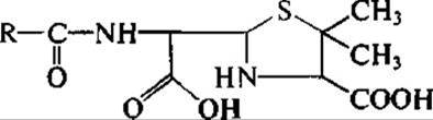
which is entirely inactive. This is easily explained, as the MECHANISM OF ACTION of beta-lactam antibiotics relies precisely on the cleavage of the beta-lactam ring and the acylation of the Serine hydroxyl group in the Active Site of target enzymes. In the case of penicillinases and cephalosporinases, the beta-lactam ring is also cleaved, and the antibiotic is rapidly released from the active site of these enzymes with The addition of hydrogen and hydroxyl atoms.
Today, penicillinases and cephalosporinases comprise a vast group of enzymes that share a common mechanism of action yet differ in substrate specificity, collectively known as "beta-lactamases." It is well established that beta-lactamases evolved from peptidoglycan transpeptidases and D,D-Carboxypeptidases—that is, from bacterial target enzymes that are irreversibly inactivated by beta-lactams.
Beta-lactamase genes, particularly those encoding cephalosporinases, are located on both the bacterial chromosome and plasmids. Plasmids lack the stringent regulatory control exerted on chromosomal genetic material within the cell and can exist in multiple copies, thereby increasing the gene dosage of beta-lactamases and the intracellular concentration of the enzymes themselves. Genetic exchange, particularly of beta-lactamase genes, frequently occurs between chromosomes and plasmids.
Crucially, plasmid-borne beta-lactamase genes can be transferred via conjugation alongside the plasmid into another cell. This means that plasmid genes spread rapidly through a cell population without even requiring cell division. Interspecies transfer of plasmid-mediated beta-lactamase genes is also possible—for instance, from an Escherichia coli cell to a Salmonella cell, and so forth.
Beta-lactamases can be either constitutive (in certain bacterial strains) or inducible (in others). Occasionally, a single cell may produce two different beta-lactamases: one synthesized continuously (constitutively), and the other expressed only when the cell encounters an environment containing a beta-lactam antibiotic.
From a medical standpoint, the capacity to induce beta-lactamases is an undesirable property of beta-lactam antibiotics. Consequently, when evaluating new beta-lactam structures, researchers assess not only their resistance to enzymatic inactivation but also their ability to induce beta-lactamases. The latter depends on which specific target, or PBPs, the beta-lactam antibiotic binds to, as PBPs act as the "sensors" that trigger the complex mechanism of beta-lactamase induction.
Schematically, this mechanism operates as follows: a beta-lactam antibiotic present in the medium interacts with one of the penicillin-binding proteins (PBPs). This interaction induces a conformational change in the protein, altering its biophysical parameters. The signal is then transmitted to a specific transmembrane protein, which spans the cytoplasmic membrane and exposes its domain on the outer surface. Subsequently, the signal is relayed sequentially to the First and Second cytoplasmic proteins—Components of the enzyme induction system—and finally to the repressor protein, which directly regulates the expression of the beta-lactamase gene. As a result, the repressor ceases to inhibit Gene Expression, triggering Transcription and the synthesis of Messenger RNA. This mRNA is then transported to the ribosomal machinery, where it serves as a template for beta-lactamase synthesis.
The beta-lactamase induction system is highly specific: at the initial stage, PBPs act as primary sensors (unlike other Membrane Proteins), while at the terminal stage, the repressor protein is specific exclusively to the beta-lactamase gene. Among beta-lactam antibiotics, both potent Inducers of beta-lactamases and weak inducers have been identified, with the latter being more preferable for clinical use.
Given the striking structural similarity between many beta-lactamases and their target enzymes, research was initiated to discover specific beta-lactamase inhibitors. Screening among natural beta-lactams and their chemical derivatives yielded inhibitors capable of targeting both beta-lactamases and peptidoglycan transpeptidases, thereby exhibiting intrinsic antibacterial activity.
The practical value of beta-lactamase inhibitors stems from their co-administration with beta-lactam-susceptible antibiotics, protecting the latter from enzymatic inactivation. Widely known Examples of such inhibitors include clavulanic acid (left) and sulbactam (right):
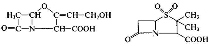
and several others. However, it must be noted that no single inhibitor can neutralize all the diverse types of beta-lactamases. THE SPECTRUM OF activity of each inhibitor is limited to only a few prevalent types of bacterial beta-lactamases.
Internationally, a 2:1 combination of the semisynthetic penicillin ampicillin with sulbactam is marketed under the trade name Unasyn, alongside sultamicillin, a mutual prodrug (chemical conjugate) of ampicillin and sulbactam. Another clinically established formulation is Augmentin, which combines amoxicillin (a semisynthetic penicillin) with clavulanic acid. When selecting combinations of beta-lactamase inhibitors and beta-lactam antibiotics, it is crucial to ensure that their pharmacokinetics are comparable. In other words, their distribution across Organs and Tissues, elimination pathways (e.g., predominantly renal or biliary), and systemic half-lives should be closely matched. An inhibitor will fail to provide adequate protection if its concentration at the site of infection is insufficient or if it is cleared from the body much faster than the companion antibiotic.
Enzymatic inactivation is the most common mechanism of resistance to aminoglycoside antibiotics. The enzymes responsible for aminoglycoside inactivation differ significantly from beta-lactamases. First, they are not Hydrolases; their activity requires more complex reaction conditions than simply an aqueous environment. Second, while some beta-lactamases in Gram-positive microorganisms are extracellular—facilitating their detection—the enzymes catalyzing aminoglycoside inactivation are almost exclusively intracellular.
According to the modern enzyme Nomenclature and Classification, enzymes that inactivate aminoglycoside antibiotics belong to the class of transferases. This means they do not cleave the aminoglycoside molecule; instead, they transfer specific chemical moieties onto it, catalyzing the replacement of hydroxyl groups with phosphate or adenylyl residues, and amino groups with acetate residues. Consequently, aminoglycoside-inactivating enzymes comprise phosphotransferases, adenylyltransferases, and acetyltransferases. A critical characteristic of these enzymes is that each catalyzes the substitution of only a single functional group within the aminoglycoside molecule. Nonetheless, this modification is typically sufficient to abolish the antibiotic's activity.
The donor of the phosphoryl and adenylyl groups transferred to aminoglycoside antibiotics is adenosine triphosphate (ATP), a well-known high-energy compound. ATP must be present in the reaction mixture; extracellular enzymes cannot exert a protective function in the medium because high concentrations of ATP are required for their catalytic activity. Similarly, The activity of aminoglycoside acetyltransferases requires the presence of acetic acid, coenzyme A, and ATP (or acetyl-CoA, which replaces the first two components).
Enzymes targeting aminoglycosides are typically localized in the bacterial cytoplasmic membrane, or in the periplasmic space (between the cell wall and the outer membrane) in Gram-negative bacteria. This strategic localization inactivates aminoglycosides as they penetrate the cell, thereby safeguarding their intracellular targets (ribosomes).
The genes encoding enzymes that catalyze the phosphorylation, Acetylation, or adenylylation of aminoglycosides are generally harbored on R-plasmids in bacteria, with chromosomal localization being extremely rare. Thus, they differ from beta-lactamases, whose genes are found in both plasmids and bacterial chromosomes.
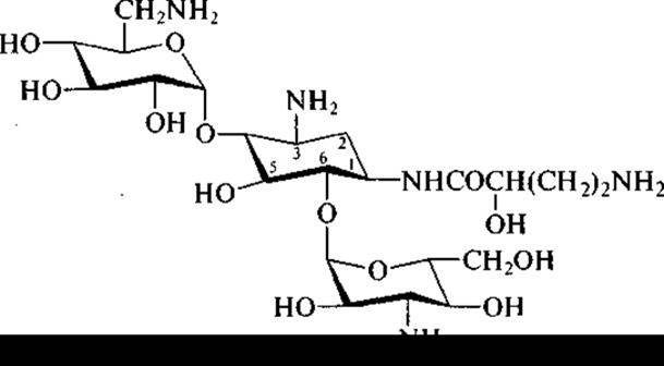
Following the elucidation of the mechanisms by which resistant bacteria enzymatically inactivate aminoglycoside antibiotics, efforts began to purposefully modify aminoglycoside structures to render them refractory to these modifying enzymes. For instance, in the kanamycin molecule, the NH2 group at position 1 of the aminocyclitol ring was substituted with an L-γ-amino-α-hydroxybutyric acid residue. This modification significantly altered the conformation of the natural molecule while preserving almost all of its functional groups. Consequently, antibacterial activity was retained, whereas susceptibility to all prevalent aminoglycoside-inactivating enzymes of resistant microbes was lost. The resulting derivative, named amikacin, proved highly effective against bacterial strains producing phosphotransferases, acetyltransferases, and adenylyltransferases that otherwise inactivate parent kanamycin.
Currently, amikacin is the most potent semisynthetic aminoglycoside antibiotic. It effectively inhibits the growth of microorganisms resistant to natural aminoglycosides due to its resistance to enzymatic inactivation, which is the most common resistance mechanism:
Microbial resistance to tetracycline antibiotics—such as tetracycline, oxytetracycline, and chlortetracycline—has become widespread due to their decades-long use in human medicine and livestock farming as growth-promoting feed additives.
At present, tetracycline is primarily used in clinical practice, whereas all three aforementioned antibiotics are employed in animal husbandry.
Research into the mechanisms of bacterial resistance to tetracyclines has yielded results that differ markedly from those established for beta-lactams and aminoglycosides. Specifically, enzymatic inactivation of tetracyclines by resistant microorganisms has not been observed.
In rare instances, resistance is associated with the protection or shielding of ribosomes (Protein Synthesis Inhibitors) from tetracyclines. Resistant strains have been found to express a protein that prevents tetracyclines from accessing their binding sites on the ribosome.
The most prevalent mechanism of tetracycline resistance stems from alterations in the cell envelope, specifically the bacterial cytoplasmic membrane. It is well established that tetracyclines do not accumulate within the cells of resistant strains. Instead, the Cytoplasmic membranes of these strains contain several novel proteins absent in tetracycline-susceptible strains. These newly appearing membrane proteins constitute an active efflux system that expels tetracyclines upon cell entry. In other words, although tetracyclines successfully penetrate the bacterial envelope and cytoplasmic membrane, they are rapidly pumped back out into the extracellular environment before they can interact with ribosomes.
Currently, several chemically modified derivatives of natural tetracyclines have been introduced into clinical practice. The most significant among them is doxycycline (6-deoxy-5-oxytetracycline), which exhibits a substantially longer half-Life in the body compared to natural tetracyclines.
Several mechanisms of resistance to erythromycin exist.
1. Ribosomes of resistant cells fail to bind erythromycin, rendering the antibiotic incapable of inhibiting protein synthesis. This is caused by the methylation of ribosomal RNA within the large subunit of the bacterial ribosome. In resistant cells, a specific methylase catalyzes the introduction of methyl groups into a strictly defined adenine residue within the rRNA molecule. Consequently, this alters the conformation of the entire large ribosomal subunit, preventing erythromycin from binding to it and to the ribosome as a whole.
2. Enzymatic cleavage of the macrocyclic lactone ring, leading to a loss of erythromycin activity.
3. Phosphorylation or hydroxylation at the OH group of one of the sugar moieties present in the antibiotic molecule.
Last update: 06/08/2026
Editorial and Educational Adaptation: This material has been compiled based on the primary/original source text. The project team performed an editorial review, corrected technical inaccuracies, structured sections, and adapted the content for an educational format.
What was processed:
- elimination of formatting defects (OCR errors, structural breaks, corrupted characters);
- editorial organization of content;
- standardization of terminology in accordance with academic sources;
- verification of factual statements against the original source text.
All mentions of the author, publication year, and origin of the primary text have been preserved in accordance with the source.