INVERTEBRATE ZOOLOGY IN THREE VOLUMES - BOOK 1 - H.I. Shcherbak - 1995
SUBKINGDOM PROTOZOA, OR UNICELLULAR ANIMALS (PROTOZOA)
PHYLUM SARCOMASTIGOPHORA (SARCOMASTIGOPHORA)
Subphylum Flagellata, or Mastigophora (Mastigophora, or Flagellata)
CLASS ANIMAL FLAGELLATES (ZOOMASTIGOPHOREA)
A significant number of species of animal flagellates inhabit fresh and salt waters, but the vast majority are parasites of animals and plants. Representatives of this Class lack chlorophyll, are heterotrophic, and feed via pinocytosis and phagocytosis. Representatives of various orders differ in the number of flagella, body shape, and other structural features. The class consists of eight orders, of which we will examine the five of greatest practical and theoretical importance: Choanoflagellida, Kinetoplastida, Diplomonadida, Trichomonadida, and Multiflagellata.
Order Choanoflagellates (Choanoflagellida)
These are very small, solitary or colonial flagellates inhabiting fresh and salt waters. A flagellum is located at the anterior end of The Cell. Its base is surrounded by a collar consisting of a wreath of very thin microvilli, visible only under an Electron microscope. A stream of Water generated by the movement of the flagellum passes between them; small food particles are filtered out and phagocytosed with the aid of pseudopodia. Among the choanoflagellates, there are swimming and attached (by the posterior end) species; some marine forms form a "lorica" (house) around the cell made of fused siliceous spicules.
Choanoflagellates bear a strong morphological resemblance to the choanocytes of Sponges, indicating THE ORIGIN OF sponges from these Protozoans. Their representatives include the colonial organisms Sphaeroeca, Proterospongia, and the solitary Salpingoeca (Fig. 7).
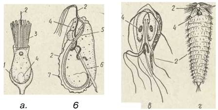
Fig. 7. Structure of animal flagellates—Salpingoeca amphoroideum in a lorica (a), Trichomonas termopsidis (б), Lamblia intestinalis (в), Teratonympha mirabilis (г): 1—lorica; 2—flagellum; 3—microvilli; 4—Nucleus; 5—costa; 6—axostyle; 7—undulating membrane
Order Kinetoplastids (Kinetoplastida)
Most members of this order are parasites, although free-living species also exist (e.g., from the genus Bodo, inhabiting fresh waters). A characteristic feature of the order is the presence, near the Base of the flagellum, of a kinetoplast located next to the basal body—the kinetosome (Fig. 8). This is an accumulation of DNA in a Specialized Part of a giant single mitochondrion.
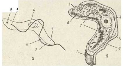
Fig. 8. Structural diagram (a) and ultrastructure (б) of Trypanosoma brucei:
1—anterior end; 2—flagellum; 3—undulating membrane; 4—nucleus; 5—kinetosome; 6—kinetoplast; 7—mitochondrion
Many species of kinetoplastids possess an undulating membrane—a thin plasma fold connecting the flagellum to the cell surface. The flagellum originates at the posterior end of the cell, runs parallel to its surface, and its free end protrudes beyond the anterior margin of the animal's body. This membrane facilitates movement in a viscous medium, such as vertebrate Blood. Kinetoplastids reproduce by division.
In many parasitic forms, The life cycle involves a change of host species and an associated change in
morphological phases, which arose in connection with adaptation to the internal environment of a particular host. The ancestral form—the so-called promastigote—is characterized by the flagellum being located at the anterior end of the cell, with no undulating membrane. This form can change in two directions: 1) the flagellum is reduced, leaving only the basal body and kinetoplast, while the cell itself shortens—the amastigote stage; 2) the locomotory function of the flagellum increases, it originates from the middle of the cell, an undulating membrane appears—the epimastigote stage, and when the flagellum elongates further, its base is located at the posterior end of the body—the trypomastigote stage (Fig. 9, a).
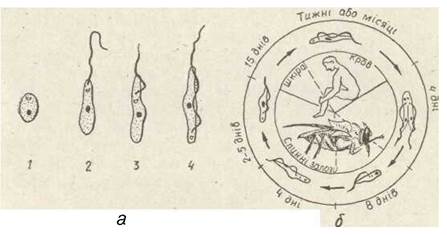
Fig. 9. Modification forms of trypanosomes (a) and developmental cycle of Trypanosoma brucei gambiense (б): 1—amastigote form; 2—promastigote; 3—epimastigote; 4—trypomastigote
Among kinetoplastids, There are many pathogens causing severe Human and Animal diseases. The species of the genera Trypanosoma and Leishmania are of the greatest medical and veterinary importance. A severe human disease, sleeping sickness, is widespread in Equatorial Africa; it is caused by two subspecies of the species Trypanosoma brucei—T. b. gambiense (chronic form of the disease) and T. b. rhodesiense (acute form).
The CAUSATIVE AGENT OF sleeping sickness inhabits the blood of antelopes (the reservoir host) without causing them noticeable harm. In antelopes, the trypanosome exists in the trypomastigote stage. Infection of antelopes and humans occurs during the bite of a blood-sucking tsetse fly (genus Glossina), in whose intestine and subsequently Salivary Glands the pathogen multiplies (epimastigote stage). The fly can also transmit the pathogen directly from one human to another. After the fly's bite, the parasite enters human blood with its saliva, where it transitions back into the trypomastigote stage (Fig. 9, б). In humans, trypanosomes parasitize Blood Plasma, Lymph, and CEREBROSPINAL FLUID, damaging the Central Nervous system. The disease is accompanied by fever, inflammation of the Lymph Nodes, weakness, mental disorders, and drowsiness; in the absence of Treatment, a person with the acute form of the disease dies within six to nine months, and with the chronic form, within several years.
In the 19th century, sleeping sickness was a terrible scourge. However, drugs (such as Germanin) have now been developed that completely cure people.
Sleeping sickness is a classic example of a vector-borne disease. Vectors are Arthropods that transport pathogens from one vertebrate animal (or human) to another. Vectors can be specific, when the parasite undergoes a certain stage of development and multiplies in the vector's body (e.g., the tsetse fly is a specific vector of sleeping sickness), or mechanical, when the pathogen retains viability in the vector's body for some time without multiplying, for example, horseflies are mechanical vectors of T. brucei brucei, which causes nagana, a disease of cattle.
Sleeping sickness is a vector-borne disease with natural foci. One of the first to discover
natural plague foci in the 1920s and 1930s was the Ukrainian scientist D. K. Zabolotny. The fundamental principles of The Doctrine of natural-focal diseases were formulated by the Russian scientist Ye. N. Pavlovsky. A natural focus of vector-borne diseases is a phenomenon in which the pathogen, its vector, and the animal reservoir coexist indefinitely in natural conditions independently of humans.
In cases where the pathogen is transmitted from one animal to another directly rather than via a vector—for example, when animals prey on each other—a natural focus of a non-vector-borne disease arises.
In Latin America, a severe human pathology known as Chagas disease is caused by another species, T. cruzi (Fig. 10). It is transmitted by blood-sucking bugs of the family Reduviidae (such as Triatoma), with the trypanosomes parasitizing the bug's hindgut. The bug feeds on blood from the mucous membranes of the eyes, Lips, or Nose. During this process, the bug's feces are deposited onto the mucosa, and the trypanosomes contained within them penetrate the body through microscopic cracks in the Skin. Trypanosomes affect the Cells of the Heart, Blood Vessels, Muscles, and Nervous Tissue, among others. The disease is accompanied by fever, enlargement of The Liver and Spleen, psychiatric and cardiac disorders, and gastrointestinal disturbances. It is most severe in children (with mortality rates reaching up to 14%). Reservoir hosts for the pathogen include various wild animals (rodents, monkeys, etc.), as well as domestic dogs and cats in human settlements.
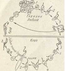
Fig. 10. Life Cycle of Trypanosoma cruzi: 1 — amastigote form; 2 — epimastigote; 3 — trypomastigote; 4 — promastigote
Several species of trypanosomes are pathogens of livestock diseases. Nagana, a disease of cattle, is caused by T. b. brucei and two other trypanosome species. It is widespread in Africa, where it is transmitted by various species of tsetse flies, and in tropical Asia, where it is spread by mechanical vectors such as horseflies. Nagana is characterized by fever, anemia, and fatal outcomes. In addition to cattle, nagana affects camels, horses, sheep, and other animals. Natural foci are maintained through populations of various wild ungulates.
In North Africa, the Mediterranean, and Central Asia, T. evansi causes surra—a disease in horses, donkeys, and camels that is clinically very similar to nagana and is mechanically transmitted by horseflies.
Dourine, a contagious venereal disease of horses that leads to death, is widespread in various PARTS OF THE world and is caused by T. equiperdum. The disease is transmitted without a vector, during sexual intercourse.
Close relatives of trypanosomes are leishmanias (genus Leishmania), which are intracellular parasites of humans and other vertebrates, existing within the host in the amastigote stage. Their vectors are blood-sucking dipterans of the sandfly family (Phlebotomidae), in whose gut and salivary glands the parasite resides in the promastigote stage; infection occurs through a bite. Leishmanias never form an undulating membrane (Fig. 11).
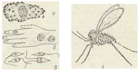
Fig. 11. Leishmanias:
a — Leishmania tropica in a cell from an ulcer; b — skin ulcers caused by L. tropica parasitism; c — L. donovani from a culture; d — Phlebotomus papatasii, the vector of leishmaniasis pathogens
L. tropica causes a cutaneous condition known by several names: cutaneous leishmaniasis, Borovsky's disease, or oriental (or Pendinsky) sore. It is prevalent in the Transcaucasia and Central Asia. The reservoir hosts of L. tropica are various rodents.
L. donovani causes visceral leishmaniasis (kala-azar), a disease widespread in Asia. Leishmanias infect cells of Internal Organs, including the liver, spleen, lymph nodes, and intestinal walls. The disease is accompanied by fever, enlargement of the affected organs, and anemia. Reservoir hosts are primarily dogs and jackals.
Representatives of the genus Leptomonas parasitize plants, locating themselves in latex-filled vessels, intercellular spaces, and cell vacuoles, often multiplying in huge numbers within plant tissues. They are among the most harmful coffee pests (especially in South America), causing infested trees to die within 3 to 12 months. The vectors are herbivorous insects with piercing-sucking mouthparts, such as certain true bugs.
Order Diplomonadida
Members of this order lead a predominantly parasitic lifestyle. These are dienergid organisms typically possessing two nuclei and eight flagella arranged in two groups associated with a specific nucleus. Each flagellum group is linked to a groove on the body surface. Diplomonads include Lamblia intestinalis (Giardia), a parasite of the upper human Small Intestine. It exhibits bilateral Symmetry, with an attachment organelle—a sucker—located on the flattened ventral side of the body. At times, lamblias multiply on the intestinal walls in such quantities that they block nutrient absorption processes. They feed by pinocytosis of nutrients from the midgut (see Fig. 7). Upon reaching the hindgut, lamblias shed their flagella and secrete a thick wall, transforming into cysts. In the encysted state, the parasite disperses and infects a new host. Diplomonads also parasitize rodents, amphibians, and other animals.
Order Trichomonadida
Members of this order are parasites. At the anterior end of the body, they possess four to six flagella, one of which is directed backward and connected to the body by an undulating membrane, beneath which a special contractile rod-like organelle, the costa, lies within the cell; the remaining flagella project freely beyond the body.
They feature a supporting structure known as the axostyle, which runs longitudinally through the cell interior. It consists of microtubule strips originating from the kinetosomes of the flagella. Mitochondria are absent in trichomonads (see Fig. 7). Trichomonads parasitize various organs, primarily the intestinal cavity of vertebrates. In humans, Trichomonas vaginalis (a parasite of the Urogenital System) and T. hominis (an intestinal parasite) are found. In the reproductive organs of cattle, T. foetus parasitizes and can cause Infertility or abortion if it enters the Uterus.
Order Hypermastigida
Members of this order are intestinal symbionts of Cellulose-digesting insects (such as termites and certain species of cockroaches). Hypermastigids possess A large number of flagella grouped at the anterior end of the body or covering the entire cell surface; they have one or several axostyles and lack mitochondria, much like trichomonads. Both mono- and polyenergid species are known; in the latter, all nuclei are morphologically identical. Hypermastigids feed primarily on wood, ingesting small fragments using pseudopodia formed only in certain Regions of the body. The relationship between termites and the hypermastigids inhabiting their gut is a classic example of Symbiosis: these flagellates can only survive within the termite gut, where the contents serve as both their habitat and food source. Conversely, termites cannot survive without these flagellates, as their own guts do not secrete Enzymes capable of breaking down cellulose. The flagellates possess this enzyme and digest cellulose, utilizing only a portion of the resulting nutrients, while the remainder is assimilated by the termites. Termite nymphs feed on the feces of adults, which contain the symbionts. Termites artificially deprived of hypermastigids die of starvation. About 200 species of hypermastigids—termite symbionts—have been described. The most widespread among them are Teratonympha mirabilis and Calonympha grossi (see Fig. 5, e).
Last update: 13/08/2026
Editorial and Educational Adaptation: This material has been compiled based on the primary/original source text. The project team performed an editorial review, corrected technical inaccuracies, structured sections, and adapted the content for an educational format.
What was processed:
- elimination of formatting defects (OCR errors, structural breaks, corrupted characters);
- editorial organization of content;
- standardization of terminology in accordance with academic sources;
- verification of factual statements against the original source text.
All mentions of the author, publication year, and origin of the primary text have been preserved in accordance with the source.