IMMUNOLOGY - Roitt I. - Mir 2000
Chapter 7. T-Cell Antigen-Recognition Receptors and MHC Molecules
MHC GENOMIC ORGANIZATION
Class I loci in the mouse
The mouse MHC is designated as H-2. The regions of H-2 containing class I and class II genes are denoted by letters; for instance, the chromosomal region housing class I MHC genes is designated H-2K. The MHC genes themselves, as well as their loci, are highly polymorphic, with different mouse strains varying both in The Structure of these genes and in the number of their alleles within the loci. Therefore, distinct H-2 complexes are also assigned superscript letter designations, such as the BALB/c mouse haplotype denoted as H-2d.
The mouse MHC (H-2) contains three class I loci, but different haplotypes vary in the number of class I genes. The haploid mouse genome contains approximately 30 class I genes, though different strains may differ in the exact copy number. Class I genes encoding classical Histocompatibility Antigens detectable by serological assays are located within the H-2K, H-2D, and H-2L loci. The Functional Significance of most other class I genes mapped to the Qa, TIa, and M loci (Fig. 7.12) remains largely unclear, although it is known that Qa molecules can induce T-Cell activation.
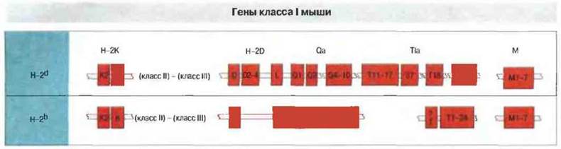
Fig. 7.12. Gene order of class I MHC genes on the chromosome of two mouse haplotypes: BALB/c (H-2d) and B10 (H-2b). Class II and class III genes are situated between the H-2K and H-2D regions. To align corresponding alleles of different haplotypes vertically, linear maps include arbitrary breaks represented by jagged edges. The TIa region harbors numerous class I genes of unknown function.
The STRUCTURE OF THE H-2K region is identical across all mouse strains studied to date. It contains two class I genes, designated K and K2 (see Fig. 7.12). The H-2K gene encodes the H-2K antigen, which is expressed and serologically detected on most cell types, whereas the expression pattern of the H-2K2 gene varies depending on the strain.
In the H-2d and H-2b haplotypes, the H-2D/H-2L region contains a variable number of class I genes (see Fig. 7.12). In BALB/c mice (H-2d haplotype), five such genes have been mapped within this region. Two of them encode the serologically defined H-2Dd and H-2Ld antigens. The remaining three class I genes are located in the chromosomal segment between the H-2Dd and H-2Ld genes (positioned proximally and distally, respectively) and are designated D2d, D3d, and D4d. Their Functions remain unknown. Conversely, in B10 mice (H-2b haplotype), only a single class I gene has been identified in the H-2D region.
GENES OF THE Qa, TIa, and M loci encode "non-classical" class I MHC molecules. The products of genes residing in the Qa, TIa, and M loci are structurally similar to the classical class I molecules described above, which is why they are sometimes referred to as "non-classical" class I molecules. The Qa region spans approximately 200 kb distally to the H-2D/L region (see Fig. 7.12) and contains genes for the serologically defined Qa-2, 3, 4, and 5 specificities, alongside a cluster comprising between 8 (BALB/c) and 10 (B10) class I genes.
The TIa locus, initially believed to house genes exclusively encoding TL (Thymus leukaemia) antigens—i.e., markers of thymocytes and T-cell leukemias—was subsequently found to contain numerous class I genes and extensive structural divergence between the H-2b and H-2d haplotypes (B10 and BALB/c strains, respectively) (see Fig. 7.12). The M region, positioned between the K and A regions, contains a series of recently identified class I genes designated M1–M7, which exhibit low polymorphism.
The human MHC contains three class I loci
In humans, the class I gene region comprises three loci termed HLA-A, HLA-B, and HLA-C, spanning a segment of over 1500 kb (Fig. 7.13). The genes at each locus encode the heavy chains of "classical" class I MHC antigens. Closer examination of this region revealed numerous additional genes encoding class I MHC-like Proteins outside these loci. These include the HLA-E, HLA-F, and HLA-G loci. The HLA-G gene product is known to be expressed on extravillous cytotrophoblast Cells of the Placenta, presumably protecting them from maternal NK cells (see Ch. 10). Other class I genes—potential homologues of the murine Qa, TIa, and M genes—are localized near the HLA-G and HLA-A loci.

Fig. 7.13. In the human chromosome, class I genes are located closer to the telomere than class II and class III genes. In addition to the classical transplantation antigen genes (HLA-A, HLA-B, and HLA-C), several "class I-like" genes are identified in this region, which are likely equivalent to the murine TIa/Qa region genes. This area also contains A number of other non-class I genes.
Class II genes are located in the H-2I region
In the mouse, the α and β chains of class II molecules are encoded by distinct genes situated within the I region of the H-2 complex (Fig. 7.14). For example, the α and β chains of the A molecule are encoded by the Aa and Ab genes, respectively, while the corresponding chains of the E molecule are encoded by Ea and Eb genes (gene nomenclature indicates the locus first, followed by the encoded chain). Protein products of several other cloned class II a and b genes have not yet been identified. One of these, Pb, is a pseudogene, whereas two others, Ob and Eb2, may possess some biological function. These latter two genes display low polymorphism and are actively transcribed, though it remains unclear whether they are translated.
The H-2I region has been mapped almost entirely. It lies proximally adjacent on the chromosome to the H-2Rk region, whose genes encode class I molecules. Mice with the b, s, f, and q haplotypes lack expression of class II molecules encoded by the I-E subregion genes. Haplotypes b and s fail to transcribe the Ea gene yet exhibit normal cytoplasmic levels of Eβ chains. Mice with the f and q haplotypes fail to synthesize both Eα and Eβ chains.
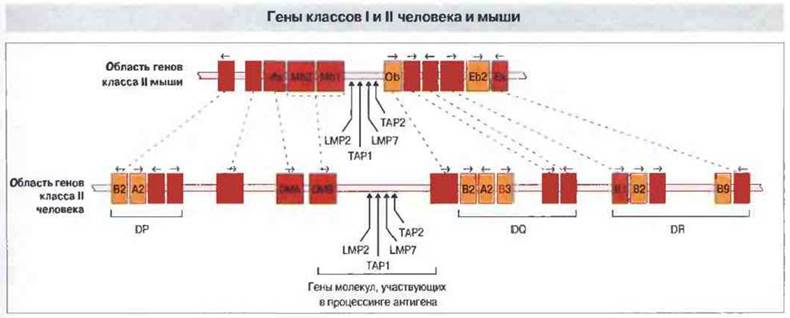
Fig. 7.14. MHC Introduction/29.html">Gene Organization in the mouse and human. Homologous genes are connected by dashed lines. Expressed class I and class II genes are highlighted in orange, and pseudogenes in yellow.
Human class II genes are localized in the HLA-D region
The genes of the HLA-D region encode at least six variants of α-chains and ten variants of β-chains that assemble into class II molecules (see Fig. 7.14). These genes span three loci: DR, DQ, and DP. Their expression products comprise the majority of human class II molecules. Additional class II genes situated outside these loci have also been identified. The DR locus contains a single α-chain gene (DRA) and up to nine β-chain genes (DRB1-9), including pseudogenes. Several organizational variants of genes within this locus are known. Each of the DQ and DP loci contains one expressed pair of α- and β-chain genes, alongside an additional pair of homologous genes that may be functional or non-functional.
As with their murine counterparts, human DR, DQ, and DP α-chains within the molecular complex associate predominantly with β-chains encoded by genes from the same locus. Using specific Antibodies, it has been shown that HLA-class II DP molecules are formed by pairing the products of the DPA1 and DPB1 genes. Similarly, the DQA1 and DQB1 genes encode HLA-DQ antigens.
The organization and extent of the DRB region vary according to haplotype (Fig. 7.15), as does the number of expressed β-chain variants. The DRB2 locus represents a pseudogene. The DRB1, DRB3, and DRB4 genes are typically functional, whereas DPA2, DPB2, and DQB3 are mostly pseudogenes and therefore unexpressed. The DNA, DOB, and DQA2 genes may be functional.
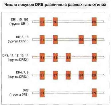
Fig. 7.15. The number of DRB loci varies across different haplotypes. Their arrangement is shown for the DR1, DR10, and DR103 haplotypes (top line), DR15 and DR16 (second line from the top), etc. Not all of these loci serve as templates for the synthesis of DRβ-chain mRNA.
The HLA-D region also contains genes for proteins involved in antigen presentation that are not expressed on The Cell surface. These genes and their products are discussed in Chapter 9.
Long-range DNA mapping techniques have made it possible to determine the linear order of all identified class II genes within the HLA-D region. This entire region spans a DNA segment of approximately 1000 kb. The linear organization and orientation of homologous class II loci are identical in humans and mice.
Polymorphic regions of MHC molecules are concentrated primarily in the antigen-binding site
A unique feature of the MHC is the extraordinary polymorphism (structural Variability) of the molecules encoded by its genes. However, not all MHC products are polymorphic to the same degree. The Qa, Tla, and M antigens, which are structurally similar to class I molecules, are much less polymorphic than classical class I and II antigens. A list of human leukocyte antigen (HLA) class I and II specificities, as well as the alleles for each HLA locus, is provided in Appendix I.
Structural differences between variants of a given class of molecules mainly involve specific regions of The polypeptide chains; for instance, in class I molecules, these are concentrated in the α1 and α2 domains. The α3 domain appears to be more conserved.
The degree of variability of class II molecules depends on the locus and can vary among different chains; for example, the DRβ and DQβ chains exhibit exceptional polymorphism, and the DPβ chains to a slightly lesser extent. The DQα chains are variable, whereas DRα is considered invariant, and DPα has only two allelic forms. In outbred populations, carriers of two MHC haplotypes can express hybrid class II molecules containing chains from different haplotypes, introducing an additional level of structural diversity.
Most of the variable amino acid residues in class I and II MHC antigens are clustered in the upper region of the molecule, where they form a deep cleft for binding foreign Peptides. Furthermore, Almost all amino acid substitution positions are gathered together at the bottom of this cleft or on the inward-facing sides of the α-helices. The functional significance of such a cluster of variable residues (regarding T-cell antigen recognition) is discussed in detail in Chapter 9.
Questions for Thought
■ The antigen-binding cleft of a class I molecule accommodates a peptide of 8–9 amino acid residues. Is there any reason why human MHC molecules could not accommodate peptides of 3 or 30 residues?
■ Determining the three-dimensional structures of class I and II molecules has provided insight into the mechanism by which they bind antigenic peptides. Consequently, it has been suggested that immune responses could be enhanced or suppressed by introducing peptides that bind to MHC molecules. How could an understanding of the binding mechanism aid in the design of therapeutic drugs based on such peptides?
■ Unlike the B-cell receptor, the TCR typically exhibits low affinity for antigen and recognizes it only in complex with class I or class II MHC molecules. Cell-surface accessory molecules also play a key role in interactions between T cells and target cells, performing a variety of functions. What processes, aside from antigen recognition, occur when a T cell comes into contact with a virus-infected target cell?
■ Compared to class I molecules, the antigen-binding cleft of class II molecules can accommodate longer peptides. What advantage does this confer?
Bentley G.A., Mariuzza R.A. 1996. The structure of the T CELL ANTIGEN receptor. Annu. Rev. Immunol. 15: 563-590.
Bjorkman P.J., Parham P. 1990. Structure, function and diversity of Class I Major Histocompatibility Complex molecules. Annu. Rev. Biochem. 59: 253-288.
Bjorkman P.J., Saper M.A., Samraoui B. et al. 1987. The structure of the human Class I histocompatibility antigen HLA-A2. Nature 329: 506-512.
Bjorkman P.J., Samraoui B., Bennett W.S. et al. 1987. The foreign antigen binding site and T-Cell Recognition regions of Class I histocompatibility antigens. Nature 329: 512-516.
Bodmer J.G., Marsh S.E., Albert E.D. et al. 1994. Nomenclature for factors of the HLA system, 1994. Tissue Antigens 44: 1-18.
Brenner M.B., MacLean J., Dialynas D.P. et al. 1986. Identification of a putative second T-cell receptor. Nature 322: 145-149.
Brown J.H., Jardetzky T.S., Gorga J.C. et al. 1993. Three-dimensional structure of the human class II histocompatibility antigen HLA-DR1. Nature 364: 33-39.
Carosella E.D., Dausset J., Kirzenbaum H. 1996. HLA-G revisited. Immunol. Today 17: 407-409.
Clevers H., Alarcon B., Wileman T. et al. 1988. The T-cell receptor/CD3 complex: a dynamic protein ensemble. Annu. Rev. Immunol. 6: 629-662.
Garcia K.C., Degano M., Stanfield R.L. et al. 1996. An αβ T cell receptor structure at 2.5Å and its orientation in the TCR-MHC complex. Science 274: 209-219.
Garratl T.P.J., Saper M.A., Bjorkman P.J. et al. 1989. Specificity pockets for the side chains of peptide antigens in HLA-w68. Nature 342: 692-696.
Green N.M. 1991. The semiotics Of charge. Nature 351: 349.
Hass W., Pereira P., Tonegawa S. 1993. Gamma/delta cells. Annu. Rev. Immunol. 11: 637-685.
Koop B.F., Hood L. 1994. Striking sequence similarity over almost 100 kilobases of human and mouse T-cell receptor DNA. Nat. Genet. 7: 48-53.
Lefranc М.-P., Rabbitts T.N. 1989. The human T-cell receptor γ (TRG) genes. Trends. Biochem. Sci. 14: 214 218.
Madden D.R., Gorga J.C., Strominger L. et al. 1991. The structure of HUX-B27 reveals nonamer self-peptides bound in an extended conformation. Nature 353: 321-325.
Manolios N., Letourneur F., Bonifacino J.S. etal. 1991. Pairwise cooperative and inhibitory interactions describe the assembly and probable structure of the T-cell antigen receptor. EMBOJ. 10: 1643-1651.
Neefjes J.J., Momburg F., Hamerling B.J. 1993. Selective and ATP-dependent translocation of peptides by the МНС-encoded transporter. Science 261: 769-771.
Powis S.H., Trowsdale J. 1994. Human major histocompatibility complex genes. Behring Inst. Mitt. 94: 17-25.
Raulet D.H. 1994. How γδ T cells make a living. Curr. Biol. 4 246-251.
Salter R.D., Benjamin R.J., Wesley P.K. et al. 1990. A binding site for the T-cell co-receptor CD8 on the otg domain of HLA-A2. Nature 345: 41-46.
Sloan-Lancaster J., Allen P.M. 1996. Altered peptide-Ligand induced partial T cell activation: molecular mechanisms and role in T cell biology. Annu. Rev. Immunol. 14: 1-27.
Stern L.J., Wiley D.C. 1994. Antigenic peptide binding by class I and class II histocompatibility proteins. Structure 2: 245-61.
Stern L.J., Brown J.H., Jardetzky T.S. et al. 1994. Crystal structure of the human class II MHC protein HLA-DR1 complexed with an Influenza virus peptide. Nature 368: 215-221.
Weiss A., Liftman D.R. 1994. Signal Transduction by lymphocyte antigen receptor. Cell 76: 263-274.
Williams A.F., Barclay A.N. 1988. The immunoglobulin superfamily - domains for cell surface recognition. Annu. Rev. Immunol. 6: 381-405.
Last update: 13/08/2026
Editorial and Educational Adaptation: This material has been compiled based on the primary/original source text. The project team performed an editorial review, corrected technical inaccuracies, structured sections, and adapted the content for an educational format.
What was processed:
- elimination of formatting defects (OCR errors, structural breaks, corrupted characters);
- editorial organization of content;
- standardization of terminology in accordance with academic sources;
- verification of factual statements against the original source text.
All mentions of the author, publication year, and origin of the primary text have been preserved in accordance with the source.