IMMUNOLOGY - Roitt I. - Mir 2000
Chapter 18. Immunity to Parasitic Infections
MECHANISMS OF DEFENSE AGAINST THE IMMUNE RESPONSE
For successful ESTABLISHMENT AND DEVELOPMENT within the host Organism, parasites must evade its defense mechanisms; consequently, parasitic Protozoa and helminths have developed various ways to bypass them. Some parasites even exploit the host's immune Cells and molecules to their own advantage: Leishmania enter macrophages via Complement receptors, thereby avoiding a respiratory burst and subsequent destruction by toxic macrophage products.
While host cytokines play a protective role in the Immune Response to many parasitic infections, they simultaneously promote parasite growth. For instance, TNFα stimulates egg-laying by adult S. mansoni, whereas IFNγ serves as a growth factor for T. brucei.
Parasites may be resistant to complement-mediated destruction
In Leishmania, such resistance correlates with virulence. Infection with L. tropica, whose cells are readily destroyed by complement, results in a localized, self-healing Skin infection, whereas L. donovani — an organism 10 times more resistant to complement action — spreads to Internal Organs, causing a disease that is frequently fatal.
The mechanisms by which parasites protect themselves against complement vary. In L. major, the protozoan's lipophosphoglycan surface coat activates complement, but the membrane-attack complex is shed from their cells, thereby avoiding lysis. Trypomastigotes of T. cruzi carry a surface glycoprotein with decay-accelerating factor (DAF) activity for C3 convertase, which limits complement reactivity (see Chapter 4). Schistosomula acquire resistance as they mature, which also correlates with the generation of surface molecules similar to DAF.
Intracellular parasites evade destruction through various pathways
Protozoa that parasitize macrophages avoid destruction by oxygen metabolites and lysosomal Enzymes through various mechanisms (Figs. 18.20 and 18.21). T. gondii cells enter macrophages not via conventional phagocytosis (Fig. 18.21) and thus do not trigger a respiratory burst; Leishmania spp. can enter macrophages by binding to complement receptors, representing another pathway to bypass the respiratory burst. In addition, Leishmania possess superoxide dismutase and Other Enzymes that protect them from highly reactive oxygen metabolites. The vacuoles in which Leishmania survive are actually Lysosomes (Fig. 18.22), yet these protozoa possess mechanisms that ensure protection against lysosomal enzymes. The lipophosphoglycan surface coat of Leishmania Functions as a "trap" for oxygen metabolites and protects against enzymatic lysis; furthermore, the contained glycoprotein Gp63 (Fig. 18.23) inhibits macrophage lysosomal enzymes. Leishmania are also capable of downregulating MHC Class II antigen expression On the surface of their host macrophages, thereby reducing the ability of macrophages to stimulate Th cells. However, these mechanisms are less effective when infecting an immune host.

Fig. 18.19. Various mechanisms of schistosome damage identified in vitro. Complement not only directly damages helminths (1) but also acts in conjunction with Antibodies (2). Th1 cells can suppress The Development of schistosomula in the Lungs (3). Antibodies sensitize neutrophils (4), macrophages (5), platelets (6), and eosinophils (7), triggering antibody-dependent Cell-mediated cytotoxicity reactions. Neutrophils and macrophages likely act via the generation of toxic oxygen and nitrogen metabolites, whereas eosinophils damage the worm tegument by releasing major basic protein. The response is enhanced by cytokines (e.g., TNFα). IgE antibodies play an important role in sensitizing eosinophils and local mast cells, which release various mediators, including those that activate eosinophils.
(ROM — reactive oxygen metabolites.)

Fig. 18.20.
Toxoplasma gondii. Live tachyzoites actively invade the host cell, localizing within a membrane-bound vacuole. In this state, they are inaccessible to enzymes because lysosomes fail to fuse with such vacuoles. However, dead tachyzoites are engulfed by phagocytes via normal phagocytosis with The formation of phagosomes (if coated with antibodies that bind to Fc receptors on the macrophage surface), where they are destroyed by enzymes following phagosome-lysosome fusion.
Trypanosoma cruzi. The survival of this parasite depends on its developmental stage: trypomastigotes escape from phagosomes and divide in the Cytoplasm, whereas amastigotes remain in phagosomes and are destroyed. The proportion of parasites present in the cytoplasm is lower if the macrophages are activated.
Leishmania spp. Leishmania multiply within phagosomes: they are resistant to lytic enzymes due to possessing a surface protease. If macrophages have been pre-activated by cytokines, the number of parasites invading the cells and multiplying within them will be lower.

Fig. 18.21. Electron micrograph showing the active invasion of a host cell by Toxoplasma gondii. Entry occurs via the formation of a tight contact with the host cell membrane (indicated by the arrow) and is accompanied by the Modification of the resulting phagosome to prevent its fusion with a lysosome. (Photo courtesy of Dr. J. Smith.)
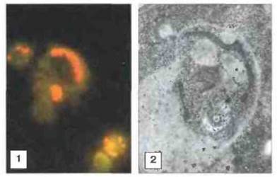
Fig. 18.22. The vacuole containing Leishmania cells is a lysosome. 1. Immunofluorescence staining of mouse macrophages infected with Leishmania mexicana using rhodamine-conjugated anti-tubulin antibodies — staining Leishmania yellow-red — and fluorescein-conjugated Monoclonal Antibodies reacting with the late endosome/lysosome marker LAMP-1 — green. 2. Electron micrograph of an infected L. mexicana mouse macrophage treated with gold-labeled anti-cathepsin D antibodies to detect lysosomal proteinase within the Leishmania-containing vacuole. (Photos courtesy of Dr. D. Russell.)

Fig. 18.23. Schematic representation of two Leishmania surface Antigens anchored in the membrane by phosphatidylinositol tails (GPI anchor).
1. The protein antigen Gp63 exhibits protease activity. In L. mexicana, it acts in concert with lipophosphoglycan to bind complement. This enables the promastigote to enter the macrophage via the complement C3 component receptor.
2. The glycolipid antigen lipophosphoglycan provides resistance to complement-mediated lysis. In L. major, it binds C3b, a fragment of the third complement component, allowing promastigotes to enter macrophages via the CR1 complement receptor. In mice, antibodies to both antigens confer protection against cutaneous leishmaniasis.
It is now well established that many parasite coat Proteins, such as the variant surface glycoprotein of T. brucei, are anchored to the surface membrane via a GPI anchor.
Extracellular parasites are capable of camouflage
Parasites susceptible to specific antibodies possess various mechanisms of resistance to the HUMORAL IMMUNE RESPONSE. African trypanosomes exhibit antigenic variation: the variant surface glycoprotein that forms the surface coat of the parasites can be expressed in novel, structurally altered forms, protecting the underlying membrane from lysis by the host's defense mechanisms. New trypanosome populations emerging during infection differ in antigenic composition from preceding ones (Figs. 18.24 and 18.25). Certain antigens of malaria parasites also display antigenic variation.
Other parasitic organisms, such as schistosomes, "dress" in a coat of host antigens so that the host defense systems fail to recognize them as foreign objects and do not distinguish them from host Tissues. When cultured in a nutrient medium containing human serum and erythrocytes, schistosomula acquire surface structures bearing Blood group A, B, and H determinants, as well as MHC antigens. However, in media devoid of host molecules, schistosomula, as noted above, also acquire resistance to antibodies and complement.

Fig. 18.24. Antigenic variation in trypanosomes. Immunofluorescence staining of trypanosomes using monoclonal antibodies specific for a particular antigenic variant (1). Micrograph (2) shows the same field of view, but with the parasite nuclei and kinetoplasts stained with a DNA-binding dye. Only some of the parasites express this variant, whereas others express distinct antigen variants. (Photographs kindly provided by Dr. M. Turner.)

Fig. 18.25. Trypanosome infection can last for months, accompanied by periodic bursts of parasitemia.
Graph 1 illustrates fluctuations in parasitemia during sleeping sickness. Although the infection originated from a single infectious agent, successive outbreaks are caused by immunologically distinct populations of the parasite (a, b, c, d), while antibodies against preceding antigenic variants fail to provide ongoing protection. There is a consistent tendency for new antigenic variants to appear in the same sequence across different host organisms. Such variation is not observed in immunocompromised animals (e.g., those treated in ways that impair specific immune functions).
Graph 2 shows The kinetics of antibody production against four variant trypanosome antigens in a rabbit following a bite by the tsetse fly vector, Trypanosoma brucei. Antibodies to a given antigenic variant appear immediately after its emergence, and their titer rises to a plateau. Antibody production prompts A change in parasite antigens and the appearance of a new immunologically distinct parasite population.
Graph 3 illustrates the kinetics of a single cycle of antigenic variation. A rat was infected with a homogeneous population of a single antigenic variant of T. brucei (a). A second wave of parasitemia occurs when a new dominant antigenic variant emerges (b).
Some extracellular parasites "hide" from immune attack
Certain protozoan species (such as Entamoeba histolytica) and helminths (such as T. spiralis) form protective cysts; adult Onchocerca volvulus residing in the skin induce the host organism to wall them off within Collagen nodules. Nematodes and tapeworms are shielded from many host immune responses simply by virtue of inhabiting the intestinal lumen.
Some extracellular parasites are able to withstand immune attack
Numerous Examples of simple physical defense mechanisms are known in parasites: nematodes possess a thick extracellular cuticle that protects them from toxic host metabolites (Fig. 18.26); the tegument of schistosomula thickens as they mature into adults, providing additional protection; the "loose" surface coat of many nematodes can be "shed" in response to an immune attack; and tapeworms prevent immune reactions by secreting an Elastase inhibitor that blocks neutrophil recruitment.
Many helminths have The ability to protect themselves against highly reactive O2 metabolites generated during the respiratory burst of immune cells. For instance, lymphatic-dwelling filarial worms can secrete surface-bound Glutathione peroxidase; schistosomes possess surface-bound glutathione S-transferases; and Onchocerca nematodes can secrete superoxide dismutase. Certain nematodes and trematodes have developed mechanisms to impair antibodies by secreting proteases that cleave IMMUNOGLOBULINS and remove the Fc fragment.

Fig. 18.26. Transmission electron micrograph of an infectious Toxocara canis larva. The surface coat (SC) is visible, associated with cationized ferritin (CF) coating the epicuticle (EC). Fixed with glutaraldehyde and osmium tetroxide. (Photograph kindly provided by Prof. R. Maizels; reproduced with permission from Exp. Parasitol. 1992; 75: 72-86.)
Most parasites counteract the immune response
Immunosuppression is a hallmark of all parasitic infections (Fig. 18.27) and affects both humoral and cellular responses. Some parasites directly induce the destruction of lymphoid Cells and Tissues (for example, juvenile T. spiralis larvae release a lymphocytotoxic factor); however, immune suppression occurs primarily through macrophage inactivation. These cells can be "overloaded" with free antigens, as many helminths secrete substantial amounts of Polysaccharides and Glycoconjugates that interfere with antigen Processing. Macrophages from mice infected with schistosomes exhibit impaired antigen presentation. Similarly, mice infected with African trypanosomes show reduced antigen presentation and IL-1 secretion. In malaria, the malaria pigment hemozoin—a Hemoglobin Digestion product—accumulates in macrophages and suppresses various functions of these cells. Many parasite metabolic products stimulate macrophages to release Prostaglandins and other suppressive molecules that inhibit inflammatory reactions. Interestingly, cestodes and nematodes are themselves capable of secreting prostaglandins, which also suppress inflammatory responses.

Fig. 18.27. Free antigens are capable of:
1) interacting with antibodies and preventing their action against parasites, i.e., acting as a smoke screen or decoy. This appears to be characteristic of the variant surface glycoprotein of Trypanosoma brucei and the soluble antigens of Plasmodium falciparum, which are polymorphic and contain repeating amino acid residue sequences;
2) blocking effector cells either directly or via immune complexes. Circulating complexes, for example, can suppress the cytotoxic activity of K cells against Schistosoma mansoni;
3) induce T- or B-cell tolerance, likely by blocking antibody-forming cells (AFCs) or depleting the pool of mature antigen-specific lymphocytes through the suppression of clonal expansion;
4) trigger polyclonal activation. Many parasite-derived products exert a mitogenic effect on B AND T cells; consequently, high concentrations of nonspecific IgM (and IgG) are often present in the serum during infections—a likely outcome of such polyclonal stimulation. This stimulation is presumed to ultimately impair B-cell functions, progressively deplete the pool of antigen-reactive B lymphocytes, and thus induce immunosuppression;
5) activate T cells (primarily Th2 cells), macrophages, or both, thereby stimulating the release of immunosuppressive factors.
Soluble parasite antigens, secreted in massive amounts, can suppress the host immune response through a mechanism known as immune distraction. For example, soluble (S- or thermostable) antigens of P. falciparum appear to neutralize circulating antibodies, creating a kind of "smoke screen" that shields the parasites from immune attack. Many shed surface antigens of parasites represent soluble forms of molecules anchored to their cell membrane via a GPI anchor; these include the variant surface glycoprotein of T. brucei, the lipophosphoglycan (or "secreted factor") of Leishmania (Fig. 18.23), and certain surface antigens of schistosomules. Such antigens are released through the action of endogenous phosphatidylinositol-specific phospholipases.
Antigen-specific suppression is also manifested by the inhibition of delayed-type hypersensitivity reactions. This suppression can be selective, affecting only the CD4+ T-cell population while shifting the Th1/Th2 balance in favor of the parasite. T cells from patients with visceral leishmaniasis (L. donovani infection) fail to secrete IL-2 or IFNγ when cultured with the specific antigen. Furthermore, their IL-1 production and MHC class II antigen expression are reduced, whereas prostaglandin secretion is elevated. The clinical condition of such patients improves following Treatment with IFNγ combined with pentavalent antimony. Similarly, in filariasis, Th1 cells fail to proliferate in response to specific antigens, although humoral Immunity remains intact. Indeed, patients with severe filariasis or Schistosomiasis exhibit high levels of IgG4 antibodies, which block the protective IgE response. In schistosomiasis, IgM and IgG2 antibodies directed against certain carbohydrate antigens of schistosomes inhibit the cytotoxic functions of granulocytes, and the titers of these antibodies correlate with susceptibility to reinfection.
Deficient secretion of IL-2—another cytokine produced by proliferating Th1 lymphocytes—has been observed in several protozoan infections, including malaria, African sleeping sickness, and Chagas disease. In mice infected with T. cruzi, a parasite-derived substance appears to suppress IL-2 receptor expression. Administration of exogenous IL-2 induces receptor expression, restores helper T-cell activity, enhances the synthesis of parasite-specific IgM and IgG antibodies, reduces parasitemia, and improves animal survival.
Some of the aforementioned mechanisms by which parasites evade host immune responses are summarized in Fig. 18.28.

Fig. 18.28. Parasites employ various mechanisms to evade host defense responses. (DAF - decay-accelerating factor for C3 convertase (see Chapter 4).)
Last update: 13/08/2026
Editorial and Educational Adaptation: This material has been compiled based on the primary/original source text. The project team performed an editorial review, corrected technical inaccuracies, structured sections, and adapted the content for an educational format.
What was processed:
- elimination of formatting defects (OCR errors, structural breaks, corrupted characters);
- editorial organization of content;
- standardization of terminology in accordance with academic sources;
- verification of factual statements against the original source text.
All mentions of the author, publication year, and origin of the primary text have been preserved in accordance with the source.