Medical Genetics - V. M. Zaporozhan 2005
Fundamentals of Oncogenetics
Regulation of the Mitotic Cycle
The Role of CYCLIN-CDKS IN THE Regulation of the MITOTIC CYCLE
The primary hallmark of tumor Cells is uncontrolled Cell Division (proliferation), which is linked to a disruption in mitotic cycle regulation.
The mitotic cycle includes the following stages:
1. Interphase: the presynthetic period of interphase (G1 phase), the synthetic period (S phase), and the postsynthetic G2 phase.
2. Mitosis (M).
Cells that exit the mitotic cycle to undergo differentiation (which typically occurs during the G1 phase) enter the so-called G0 phase.
Cells that exit the mitotic cycle to undergo differentiation (which typically occurs during the G1 phase) enter the so-called G0 phase.
Two Protein Families play a key role in the sequential transition of mitotic cycle phases:
— specific protein kinase Enzymes known as cyclin-dependent Kinases (Cdks). Protein kinases are enzymes that phosphorylate specific Proteins, thereby altering their Functions;
— cyclin proteins (A, B, D, E), which bind to Cdks, not only activating them but also conferring substrate Specificity toward particular target proteins.
Active complexes consist of cyclin-Cdk pairs, where the cyclin serves as the activator and the Cdk as the catalytic subunit. Each phase of the mitotic cycle is characterized by The activity of specific cyclin-Cdks. The mitotic cycle is initiated by cyclin D-Cdk4 and/or cyclin D-Cdk6 complexes, which function during the early stage of the G1 phase. These same complexes drive the reentry of quiescent G0 cells into the mitotic cycle. The second half of the G1 phase is governed by the cyclin E-Cdk2 complex. Cyclin A-Cdk2 and cyclin B-Cdk2 operate during the synthetic period. The postsynthetic period features the cyclin B-Cdk1 complex, which drives The Cell into mitosis and orchestrates the process, earning it the designation of mitosis-promoting factor (MPF).
Nearly all signaling pathways that regulate cell proliferation target the G1 phase complexes—primarily cyclin D-Cdk4,6 and, to a lesser extent, cyclin E-Cdk2.
Principles of Mitogenic Signal Transduction
The transmission of a mitogenic signal from the cell periphery to its nucleus (the genetic apparatus) is carried out through a cascade of phosphorylation reactions mediated by protein kinase enzymes. Depending on their ability to phosphorylate specific Amino Acids, there are Three types of protein kinases: Tyrosine, Serine, and Threonine kinases. Phosphate groups act as molecular switches: by altering the conformation of specific protein structures (domains), they can "turn on" and "turn off" activities such as Enzymatic Function, DNA-binding capacity, and The ability to form protein-Structure/178.html">Protein Complexes. In a simplified view, the cell proliferation signal is relayed from membrane structures via a phosphate group passed from one protein kinase to another, much like a baton in a Relay race.
Signals for cell proliferation can be triggered by the binding of a growth factor to a membrane receptor, the interaction of specialized Membrane Proteins called Integrins with the Extracellular matrix (basement membrane, Collagen fibers), and other extracellular cues.
Growth factors are proteins secreted by certain cells that affect other cells by either stimulating or inhibiting their division. Examples include epidermal growth factor (which stimulates epithelial cell proliferation), fibroblast growth factor, nerve growth factor, platelet-derived growth factor, and Insulin-like growth factor, among others. All growth factors, with the exception of insulin-like growth factor, facilitate the transition of cells from the G0 phase to the G1 phase, whereas the latter promotes the transition from the G1 phase to the S phase.
Membrane Receptors for growth factors possess tyrosine kinase activity or activate cytoplasmic tyrosine kinases. For instance, the binding of epidermal growth factor to its receptor triggers its autophosphorylation. Specific cytoplasmic signaling proteins, which also function as protein kinases (synonyms: adapter proteins, transduction factors), bind to the phosphotyrosines of the receptor. This initiates a phosphorylation cascade within the Cytoplasm. Ultimately, this activates a cascade of cytoplasmic mitogen-activated protein kinases (MAP kinases). Thus, adapter proteins relay the mitotic signal from the receptors to the MAP kinases. The latter then translocate into the Cell Nucleus and phosphorylate a group of METABOLISM/31.html">Transcription factors. The products of these genes, in turn, activate the genes for cyclin D, Cdk4, and Cdk6, as well as the MYC Gene. The Myc protein inactivates Cdk inhibitors and activates Cdk2 and Cdk4. It also activates telomerase, the enzyme responsible for catalyzing the Replication of telomeric chromosome ends. The cell then begins to divide. A schematic diagram of growth factor signaling pathways is shown in Fig. 8.2.
Many cells are capable of dividing only if they are anchored to an extracellular structure—such as the basement membrane (epithelial cells), collagen fibers (fibroblasts), or The surface of a culture dish (during in vitro cultivation). Information regarding the cell's attachment to such a structure is relayed by transmembrane proteins known as integrins. When an integrin binds to the extracellular matrix, it triggers a signaling cascade in the cell analogous to that initiated by growth factor receptors.
Class="center">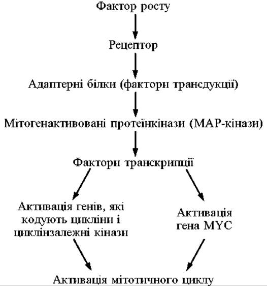
Fig. 8.2. Signaling pathways originating from growth factor receptors
Contact inhibition plays a crucial role in regulating proliferation. If a cell establishes contacts with neighboring cells rather than the extracellular matrix, its division halts. It is believed that the signal regarding intercellular contact originates from membrane proteins known as cadherins.
Cells possess other signaling pathways that converge on the cyclin-Cdk complex cascade, which acts as the engine of the mitotic cycle. Only the coordinated action of all these factors allows a cell to choose between the path of differentiation and that of division. All genes encoding growth factors, adapter proteins, and Other components of signaling pathways that promote cell mitosis are referred to as Proto-oncogenes. Mutations that enhance the activity of any of these factors can lead to uncontrolled cell division; such mutant alleles are known as oncogenes.
Checkpoints
The Cell Cycle features regulatory checkpoints, the passage of which is permitted only upon the successful completion of preceding stages and the absence of damage to the genetic apparatus. During these pauses, cellular protein systems are activated to: 1) detect DNA damage; 2) arrest the mitotic cycle; 3) activate DNA Repair Mechanisms; and 4) initiate apoptosis if the damage is irreparable. Defects in the proteins responsible for these mechanisms can lead to the generation of daughter cells with a compromised genome.
At least four such checkpoints are recognized: in the G1, S, and G2 phases, and the spindle assembly checkpoint during mitosis.
1. The G1 checkpoint. A primary requirement for a cell entering the S phase is the structural integrity of its DNA, as replicating damaged DNA would transmit genetic abnormalities to progeny. Consequently, cells exposed to mutagenic influences arrest at the G1 stage and fail to enter the S phase.
2. The checkpoint in the S phase monitors the accuracy of DNA replication.
3. The checkpoint in the G1 phase. It detects DNA damage that was missed at previous checkpoints or acquired during later stages. It also checks for the completion of DNA replication within the cell.
4. The spindle assembly checkpoint in metaphase of mitosis. To prevent improper chromosome segregation, cells are arrested in metaphase until all kinetochores are attached to microtubules.
A large group of proteins, including pRb, p53, and others, is involved in the checkpoint mechanism. Together, they form a system of cellular "brakes" that prevent the cell from dividing if its genetic material is damaged or under other adverse stimuli. The genes encoding these proteins are known as tumor suppressor genes. The Transformation of a normal cell into a malignant tumor cell can only occur when the activity of the tumor suppressor genes involved in regulating checkpoint mechanisms is impaired.
Last update: 11/08/2026
Editorial and Educational Adaptation: This material has been compiled based on the primary/original source text. The project team performed an editorial review, corrected technical inaccuracies, structured sections, and adapted the content for an educational format.
What was processed:
- elimination of formatting defects (OCR errors, structural breaks, corrupted characters);
- editorial organization of content;
- standardization of terminology in accordance with academic sources;
- verification of factual statements against the original source text.
All mentions of the author, publication year, and origin of the primary text have been preserved in accordance with the source.