Tuberculosis - I.T. Pyatnochka 2005
Extrapulmonary tuberculosis
Tuberculous pleurisy (empyema)
Tuberculous Pleurisy is a specific inflammation of the Pleura that develops either as an independent clinical form or as a complication of pulmonary or Extrapulmonary tuberculosis (Fig. 26). Among patients with newly diagnosed Pulmonary Tuberculosis, tuberculous pleurisy accounts for 3–6%.
Pathomorphology. Most commonly, pleurisy is a complication of the Primary tuberculosis complex, Tuberculosis of the intrathoracic Lymph Nodes, and Disseminated pulmonary tuberculosis. Occasionally, pleurisy presents as an independent form of tuberculosis without apparent involvement of other Organs. MBT can invade the pleura via lymphogenous, hematogenous, or contact routes.
It should be noted that under normal conditions, the pleural cavity contains approximately 20 ml of fluid (lubricant). The parietal pleura produces fluid (approximately 100
ml per hour), while the visceral pleura absorbs it.
Based on The Mechanism of development, pleurisy is classified into allergic, perifocal, and tuberculosis of the pleura.
Allergic pleurisy most frequently occurs in primary tuberculosis and is a manifestation of pleural hypersensitization by MBT decay products. Provoking factors include chest trauma or hypothermia.
Perifocal pleurisy arises As a result of the spread of tuberculous inflammation to the pleura and may be localized in nature.
Tuberculous pleurisy more commonly develops as a complication of primary or disseminated pulmonary tuberculosis. MBT penetrate the pleura via lymphogenous and hematogenous pathways. Tuberculous tubercles and exudate appear on the pleura.
Class="center">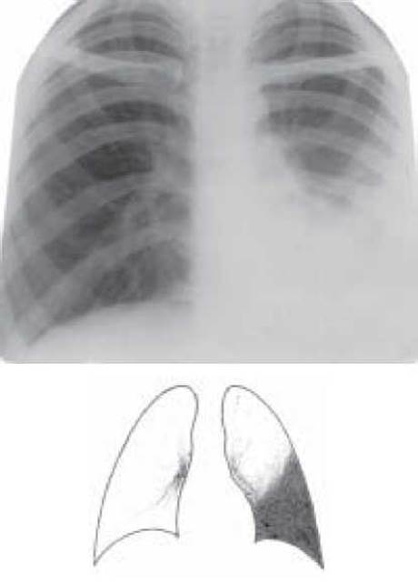
Fig. 26. Left-sided exudative pleurisy. Overview radiograph
Tuberculosis of the pleura accompanied by the accumulation of purulent exudate and a protracted course is termed Pleural Empyema. Pleural empyema develops in cases of widespread caseous lesions of the pleura, as well as a result of the perforation of a subpleurally located focus or cavity. Chronic pleural empyema is characterized by an undulating clinical course. The pleura is significantly thickened due to inflammatory, necrotic-caseous processes, The Development of specific granulation tissue, and scarring changes, resulting in the complete loss of its function.
Depending on The Nature of the inflammation, tuberculous pleurisy is divided into two main forms: dry (fibrinous) and exudative (effusion). Depending on the Nature of the exudate, pleurisy can be serous, purulent, hemorrhagic, mixed, or chylous. According to its localization, tuberculous pleurisy may be costal, costodiaphragmatic, subdiaphragmatic, mediastinal, interlobar, or apical.
Clinical presentation. The clinical picture of tuberculous pleurisy is diverse. Dry pleurisy begins gradually; the deterioration of the patient's general condition is accompanied by chest pain, dry cough, and intermittent low-grade fever. The localization of pain depends on the site of pleural involvement. Left-sided costal pleurisy sometimes mimics angina pectoris; in apical pleurism, pain radiates along the brachial nerve plexus; in interlobar pleurism, it radiates to the interscapular space. In lower costal or subdiaphragmatic pleurisy, the pain resembles that of Calculous Cholecystitis or urolithiasis. Physical examination reveals lagging of the affected side of the chest and pleural friction rub upon Auscultation.
Exudative pleurisy typically begins acutely with pronounced signs of intoxication. The appearance of exudate in the pleural cavity is accompanied by chest pain, dyspnea, cough, and elevated body Temperature. Patients tend to lie on the affected side. The affected hemithorax lags during Respiration; a dull Percussion tone, diminished vocal fremitus, and markedly decreased breath sounds are detected over the exudate.
The acute phase of pleurisy lasts 2–4 weeks, after which the process subsides, and signs of resorption or, less commonly, Organization OF THE exudate appear.
Analysis of the exudate is of paramount importance for Diagnosis. In tuberculous pleurisy, the exudate is predominantly light yellow, with a relative density exceeding 1015, a protein content of more than 30 g/l, and a positive Rivalta test. At the onset of the disease, neutrophils predominate in the exudate (50–60%); as the process resolves, lymphocytes become predominant (90–95%). In specific etiologies of the exudate, glucose concentration is elevated (up to 0.8 g/l), which can serve as a diagnostic marker.
Peripheral Blood changes are characterized by moderate leukocytosis, an increased percentage of band neutrophils, lymphopenia, and a significantly accelerated ESR. For the verification of tuberculous pleurisy, the definitive detection of MBT in the exudate or histological examination of a pleural biopsy specimen obtained during pleuroscopy (see "Instrumental Methods of Investigation and Treatment") is essential. The Mantoux tuberculin Skin test is positive or hyperergic.
The radiograph characteristically reveals an intense, homogeneous opacification of a specific configuration, depending on the localization of the exudate and the patient's body position.
Cytology/practical/136.html">Differential diagnosis OF tuberculous pleurisy is performed with pleurisy of other origins (parapneumonic, neoplastic), Croupous Pneumonia, intercostal neuralgia, and cardiogenic transudate (or transudate of other origins).
Treatment of tuberculous pleurisy must be comprehensive, utilizing antimycobacterial drugs (isoniazid, rifampicin, streptomycin for 2 months, followed by isoniazid and rifampicin or ethambutol for up to 4 months), Vitamins B1, B6, C, along with desensitizing, corticosteroid, and symptomatic therapy. Pleural punctures are performed systematically (at least twice a week) with the subsequent instillation of kanamycin (or streptomycin) with hydrocortisone into the pleural cavity. In cases of purulent effusion, aspiration via puncture or drainage of the pleural cavity must be performed daily; in cases of transition to a chronic form, surgical intervention is indicated (pleurectomy, decortication).
In general, isolated tuberculous pleurisy (without pulmonary involvement) in newly diagnosed patients is treated according to clinical category 3. In combined pleural and pulmonary involvement, tuberculosis is treated under dispensary registration category 1, 2, 3, or 4, depending on the type and extent of pulmonary lesions, the presence of bacterial excretion, and mycobacterial resistance. The duration of the main course of Chemotherapy for patients with tuberculous pleurisy is 6 months. Chemoprophylaxis is not used for tuberculous pleurisy.
CONTROL QUESTIONS
1. Pathogenesis and pathomorphology of tuberculous pleurisy.
2. Classification of tuberculous pleurisy according to localization and the nature of the exudate.
3. Clinical Presentation and diagnosis of tuberculous pleurisy.
4. The Significance of pleural puncture and pleuroscopy in the diagnosis of tuberculous pleurisy.
5. Differential diagnosis of tuberculous pleurisy.
6. Management and treatment of tuberculous pleurisy.
Last update: 10/08/2026
Editorial and Educational Adaptation: This material has been compiled based on the primary/original source text. The project team performed an editorial review, corrected technical inaccuracies, structured sections, and adapted the content for an educational format.
What was processed:
- elimination of formatting defects (OCR errors, structural breaks, corrupted characters);
- editorial organization of content;
- standardization of terminology in accordance with academic sources;
- verification of factual statements against the original source text.
All mentions of the author, publication year, and origin of the primary text have been preserved in accordance with the source.