Tuberculosis - I.T. Pyatnochka 2005
Extrapulmonary tuberculosis
Tuberculosis of the intrathoracic lymph nodes
This is one of the most common Clinical forms of Primary tuberculosis, characterized by specific involvement of the intrathoracic Lymph Nodes (the ROOT of the lung and the Mediastinum) (Fig. 25).
Pathogenesis. Infection mainly occurs via the airborne-droplet and dust route; MBT penetrate through the mucous membrane of the Tonsils and Bronchi into the Lymphatic vessels and nodes, where the specific process develops. Depending on the state of the micro- and macroorganism, infiltrative-inflammatory or necrotic changes prevail in the lymph nodes.
Pathomorphology. In tuberculosis, one or several groups of lymph nodes can be affected. Paratracheal, tracheobronchial, bronchopulmonary, bifurcation, and other lymph nodes are involved. The process can be unilateral or bilateral, predominantly asymmetric.
Based on The Nature of pathomorphological changes, hyperplastic and caseous forms of intrathoracic lymph node tuberculosis are distinguished. The caseous form is characterized by a severe course and is more difficult to treat.
The Clinical presentation of uncomplicated intrathoracic lymph node tuberculosis is similar to that of the Primary tuberculous complex.
Clinically and radiologically, three forms of intrathoracic lymph node tuberculosis are distinguished: the "minor", infiltrative, and tumor-like (hyperplastic, indurative) forms, depending on the morphological substrate, primarily caseation. The "minor" form is characterized by an asymptomatic or mildly symptomatic course. A feature of infiltrative bronchial adenitis is a slight enlargement of the lymph nodes with perifocal inflammation in the lung tissue, which radiologically manifests as widening of the root shadow, protrusion, and blurred contours; clinically, symptoms of intoxication are present. Tumor-like bronchial adenitis is characterized by the enlargement of one or several groups of intrathoracic mediastinal or lung root lymph nodes with polycyclic, well-defined outer contours. Signs of intoxication are more pronounced, with a tendency toward a complicated course.
Class="center">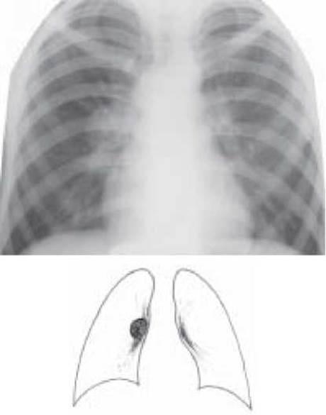
Fig. 25. Right-sided tumorous bronchial adenitis. Overview radiograph
The "minor" form of tuberculosis is diagnosed based on indirect signs of intrathoracic lymph node enlargement, in particular, local enhancement and deformation of the perihilar pulmonary pattern, decreased Structure OF THE root shadow, double contour of the mediastinal shadow at a certain level, and disappearance of the intermediate bronchus shadow.
The course and complications of intrathoracic lymph node tuberculosis are nearly identical to those of the primary tuberculous complex and are associated with specific involvement of the lymph nodes with corresponding clinical and radiological signs: hematogenous and lymphogenous dissemination, exudative Pleurisy, involvement of the adjacent bronchus with subsequent bronchogenic dissemination, or impaired bronchial patency and Atelectasis.
Diagnosis is established based on the Anamnesis, clinical and radiological picture during the tuberculin Skin test conversion period, as well as laboratory, bacteriological, and instrumental examinations.
Differential diagnosis is performed with sarcoidosis (stage I), lymphogranulomatosis, lymphosarcoma, Lymphocytic Leukemia, central Cancer, and nonspecific adenopathies.
For this purpose, the following are performed: radiography in direct and lateral projections, tomography at the level of the bifurcation (median slice), computed tomography, tuberculin tests, bronchoscopy, and puncture or surgical biopsy.
Treatment. 3-4 anti-tuberculosis drugs are prescribed: isoniazid, rifampicin, pyrazinamide, streptomycin. After two months of effective Chemotherapy: isoniazid + rifampicin (ethambutol) daily or every other day. The duration of chemotherapy is 6-8 months. Nonspecific therapy includes Vitamins B1, B6, C, desensitizing, hormonal, and symptomatic agents.
Overall, intrathoracic lymph node tuberculosis is treated under category 1 or 3 of dispensary registration, depending on the extent of tuberculosis and the presence of bacterial excretion.
Last update: 10/08/2026
Editorial and Educational Adaptation: This material has been compiled based on the primary/original source text. The project team performed an editorial review, corrected technical inaccuracies, structured sections, and adapted the content for an educational format.
What was processed:
- elimination of formatting defects (OCR errors, structural breaks, corrupted characters);
- editorial organization of content;
- standardization of terminology in accordance with academic sources;
- verification of factual statements against the original source text.
All mentions of the author, publication year, and origin of the primary text have been preserved in accordance with the source.