Review of Medical Physiology - William F. Ganong 2002
Respiration
Regulation of Respiration
Chemical Regulation of Respiration
Chemical regulatory mechanisms adjust pulmonary ventilation in such a way that alveolar PCO2 normally remains constant, the effects of excess Blood H+ are mitigated, and PO2 increases whenever its decline reaches a potentially dangerous level. Minute ventilation is proportional to the metabolic rate; however, The Link Between METABOLISM and Respiration is determined by CO2 rather than O2. Receptors in the carotid and aortic bodies are stimulated by an increase in arterial PCO2 or H+ concentration, or by a decrease in PO2. Following denervation of the carotid chemoreceptors, the response to decreased PO2 is abolished. The predominant effect of Hypoxia after carotid body denervation is direct depression of the respiratory center. The response to changes in arterial H+ concentration within the pH range of 7.3–7.5 is also largely eliminated, although larger changes still exert a partial effect. On the other hand, the response to changes in arterial PCO2 persists, though it is slower and decreases by no more than 30–35%.
Class="center">Table 36-1. Stimuli Affecting the Respiratory Center
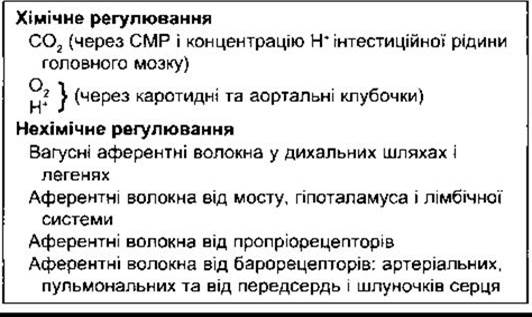
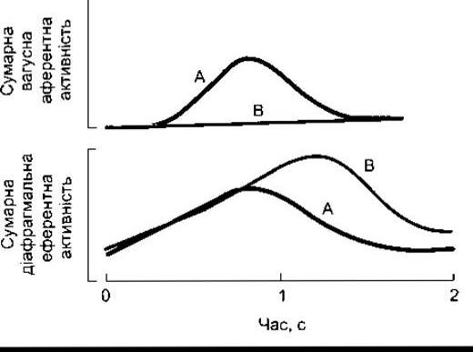
Fig. 36-3. Tracing of two breathing patterns driven by pulmonary stretch receptors: with (A) and without (B) vagal feedback. Note that The rate of increase in phrenic nerve activity driving the Diaphragm is unchanged, but the impulse duration is prolonged in the absence of vagal input.
Carotid and Aortic Bodies
The carotid bodies are located near the carotid bifurcation on each side, and typically two or more aortic bodies are situated near the aortic arch (Fig. 36-4). Each carotid or aortic body (glomus) contains islets of two Cell types: Type I and Type II, surrounded by fenestrated sinusoidal capillaries. Cup-shaped afferent nerve endings lie in close apposition to Type I, or glomus, Cells (Fig. 36-5). Glomus cells resemble the chromaffin cells of The adrenal medulla, containing dense-core vesicles filled with catecholamines that are released in response to hypoxia or cyanide (see below). These cells are stimulated by hypoxia, and the primary emerging neurotransmitter is dopamine, which excites the nerve endings via O2 receptors. Type II cells are glial-like cells, each enclosing four to six Type I cells. The precise Functions of Type II cells remain unclear.
Outside the capsule of each body, the nerve fibers are myelinated. These fibers have a diameter of 2-5 µm and a conduction velocity of 7-12 m/s. Afferent fibers from the carotid bodies ascend to the Medulla Oblongata via the carotid sinus and glossopharyngeal nerves, whereas fibers from the aortic bodies travel via the vagus nerves. In studies where a single carotid body was isolated and perfused, a graded increase in impulse frequency was observed in active afferent nerve fibers when PO2 in the perfused blood decreased (Fig. 36-6) or PCO2 increased.
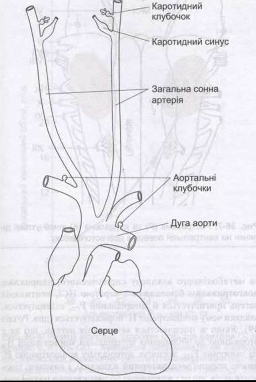
Fig. 36-4. Location OF THE carotid and aortic bodies
Type I glomus cells possess O2-sensitive K+ channels, in which the open probability decreases in proportion to the severity of hypoxia to which they are exposed. This reduces K+ efflux, depolarizes The Cell, and triggers Ca2+ influx primarily through L-type Ca2+ channels. Ca2+ influx initiates Action Potential generation and neurotransmitter release, resulting in the excitation of afferent nerve endings. Pulmonary artery smooth Muscle cells contain similar O2-sensitive K+ channels that initiate hypoxia-induced vasoconstriction (see Chapter 37). This contrasts with systemic Arteries, which contain ATP-dependent K+ channels that facilitate greater K+ efflux during hypoxia, subsequently leading to vasodilation rather than vasoconstriction. Blood flow through each 2-mg carotid body is approximately 0.04 mL/min, or 2000 mL/100 g of tissue/min, which compares with a blood flow of 54 mL/100 g/min in the Brain and 420 mL/100 g/min in the Kidneys (see Table 32-1). Because tissue blood flow per unit mass is exceptionally high, the cells consume more O2 than can be supplied by dissolved O2 alone. Consequently, receptor stimulation does not occur in conditions such as anemia or Carbon monoxide poisoning, where The amount of dissolved oxygen in the blood reaching the receptors is normal, but the total O2 content bound to Hemoglobin is significantly reduced. Receptor stimulation occurs when arterial PO2 is low or during vascular stasis, situations where the volume of O2 delivered to the receptors per minute is diminished. Strong stimulation is also caused by pharmacological agents such as cyanide, which blocks tissue-level O2 utilization. In sufficient doses, nicotine and lobeline activate chemoreceptors. Furthermore, infusion of K+ has been shown to increase the firing rate in afferent chemoreceptor fibers; because plasma K+ levels rise during Physical Exercise, this mechanism may contribute to exercise-induced hyperventilation.

Fig. 36-5. Organization OF THE carotid body. Type I (glomus) cells contain catecholamines. Upon exposure to hypoxia, they release these catecholamines, which stimulate cup-shaped nerve fiber endings of the Glossopharyngeal nerve within the carotid sinus. Glomus-supporting Type II glial-like cells surround Type I cells and presumably provide structural and metabolic support.
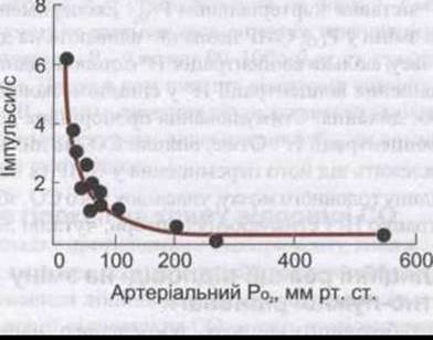
Fig. 36-6. Changes in the impulse firing rate in a single afferent fiber originating from the carotid body as PO2 decreases (courtesy of S. Sampson).
Due to their anatomical location, aortic bodies cannot be studied in as much detail as carotid bodies. Their responses are presumably similar, albeit of lesser amplitude. In human subjects whose carotid bodies have been surgically removed while the aortic bodies remain intact, responses are preserved (as is also observed in animals with sequentially denervated carotid and aortic bodies). Such individuals exhibit minor changes in resting pulmonary ventilation, but the ventilatory response to hypoxia is abolished, and the ventilatory responses to CO2 are reduced by about 30%.
Neuroepithelial bodies, composed of innervated clusters of amine-containing cells, are found in the Airways of both humans and animals. These cells exhibit an outward K+ current that decreases during hypoxia, which can trigger cellular depolarization. However, the precise Physiological Role of these hypoxia-sensitive cells remains incompletely understood, given that ablation of the carotid bodies alone virtually abolishes the respiratory response to systemic hypoxia.
Chemoreceptors in the Brainstem
The chemoreceptors responsible for initiating the hyperventilation caused by elevated arterial PCO2 following denervation of the carotid and aortic bodies reside within the medulla oblongata and are thus referred to as medullary chemoreceptors. They are anatomically distinct from the dorsal and ventral respiratory group Neurons and are localized on the ventral surface of the medulla oblongata (Fig. 36-7).
The monitoring of H+ concentration in CEREBROSPINAL FLUID (CSF) and brain interstitial fluid is performed by these chemoreceptors. CO2 crosses Introduction/36.html">Biological Membranes, such as the blood-brain barrier, rapidly, whereas H+ and HCO3- diffuse slowly. Upon entering the brain parenchyma and CSF, CO2 is rapidly hydrated. Subsequent dissociation of H2CO3 increases the local H+ concentration. In the brain interstitial fluid, H+ concentration closely mirrors arterial PCO2. Experimentally induced changes in CSF PCO2 have little effect on respiration as long as the H+ concentration remains constant, whereas any increase in CSF H+ concentration strongly stimulates breathing. This stimulatory effect is proportional to the magnitude of the H+ concentration increase. Consequently, the effects of CO2 on respiration are primarily mediated through its diffusion into the CSF and brain interstitial fluid, where CO2 raises the H+ concentration and stimulates H+-sensitive receptors.
Ventilatory Responses to Acid-Base Disturbances
In metabolic acidosis caused, for example, by the accumulation of ketoacidotic products in Diabetes Mellitus, respiratory stimulation occurs (Kussmaul breathing; see Chapter 39). Conversely, in metabolic alkalosis resulting, for example, from prolonged vomiting with loss of HCl, pulmonary ventilation is depressed, causing arterial PCO2 to rise, which helps restore the H+ concentration toward normal (see Chapter 39). If pulmonary hyperventilation occurs independently of an increase in arterial H+ concentration, the resulting decrease in PCO2 lowers the arterial H+ concentration below normal (respiratory alkalosis). Conversely, hypoventilation occurring independently of a drop in plasma H+ concentration leads to respiratory acidosis.
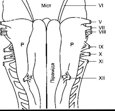
Fig. 36-7. Rostral (R) and caudal (C) chemosensitive areas on the ventral surface of the medulla oblongata.
Ventilatory Responses to CO2
Normally, arterial PCО2 is maintained at approximately 40 mmHg. If tissue metabolism accelerates and increases its level, pulmonary ventilation is stepped up, and pulmonary excretion of СО2 rises until arterial РСО2 returns to normal. This negative feedback mechanism keeps both the production and excretion of СО2 in precise balance.
When breathing a gas mixture containing СО2, both alveolar and arterial РСО2 rise, and ventilatory stimulation begins as soon as the blood carrying the elevated СО2 level reaches the medulla oblongata. СО2 elimination increases, and alveolar РСО2 is driven back down toward normal. This explains why relatively large increases in inspired РСО2 (e.g., 15 mmHg) cause only a minor rise in alveolar РСО2 (e.g., 3 mmHg). Consequently, РСО2 is not fully normalized; a new equilibrium is established once alveolar РСО2 increases slightly, and hyperventilation persists for as long as the inhalation of СО2 continues. The true linear relationship between minute ventilation and alveolar РСO2 is illustrated in Fig. 36-8. However, this linearity has its limits. When the РСО2 of the expired gas mixture approaches alveolar РСО2, СО2 elimination becomes impaired. Furthermore, if the СО2 content in the inspired gas mixture exceeds 7%, alveolar and arterial РСО2 begin to rise sharply despite ongoing hyperventilation. This leads to an accumulation of СО2 in the body (hypercapnia), which depresses the Central Nervous system—including the respiratory center—and results in headache, restlessness, and ultimately coma (СО2 narcosis).
,
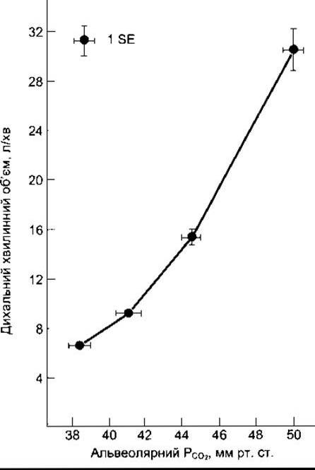
Fig. 36-8. Ventilatory responses of healthy subjects breathing 02 and approximately 2, 4, and 6% СО2. Minute ventilation increases through a combination of increased respiratory rate and tidal volume (reprinted with permission from Lambertsen CJ in: Medical Physiology, 13th ed. Mountcastle VB [editor]. Mosby, 1974).
Ventilatory Responses to Oxygen Deficiency
A decrease in the oxygen content of inspired air triggers an increase in minute ventilation. This stimulatory effect is negligible as long as inspired РCО2 remains above 60 mmHg, becoming pronounced only at lower РCО2 values (Fig. 36-9). Thus, any reduction in arterial РCО2 toward 100 mmHg leads to increased afferent nerve firing from the carotid and aortic chemoreceptors. Two hypotheses attempt to explain why this elevated impulse frequency does not normally increase pulmonary ventilation in healthy individuals until РО2 drops below 60 mmHg. Because Hb is a weaker acid than HbО2 (see Chapter 35), There is a slight increase in arterial H+ concentration as РО2 decreases and hemoglobin becomes less saturated with О2. This reduction in H+ concentration tends to inhibit respiration. Additionally, any increase in pulmonary ventilation that lowers alveolar РСО2 also exerts a depressing effect on breathing. Therefore, the stimulatory effects of hypoxia on pulmonary ventilation are not fully apparent until they become marked enough to overcome these counterbalancing inhibitory influences associated with reduced arterial H+ concentration and РСО2.
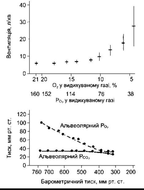
Fig. 36-9. Top: Mean minute ventilation values following exposure to gas mixtures with varying О2 content. The horizontal line in each case represents the mean value, and the vertical bar denotes one standard deviation. Bottom: Alveolar PО2 and РСО2 values during air breathing at various barometric pressures. The two graphs are aligned so that the О2 in the inspired gas mixtures (top graph) corresponds to the РО2 at different barometric pressures (bottom graph) (data provided by RH Kellogg).
The Effect of a reduction in alveolar РO2 on ventilation at a constant alveolar РСО2 is shown in Fig. 36-10. When alveolar РСО2 is stabilized at 2–3 mmHg above normal, the relationship between pulmonary ventilation and alveolar РО2 within the 90–100 mmHg range is reversed. However, at subnormal levels of alveolar PCО2, hypoxic stimulation of pulmonary ventilation does not occur until alveolar РО2 drops to 60 mmHg or lower.
Effect of Hypoxia on the СО2 Response Curve
In the reverse experiment, when alveolar РО2 is held constant, the ventilatory responses to varying amounts of СО2 follow a linear relationship (Fig. 36-11). When СО2 response curves are obtained at different РО2 levels, the slope of the response curve changes, becoming steeper as alveolar РО2 decreases. In other words, hypoxia makes an individual more sensitive to increases in arterial РСО2, although the level of alveolar РСО2 at which the curves in Fig. 36-11 intersect remains constant. In normal individuals, this threshold value lies below the normal resting alveolar РСО2, indicating that there is normally a very small yet perceptible "СО2 drive" acting on the respiratory center.

Fig. 36-10. Pulmonary ventilation at various alveolar РО2 levels with РСО2 tightly maintained at 49, 44, or 37 mmHg (data provided by HH Loeschke and KH Gertz).
Effect of H+ on the СО2 Response
The stimulatory effects of H+ and СО2 on respiration interact in much the same way as СО2 and О2. In metabolic acidosis, the СО2 response curve resembles those in Fig. 36-11, except that the curves are shifted to the left. In other words, respiratory stimulation occurs even at lower arterial levels of РСО2. It has been calculated that the СО2 response curve shifts to the left by 0.8 mmHg for each nanomole increase in arterial H+ content. More than 40% of the ventilatory response to СО2 is abolished if the rise in arterial H+ generated by СО2 is prevented. As noted above, the remaining 60% is likely attributable to the direct effect of СО2 on H+ concentration in the cerebrospinal fluid or brain interstitial fluid.
Breath-Holding
Respiration can be voluntarily suspended for a period, though conscious control is ultimately limited and rarely a major factor. The point beyond which breathing can no longer be suppressed is termed the breaking point. It is governed by the rise in arterial РСО2 and the fall in РО2. Individuals can hold their breath longer following surgical removal of the carotid bodies. Breathing 100% oxygen prior to breath-holding raises alveolar РО2, thereby delaying the breaking point. The same holds true after hyperventilating with room air, since СО2 is blown off and arterial РСО2 is initially low. Reflex or mechanical factors also influence the breaking point; thus, subjects who hold their breath to the maximum and then inhale a gas mixture low in О2 and high in СО2 can prolong their breath-holding time by an additional 20 seconds or more. Psychological factors play a role as well—individuals who can maintain breath-holding longer while speaking demonstrate better performance than when silent.

Fig. 36-11. Graphical linear family ("fan") of СО2 responses at various constant levels of alveolar РО2.
Hormonal Influences on Respiration
Pulmonary ventilation increases during the luteal phase of the Menstrual cycle as well as during Pregnancy (see Chapter 23). Animal experiments indicate that this is driven by the activation of estrogen-dependent progesterone receptors in the Hypothalamus. However, the exact physiological significance of this increase remains unclear.
Last update: 10/08/2026
Editorial and Educational Adaptation: This material has been compiled based on the primary/original source text. The project team performed an editorial review, corrected technical inaccuracies, structured sections, and adapted the content for an educational format.
What was processed:
- elimination of formatting defects (OCR errors, structural breaks, corrupted characters);
- editorial organization of content;
- standardization of terminology in accordance with academic sources;
- verification of factual statements against the original source text.
All mentions of the author, publication year, and origin of the primary text have been preserved in accordance with the source.