Orthopedics - Oleksa A.P. 2006
Bone and Joint Tuberculosis
Tuberculosis of the Bones and joints is caused by the tubercle bacillus, which enters the bloodstream from a primary focus in the Lungs (90%) or the gastrointestinal tract and is subsequently carried into the spongy Bone Structure. As a rule, The Musculoskeletal System is affected by the human Koch's bacillus, and very rarely by the bovine bacillus (which enters the body via raw milk, cheese, or meat), widely considered to be osteotropic.
Numerous cases of skeletal tuberculosis following BCG Vaccination in children have been documented (Auckland E.J., 1966; Dahl H., Halvorson K., 1967, and others).
There is a direct etiological correlation between active Pulmonary Tuberculosis and Osteoarticular Tuberculosis, particularly in children and adolescents. Müller and Woodruff report such a coexistence of tuberculosis in 71% of cases, Johanson in 61%, and Simon and Redeker in 65% of patients. Furthermore, it is established that the younger the child, the more frequently this combination occurs, though it does not significantly alter the mutual progression of the tuberculous process.
In adults, the coexistence of pulmonary and osteoarticular tuberculosis accounts for no more than 20%, which is attributed to the more rapid healing of the pulmonary process, while Bone and joint Tuberculosis frequently manifests at a later stage.
Osteoarticular tuberculosis accounts for nearly 10% of all forms of tuberculosis. The spine is most commonly affected—comprising one-third of all patients—followed somewhat less frequently by the hip (18.7%) and knee (18.0%) joints. The remaining cases involve other anatomical sites.
Historically, bone and joint tuberculosis predominantly affected children, whereas today it is primarily a disease of adults (70% of cases).
Pathogenesis. The modern understanding of the disease's pathogenesis has evolved gradually, highlighting The Significance of the primary focus, which exerts a specific immunizing effect on the subsequent development of tuberculosis. Two critical factors govern the clinical course of the disease: on the one hand, the body's natural specific defense mechanisms, and on the other, the virulence of the bacillus.
The progression of a tuberculous infection within the body depends on the balance between susceptibility and resistance. The dominance of resistance over susceptibility establishes favorable conditions for defense, whereas the prevalence of susceptibility creates adverse conditions dominated by exudative-inflammatory processes, edema, tissue breakdown, and necrosis. When susceptibility and resistance are nearly balanced, a state of homodynamics ensues, which is typically characteristic of a chronic clinical course.
The body's defense response can be either exudative or cellular. An exudative response creates a barrier that prevents the bacilli from spreading beyond their initial site of entry and dissemination. The cellular defense response involves surrounding and clustering the accumulated bacilli, resulting in The formation of a specific granuloma.
The diagram (after Malawski S., 1976) illustrates the dependence of pathological changes on the virulence of the bacillus and the host's resistance.
This diagram reflects various clinical courses of osteoarticular tuberculosis. Between the extreme states of the Organism lies a wide spectrum of intermediate and mixed manifestations of the disease, representing a highly complex immunobiological phenomenon.
Regarding the pathogenicity of the tubercle bacillus, it varies widely—ranging from low to pronounced virulence coupled with diverse antigenic properties.
The same holds true for the patient's body, whose level of resistance may be naturally normal, thus providing protection, or diminished, and in some cases, immune defense against tuberculosis may be entirely absent. The Clinical presentation and course of osteoarticular tuberculosis will unfold accordingly.
According to A.I. Abrikosov, tuberculosis is classified into primary, secondary, and hematogenously disseminated forms.
From this perspective, Mycobacterium tuberculosis spreads hematogenously from the primary complex into the spongy bone substance, where Bone Marrow resides, forming lesions known as endogenous reinfections. Healthy Bone tissue and yellow bone marrow are inherently resistant to mycobacterial colonization prior to invasion. At the site of M. tuberculosis deposition, a local inflammatory reaction develops—either exudative or cellular in nature—manifesting as a specific granuloma. Bone trabeculae encompassed by the granulomatous Cells undergo necrosis (forming caseous necrosis) and are secondarily destroyed through lacunar resorption by osteocytes, which proliferate rapidly during inflammation. This gives rise to a primary, relatively small specific osteitis resembling a cheese-like mass. It may localize centrally within the metaepiphyseal region of the bone or peripherally, either subchondrally or subperiosteally.
Progressing primary osteitis eventually breaches the bone cortex, rupturing either into the joint cavity or extra-articularly. Rupture into the joint results in tuberculous Arthritis, which initially involves the synovial membrane and progresses to a purulent-destructive process affecting all joint components, including bones and soft Tissues. The articular Cartilage remains unaffected for a prolonged period, suffering damage only secondarily. If the process breaks through into the soft tissues—most commonly observed in exudative infections—an abscess forms. As it enlarges, this abscess can dissect along interfascial planes far beyond the primary focus as a cold, migratory (cold abscess). A diagnostic puncture yields white pus. The volume and character of the pus depend on the host's resistance and the virulence of the tubercle bacilli. The lower the host resistance and the higher the pathogen's virulence (exudative process), the thinner the abscess contents, resembling a non-specific abscess that gradually thickens and becomes heterogeneous. The older the abscess, the denser the pus. Such an abscess may eventually rupture onto the Skin surface, forming a persistent, chronic sinus tract. Naturally, secondary bacterial flora frequently supervenes, complicating both the inflammatory process and its management.
Class="center">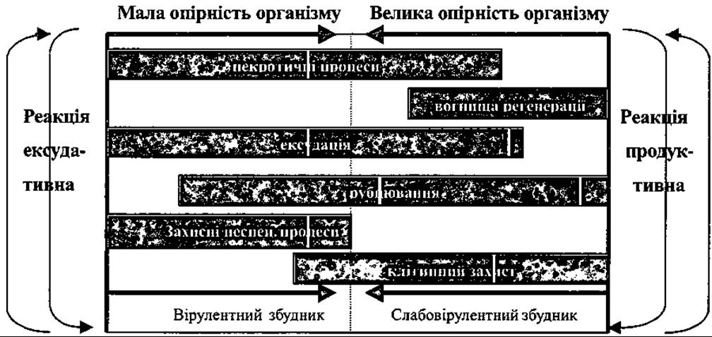
Diagram of pathological changes depending on bacillus virulence and host resistance.
Under cellular-mediated immune responses, in cases of less active tuberculous disease, caseous cheese-like masses may become encapsulated and calcified.
Very rarely, specific synovial forms of the disease occur, manifesting as synovitis (affecting one or two joints, most commonly the knees) or Poncet's disease (named after the author who described it). This condition is of a tubercular-allergic origin, and M. tuberculosis cannot be isolated from the exudate. Typically, the joints are swollen, accompanied by local hyperthermia and joint effusion (hydroarthrosis). Patients diagnosed with Rheumatoid Polyarthritis are often initially treated by internists and subsequently referred to orthopedic surgeons due to the inefficacy of antirheumatic therapy.
Osteoarticular tuberculosis develops in three distinct phases: 1) pre-arthritic — the Formation of primary osteitis; 2) arthritic — the spread of the process into the joint; 3) post-arthritic — The Development of residual structural sequelae in the bones and joints (arthrosis, scarring, ankylosis, etc.).
Tuberculous Spondylitis also comprises three phases: pre-spondylitic, spondylitic, and post-spondylitic (Fig. 381). Additionally, the arthritic phase of the tuberculous process is clinically subdivided into three stages: initial, acute (manifestation), and resolution (fading).
The cyclical progression of the tuberculous process is driven by immunobiological phenomena, which essentially encompass two phases: disease progression and regression (resolution).
During the progressive phase, due to the pathogenic dominance of the tubercle bacilli over the patient's immune resistance, the disease advances and specific bone destruction spreads. Simultaneously, the body mobilizes its defense mechanisms.
Once equilibrium is established between the host's defense forces and microbial virulence, the reversal process begins. As host resistance increasingly surpasses microbial virulence, the pathological process subsides and healing takes place.
All of this is observed during the course of osteoarticular tuberculosis when comprehensive Treatment is applied, including antimicrobial Chemotherapy, agents that enhance the patient's immune defenses, and immobilization of the affected area.
If the Diagnosis is not established in the early stage of the disease and timely treatment is not initiated, the condition becomes chronic with alternating periods of exacerbation and remission, depending on microbial virulence and host resistance.
Regarding the Classification of osteoarticular tuberculosis accepted abroad, the following is commonly used:
1. Primary tuberculous osteitis.
2. Arthritic phase:
a) active stage;
b) stage of decreasing activity;
c) stage of remission.
3. Post-arthritic phase:
a) post-tuberculous osteoarthritis;
b) contractures; .
c) ankylosis.
4. Relapse of osteoarticular tuberculosis.
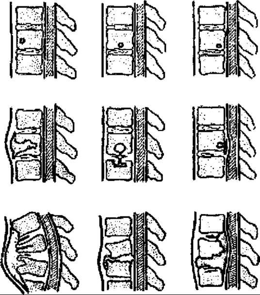
Fig. 381. Schematic drawing of a tuberculous vertebral lesion and the directions of destructive spread depending on the Location OF THE primary osteitis within the vertebra.
Diagnosing advanced cases of osteoarticular tuberculosis is straightforward, as pathological changes are clearly visible on standard plain radiographs and the diagnosis is supported by clinical symptoms.
However, diagnosing tuberculous osteitis is considerably more challenging and therefore requires supplementary diagnostic Methods. These methods can be divided into those that directly confirm a tuberculous process (bacteriological and histological) and those that indicate the patient's general physiological status.
In addition, it is necessary to perform a chest X-ray and Bacteriological examination OF sputum or urine, which helps to identify or rule out concomitant tuberculosis.
The patient's condition and systemic response are assessed through Blood tests (ESR, protein fractions) and Cytology.
Bacteriological studies are performed on material obtained via needle aspiration from the tuberculous lesion or by intraperitoneal inoculation in guinea pigs. These methods are quite complex and time-consuming. Furthermore, obtaining a sample via aspiration is difficult, and in some anatomical locations impossible. Incisional biopsy in such cases is also unjustified.
Histological and bacteriological examinations are performed exclusively in operated patients.
Alongside clinical and radiological findings, tuberculin skin tests are quite valuable. According to World Health Organization recommendations, purified protein derivative known as RT 23 is used in clinical and epidemiological studies.
The Mantoux tuberculin skin test involves the appearance of skin redness on the forearm (early reaction), followed by Swelling (delayed reaction) at the site of intradermal injection of 0,1 ml of tuberculin in an individual infected with tuberculosis. Occasionally, a small vesicle may appear at the center of the papule. The result of the Mantoux test is read in millimeters after 72 hours.
A papule size up to 5 mm in diameter is considered normal, 6 to 9 mm is considered doubtful, and greater than 10 mm is considered positive.
However, interpreting the test results can be difficult in children vaccinated with BCG, as it is still impossible to differentiate between a post-vaccination reaction and a true infection. Therefore, in the case of a positive reaction, despite the absence of primary osteitis, anti-tuberculosis chemotherapy should be prescribed, although this reaction to some extent depends on the patient's age and the time elapsed since infection with the tubercle bacillus and BCG vaccination.
The reaction is strongly positive in young individuals and immediately following tuberculosis infection. It may be indeterminate or doubtful in patients receiving corticosteroid therapy or those suffering from certain infectious diseases (such as pertussis). However, a positive Mantoux test still points toward an active tuberculous process.
Given the similarity—and occasionally identity—of clinical and radiological manifestations, bone and joint tuberculosis must be differentiated from acute, subacute, and chronic inflammations, Brodie's abscess, aseptic necrosis, and even tumors.
Acute non-specific inflammation differs from tuberculosis by an acute onset accompanied by high fever and the rapid development of an abscess. Radiologically, within 3 to 5 weeks, periostitis appears in the focal area, alongside osteonecrosis in the bone surrounded by an osteoblastic sclerotic rim, which indicates a reparative process.
This non-specific process is accompanied by neutrophilic leukocytosis and an elevated ESR. Puncture-derived pus is subjected to cytological and bacteriological analysis to clarify the diagnosis.
In chronic non-specific processes, the disease follows a course characterized by prolonged subsidence and pronounced proliferative changes, particularly in cases of Garré's osteomyelitis.
Sometimes it is necessary to differentiate tuberculosis (primary osteitis) from Brodie's abscess, which has an indolent course and localized involvement of the cancellous bone structure within the metaepiphyseal region. Radiologically, the abscess focus is invariably accompanied by a rim of osteosclerosis, which does not occur in tuberculous lesions. The diagnosis can be refined through histological and bacteriological examinations.
In cases of synovitis and arthritis, particularly rheumatoid arthritis, the diagnosis is clarified through joint aspiration combined with cytological and bacteriological examination of the retrieved fluid, and subsequently by histological analysis following surgery.
Aseptic bone necroses typically occur in children and exhibit characteristic localizations. They differ from the tuberculous process by the absence of inflammatory signs and normal blood test results. Joint function also remains normal for a prolonged period. Radiologically, aseptic necroses localize in the epiphyses and metaepiphyseal regions, initially manifesting as increased bone density followed by fragmentation of the osteonecrotic area, appearing as radiolucent clefts due to the resorption of dead bone and necrotic fragments. Adjacent bones are not affected, unlike in tuberculosis.
In the case of tumors, particularly malignant ones, the process progresses more rapidly and is accompanied by persistent pain. Benign tumors grow slowly over years and remain clinically silent for a long time. Neither clinical nor laboratory findings reveal an inflammatory process.
Radiologically, tumor involvement in the bone varies depending on its nature; in cases of destructive tumor processes, the diagnosis must be verified cytologically and histologically.
Clinical symptoms depend on the phase of the tuberculous process. The prearthritic phase is characterized by sparse and vague symptoms. During this period, signs of tubercular intoxication emerge: a general feeling of weakness, children stop running, spare their limbs, periodically complain of dull, diffuse pain in the limb, and cry out at night in cases of spondylitis. Subfebrile Temperature develops. Blood tests reveal an elevated ERYTHROCYTE SEDIMENTATION RATE and lymphocytosis. Tuberculin tests (von Pirquet, Mantoux), if previously negative, become positive. The diagnosis is refined radiologically, revealing destructive foci of varying size and shape, most commonly in the cortical and subchondral Regions of the epiphyses. As the process progresses, all these symptoms intensify, and pain increases to the point where protective Muscle guarding, limping, and muscle atrophy develop.
The arthritic phase drastically alters the symptomatology of the tuberculous process. Initially unsteady pain symptoms become constant and gradually intensify, passing through clinical stages of peak activity and gradual resolution. The initial stage begins with involvement of the synovial membrane, accompanied by the development of specific exudative and proliferative changes, the proliferation of pannus from the margins, and subsequent destruction of other joint elements. Pain intensifies, leading to protective muscle guarding and atrophy. In the peak stage of tuberculous arthritis, alongside the aforementioned changes, significant swelling and joint deformity appear, along with a marked restriction of motion, contractures, and other complications (paraarticular abscesses and cold abscesses). The resolution stage is characterized by a gradual decrease in process activity and a reduction in clinical signs of inflammation. However, all destructive changes that occurred in the joint and limb persist. The severity of joint impairment is determined using radiographs.
The postarthritic phase completes the stage of resolution. Reparative processes take place, including scarring and compensatory marginal bone proliferation (depending on the degree of joint destruction). This leads to deforming arthrosis or ankylosis, frequently resulting in a functionally unfavorable position for the limb. Muscle atrophy and joint disfiguration persist long-term, and patients may become disabled.
Treatment. It is crucial to establish the diagnosis of tuberculosis in a timely manner (during the prearthritic phase) and administer comprehensive treatment to prevent joint involvement and preserve normal joint function.
Patients with osteoarticular tuberculosis are treated in specialized hospital departments in regional centers and in sanatoria.
Treatment includes: 1) general strengthening measures (regimen, balanced Nutrition, nursing care); 2) antibacterial therapy; 3) immobilization of the affected skeletal area using plaster casts, traction devices, etc.; 4) surgical phthisio-Surgical treatment (radical necrectomy for primary osteitis, resection of joints and vertebral bodies for early arthrodesis and ankylosis, fistulo- and abscess necrectomy, etc.); 5) sanatorium-resort treatment, which is prescribed to all patients to consolidate the results achieved in the hospital.
Children are generally referred immediately to specialized sanatoria, where they undergo comprehensive treatment, including surgery.
Last update: 10/08/2026
Editorial and Educational Adaptation: This material has been compiled based on the primary/original source text. The project team performed an editorial review, corrected technical inaccuracies, structured sections, and adapted the content for an educational format.
What was processed:
- elimination of formatting defects (OCR errors, structural breaks, corrupted characters);
- editorial organization of content;
- standardization of terminology in accordance with academic sources;
- verification of factual statements against the original source text.
All mentions of the author, publication year, and origin of the primary text have been preserved in accordance with the source.