Human Anatomy - Kotsan I. Ya. 2009
Peripheral nervous system
Cranial nerves
Trigeminal nerve (V)
The Trigeminal nerve (n. trigeminal) is a mixed nerve and the thickest of all Cranial Nerves (Figs. 239, 256). Both motor and sensory nuclei of the trigeminal nerve are distinguished.
The motor Nucleus of the trigeminal nerve (nucleus motorius n. trigeminalis) is located in the Pons.
Sensory nuclei:
1. The mesencephalic nucleus of the trigeminal nerve (nucleus mesencephalicus n. trigeminalis) is located in the Midbrain. This nucleus contains the center for proprioceptive sensitivity.
2. The pontine nucleus of the trigeminal nerve (nucleus pontinus n. trigeminalis) is located in the posterior part of the pons. It contains the center for Temperature and pain sensitivity.
3. The spinal nucleus of the trigeminal nerve (nucleus spinalis n. trigeminalis) is located in the Medulla Oblongata and Spinal Cord. It contains the center for temperature and pain sensitivity.
The trigeminal nerve emerges from the Brain substance at the border between the pons and the middle cerebellar peduncles by two roots: a motor ROOT (smaller) and a sensory root (larger). The sensory root appears on the brain surface more caudally than the motor one.
Both roots of the nerve run forward and slightly laterally, passing into the slit of the dura mater—the trigeminal cavity, which lies in the region of the trigeminal impression on the anterior surface of the petrous part of the Temporal bone. This cavity contains the thickening of the trigeminal nerve—the trigeminal ganglion (ganglion trigeminale).
Class="center">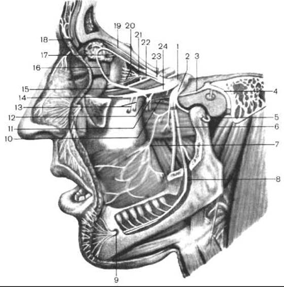
Fig. 256. Trigeminal nerve (according to M. R. Sapin)
1 — trigeminal ganglion; 2 — mandibular nerve; 3 — greater petrosal nerve; 4 — Facial Nerve; 5 — auriculotemporal nerve; 6 — lingual nerve; 7 — buccal nerve; 8 — inferior alveolar nerve; 9 — mental nerve; 10 — nerve of the pterygoid canal; 11 — pterygopalatine ganglion; 12 — ganglionic branches; 13 — superior alveolar nerves; 14 — infraorbital nerve; 15 — zygomatic nerve; 16 — communicating branch (with the zygomatic nerve); 17 — lacrimal nerve; 18 — supraorbital nerve; 19 — frontal nerve; 20 — short ciliary nerves; 21 — ciliary ganglion; 22 — nasociliary root; 23 — ophthalmic nerve; 24 — maxillary nerve
The trigeminal ganglion (Gasserian ganglion) is semilunar in shape and relatively large in size (15 to 18 mm in length). Its convex margin faces forward, and its concave margin faces backward. The ganglion is formed by The Cell bodies of pseudounipolar sensory Neurons, whose central processes form the sensory root and lead to its sensory nuclei. The peripheral processes of these Cells run within the Branches of the trigeminal nerve and terminate in receptors in the Skin, mucous membranes, and other structures of the HEAD. The motor root of the trigeminal nerve adjoins the trigeminal ganglion from below, and its fibers participate in The formation of the third (III) branch of this nerve.
From the anterior convex margin of the trigeminal ganglion, the three main branches of the trigeminal nerve depart: the ophthalmic nerve (branch I), the maxillary nerve (branch II), and the mandibular nerve (branch III).
The ophthalmic and maxillary nerves are sensory, whereas the mandibular nerve is mixed, containing both sensory and motor fibers. At its origin, each branch of the trigeminal nerve gives off a sensory branch to the dura mater of the brain.
1. The ophthalmic nerve (n. ophthalmicus) originates from the trigeminal nerve in the area of its ganglion, is located within the lateral wall of the cavernous sinus, and enters the Orbit through the superior orbital fissure. Even before entering the orbit, the ophthalmic nerve gives off the tentorial branch (meningeal branch) (r. tentorii (meningeus)), which turns posteriorly, reaches the tentorium cerebelli, and innervates it along with the falx cerebri. Within the orbit, the ophthalmic nerve gives off three branches: the lacrimal, frontal, and nasociliary nerves.
1) The lacrimal nerve (n. lacrimalis) runs along the lateral wall of the orbit toward the lacrimal gland. Before entering the lacrimal gland, the lacrimal nerve is connected by a communicating branch (ramus communicans cum n. zygomatico) to the zygomatic nerve (a branch of the maxillary nerve). The terminal branches of the lacrimal nerve innervate the skin and conjunctiva of the upper eyelid in the region of the lateral angle of the eye.
2) The frontal nerve (n. frontalis) runs forward along the superior wall of the orbit above the levator palpebrae superioris Muscle and divides into two branches: the supraorbital and supratrochlear nerves. The supraorbital nerve (n. supraorbitalis) emerges from the orbital cavity through the supraorbital notch, giving off medial and lateral branches that terminate in the skin of the forehead. The supratrochlear nerve (n. supratrochlearis) runs above the pulley (trochlea) of the superior oblique muscle of THE EYE AND terminates in the skin of the root of the Nose, the lower part of the forehead, as well as the skin and conjunctiva of the upper eyelid in the region of the medial angle of the eye.
3) The nasociliary nerve (n. nasociliaris) runs along the medial wall of the orbit between the medial rectus and superior oblique Muscles of the eye, giving off the following branches: the communicating branch, anterior ethmoidal nerve, posterior ethmoidal nerve, infratrochlear nerve, and long ciliary nerves.
The communicating branch (with the ciliary ganglion) (r. communicans cum ganglio ciliari) consists of afferent fibers originating from the Eyeball, passing transit through the ciliary ganglion, and merging with the nasociliary nerve.
The anterior ethmoidal nerve (nasal branches) (n. ethmoidalis anterior (rami rnsales)), together with the accompanying artery and vein, passes through the anterior ethmoidal foramen into the cranial cavity and, without piercing the dura mater, enters the Nasal cavity through one of the openings of the cribriform plate; there it gives off branches to the mucosa of the anterior ethmoidal sinus (internal nasal branches, rr. rnsales interni), the skin of the apex and alae of the nose (external nasal branch, r. rnsalis externus), and the mucosa of the anterior part of the lateral wall of the nasal cavity (lateral nasal branches, rr. rnsales laterales).
The posterior ethmoidal nerve (n. ethmoidalis posterior), along with its accompanying artery and vein, passes through the posterior ethmoidal foramen and innervates the mucous membrane of the posterior ethmoidal cells and the sphenoid sinus.
The infratrochlear nerve (n. infratrochlearis) runs forward below the pulley of the superior oblique muscle of the eyeball and innervates the skin of the medial angle of the eye, the lacrimal caruncle, and the lacrimal sac. The infratrochlear nerve gives off palpebral branches (rr. palpebrales) that ramify within the medial portions of the upper and lower eyelids.
The long ciliary nerves (nn. ciliares longi)—consisting of 2–3 branches—run forward toward the eyeball, occupying a medial position relative to the Optic nerve. Alongside the long ciliary nerves, 15–20 short ciliary nerves extend from the ciliary ganglion to the eyeball. The long ciliary nerves consist of afferent (sensory) fibers from the sclera, ciliary body, and iris, as well as efferent (sympathetic) fibers that reach the dilator pupillae muscle and the vascular tunic (uvea) of the eyeball.
The ciliary ganglion (ganglion ciliare) belongs to the peripheral part of the Autonomic Nervous system. In addition to the parasympathetic (oculomotor) root (a branch of the III pair of cranial nerves), the ciliary ganglion also has a sympathetic root (radix sympathetica), which consists of fibers from the internal carotid plexus, and a sensory (nasociliary) root (radix sensoria (nasociliaris)), which joins the nasociliary nerve.
II. The maxillary nerve (n. maxillaris) exits the cranial cavity through the foramen rotundum into the pterygopalatine fossa. Even before exiting the Skull, the nerve gives off the (middle) meningeal branch (r. meningeus (medius)), which heads toward the middle cranial fossa and ramifies within the dura mater of this fossa along with branches of the middle meningeal artery. Within the pterygopalatine fossa, the maxillary nerve divides into the following branches: the infraorbital nerve, zygomatic nerve, and ganglionic branches to the pterygopalatine ganglion.
1) The infraorbital nerve (n. infraorbitalis) is a direct continuation of the maxillary nerve. It enters the orbit through the superior orbital fissure, resting upon the inferior orbital wall within the infraorbital groove. Continuing through the infraorbital canal and the infraorbital foramen, the nerve branches out in the region of the canine fossa into its terminal branches, forming the so-called "minor goose's FOOT" (pes anserinus minor). These terminal branches include the inferior palpebral branches (rr. palpebrales inferiores), extending to the skin of the lower eyelid; the external and internal nasal branches (rr. nasales externi et interni), passing to the skin of the outer surface of the nasal ala and the nasal vestibule, respectively; and the superior labial branches (rr. labiales superiores), which supply the skin and mucous membrane of the upper lip.
Along its course, the infraorbital nerve gives off superior alveolar branches (rr. alveolares superiores) to the Gums and Teeth of the Maxilla. Above the dental roots, the superior alveolar branches form the superior dental plexus (plexus dentalis superior), from which branches arise supplying the upper teeth and upper gums.
2) The zygomatic nerve (n. zygomaticus) originates from the maxillary nerve within the pterygopalatine fossa, passes through the inferior orbital fissure into the orbit, and runs along the lateral orbital wall to the zygomaticoorbital foramen in the Zygomatic bone, entering it. Within the substance of the zygomatic bone, the zygomatic nerve divides into two branches: the zygomaticotemporal (r. zygomaticotemporalis) and zygomaticofacial (r. zygomaticofacialis) branches, which exit the zygomatic bone through correspondingly named foramina and distribute to the skin of the temporal and zygomatic regions. Within the orbit, a communicating branch (r. communicans) passes from the zygomatic nerve to the lacrimal nerve and lacrimal gland.
3) Ganglionic branches (rr. ganglionares), containing sensory and autonomic fibers, extend from the maxillary nerve to the pterygopalatine ganglion and its associated branches.
The pterygopalatine ganglion (ganglion pterygopalatinum) belongs to the Parasympathetic part of the autonomic nervous system and is located in the pterygopalatine fossa, medial and inferior to the maxillary nerve. The pterygopalatine ganglion gives rise to the following branches:
Posterior superior lateral and medial nasal branches (rr. nasales posteriores superiores laterales et mediales), which enter the nasal cavity and innervate the nasal mucosa and its glands. The largest of the superior medial branches is the nasopalatine nerve (n. nasopalatinus), which runs along the nasal septum and then passes through the incisive canal to the mucosa of the hard palate.
Posterior inferior nasal branches (rr. nasales posteriores inferiores) pass through the palatine canal and innervate the mucosa of the lower PARTS OF THE nasal cavity.
The greater and lesser palatine nerves (n. palatinus major et nn. palatini minores) course through the corresponding canals to supply the mucosa of the Hard and Soft palates.
III. The mandibular nerve (n. mandibularis) exits the cranial cavity through the oval foramen. Functionally, it is a mixed nerve, containing both sensory and motor nerve fibers. The former are Components of the nerves extending from the mandibular nerve to the Tongue, lower teeth, and skin of the lower face, while the latter contribute to the nerves supplying the Muscles of Mastication.
The sensory branches of the mandibular nerve include: the meningeal branch, the buccal nerve, the auriculotemporal, lingual, and inferior alveolar nerves.
1) The meningeal branch (r. meningeus) re-enters the cranial cavity through the spinous foramen, innervating the dura mater of the middle cranial fossa and the mucosa of the sphenoid sinus.
2) The buccal nerve (n. buccalis) arises from the anterior trunk of the mandibular nerve, rests upon the outer surface of the buccinator muscle, pierces it (without innervating it), and branches out within the buccal mucosa. The buccal nerve innervates the skin and mucosa of the cheek and angle of the Mouth, as well as the buccal surface of the gums at the level of the first molar.
3) The auriculotemporal nerve (n. auriculotemporalis) originates from the mandibular nerve by two roots that encircle the middle meningeal artery before uniting into a single trunk. It ascends along the inner surface of the mandibular condylar process and the capsule of the temporomandibular joint, situated deep to the parotid gland, and terminates in the skin of the temporal region via superficial temporal terminal branches (rr. temporales superficiales) that supply the temporal skin. Along its course, the auriculotemporal nerve gives off the following branches: the nerve of the external acoustic meatus (n. meatus acustici externi), which innervates the skin of the external acoustic meatus; tympanic membrane branches (rr. membranae tympani), supplying the tympanic membrane; parotid branches (rr. parotidei), which reach the parotid gland by joining branches of the facial nerve via communicating branches and innervate it; and anterior auricular nerves (nn. auriculares anteriores), which supply the skin of the anterior auricle.
4) The lingual nerve (n. lingualis) descends between the medial and lateral pterygoid muscles, then curves forward and rests upon the inner surface of the Mandible, lying superficially beneath the mucous membrane. The lingual nerve is formed by fibers that mediate general sensation (pain, Touch, temperature) from the mucosa of the anterior two-thirds of the tongue, the oral mucosa, the anterior Regions of the lower gums, the palatoglossal arch, and the palatine tonsil.
Functionally, the lingual nerve is a mixed nerve joined by the chorda tympani (a branch of the facial nerve), which carries autonomic parasympathetic secretory fibers to the sublingual and submandibular glands.
5) The inferior alveolar nerve (n. alveolaris inferior) is the largest of all mandibular nerve branches, lying adjacent to the outer surface of the lateral pterygoid muscle. It enters the mandibular canal, travels through it alongside the accompanying artery and vein, and exits the canal through the mental foramen at the level of the 2nd premolar, where it is known as the mental nerve. The mental nerve (n. mentalis) supplies the skin of the chin, the skin and mucosa of the lower lip, and the buccal surface of the gums. Along its course within the mandibular canal, the inferior alveolar nerve gives off interconnecting branches that form the inferior dental plexus (plexus dentalis inferior), whose branches innervate the teeth and buccal gums of the mandible.
Functionally, the inferior alveolar nerve is mixed. In addition to sensory fibers, it also contains a small number of motor fibers. These motor fibers form the first branch of the inferior alveolar nerve—the mylohyoid nerve (n. mylohyoideus), which initially runs in the mylohyoid groove of the mandible, then courses along the Inferior surface of the mylohyoid muscle to innervate it as well as the anterior belly of the digastric muscle.
The motor branches of the mandibular nerve include: the masseteric nerve (n. massetericus); deep temporal nerves (nn. temporales profundi); and lateral and medial pterygoid nerves (nn. pterigoidei lateralis et medialis). The mandibular nerve also emits motor branches to the tensor veli palatini muscle (n. musculi tensorius veli palatini) and the tensor tympani muscle (n. musculi tensorius tympani).
Last update: 08/08/2026
Editorial and Educational Adaptation: This material has been compiled based on the primary/original source text. The project team performed an editorial review, corrected technical inaccuracies, structured sections, and adapted the content for an educational format.
What was processed:
- elimination of formatting defects (OCR errors, structural breaks, corrupted characters);
- editorial organization of content;
- standardization of terminology in accordance with academic sources;
- verification of factual statements against the original source text.
All mentions of the author, publication year, and origin of the primary text have been preserved in accordance with the source.