Human Anatomy and Physiology - N. I. Fedyukovich 2003
Internal Organs
Urogenital System
Kidney
The Kidney (Latin: ren; Greek: nephros) is a paired excretory organ responsible for urine production. Weighing 100–200 g, the Kidneys are located on either side of THE Vertebral Column at the level of the 11th thoracic and 2nd to 3rd lumbar vertebrae. The right kidney (Fig. 82) lies slightly lower than the left one.
Class="center">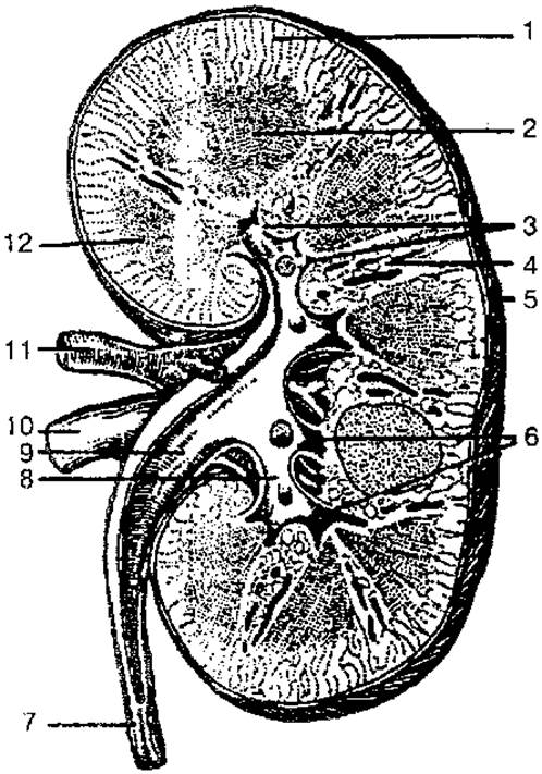
Fig. 82. Structure OF THE right kidney (frontal section):
1 — renal cortex; 2 — renal medulla; 3 — renal papillae; 4 — renal columns; 5 — fibrous capsule; 6 — minor renal calyces; 7 — Ureter; 8 — major renal calyces; 9 — renal pelvis; 10 — renal vein; 11 — renal artery; 12 — renal pyramid
The kidneys are bean-shaped, featuring upper and lower poles, an outer convex and inner concave margin, and anterior and posterior surfaces. The posterior surface lies against the Diaphragm, the quadratus lumborum Muscle, and the psoas major muscle, which form depressions known as the renal beds. Anteriorly, the right kidney is adjacent to the descending part of the duodenum and the colon, while its superior aspect contacts the Inferior surface of the Liver. The anterior surface of the left kidney is related to The Stomach, the tail of the Pancreas, and loops of the Small Intestine. The kidneys are covered by the Peritoneum only on their anterior surface (extraperitoneally) and are anchored in place by the renal fascia and Blood Vessels.
The kidneys are enveloped in three layers: the renal fascia, the fibrous capsule, and the adipose capsule. The adipose capsule is more prominent on the posterior surface, where it forms the paranephric fat body. The renal fascia consists of anterior and posterior layers. The anterior layer covers the front of the left kidney, the renal vessels, the Abdominal Aorta, and the INFERIOR VENA CAVA, extending along the vertebral column to the right kidney; the posterior layer passes behind the kidneys and attaches laterally to the vertebral column. Superiorly, the two layers fuse, while inferiorly they remain open. The parietal peritoneum lies anterior to the anterior layer of the renal fascia. The medial concave margin features the renal hilum, through which the renal artery and NERVES OF THE renal plexus enter the kidney, while the renal vein, ureter, and Lymphatic vessels exit. The hilum opens into the renal sinus, which contains the minor and major renal calyces and the renal pelvis.
The kidney consists of two layers: an outer, lighter cortex and an inner, darker medulla, which forms the renal pyramids. Each renal pyramid has a base facing the cortex and an apex terminating as a renal papilla directed toward the renal sinus. The renal pyramid is composed of straight tubules forming the loop of the nephron and collecting ducts that merge near the renal papilla to form 15–20 short papillary ducts, which open onto the papillary surface via papillary foramina.
The renal cortex consists of alternating light and dark regions. The light regions are cone-shaped and resemble rays extending from the medulla, forming the pars radiata (cortical labyrinth/medullary rays), which contain the renal tubules. These tubules continue into the medulla and the initial segments of the collecting ducts. The dark Regions of the cortex contain the renal corpuscles along with the proximal and distal convoluted tubules.
The main Structural and functional unit of the kidney is the nephron (there are about 1.5 million of them). The nephron (Fig. 83) consists of a renal corpuscle, which includes the glomerulus. The corpuscle is enclosed in a double-walled capsule (Bowman's capsule). The capsule cavity is lined with a simple cuboidal epithelium and transitions into the proximal tubule of the nephron, followed by the nephron loop (Loop of Henle). The loop extends into the medulla and then back into the cortex, leading into the distal segment of the nephron. Via the connecting tubule, it empties into the collecting ducts, which converge into papillary ducts that ultimately open into a minor renal calyx.
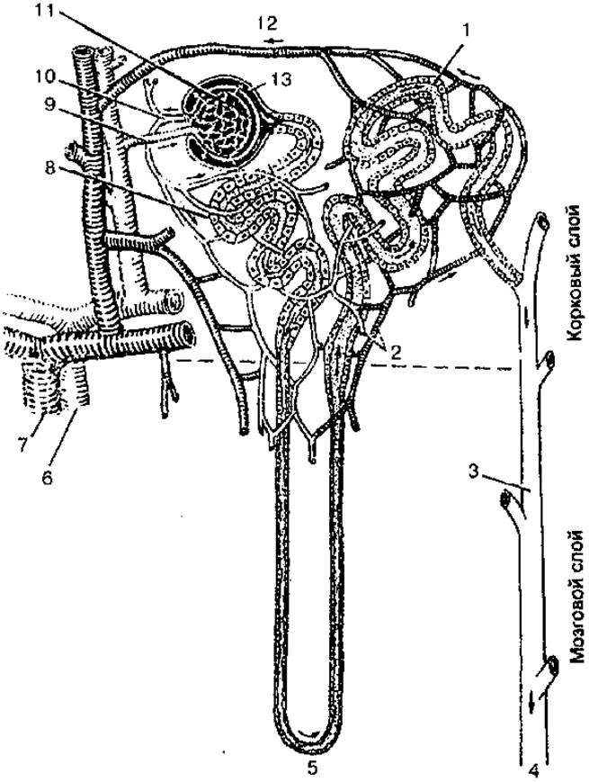
Fig. 83. Diagram of nephron structure and blood supply:
1 — Distal convoluted tubule; 2 — capillary network; 3 — collecting duct; 4 — urine flow toward the renal pelvis; 5 — loop of Henle; 6 — renal artery; 7 — renal vein; 8 — proximal convoluted tubule; 9 — afferent arteriole; 10 — efferent arteriole; 11 — renal glomerulus; 12 — venule; 13 — Bowman's capsule
The merger of two to three minor calyces forms a major renal calyx, and the confluence of two to three major calyces forms the renal pelvis. Approximately 80% of nephrons are located in the outer cortex (cortical nephrons), while 18–20% are situated deep in the renal medulla (juxtamedullary nephrons).
The Blood supply to the kidney is provided by an extensive network of blood vessels. Blood enters the kidney via the renal artery, which divides at the hilum into anterior and posterior branches that give rise to segmental Arteries. These branch into interlobar arteries running between adjacent renal pyramids and columns. At the corticomedullary junction, the interlobar arteries form arcuate arteries arching over the pyramids, which give off numerous interlobular arteries. The interlobular arteries further divide into afferent glomerular arterioles that branch into capillaries, forming the glomerulus within the renal corpuscle. An efferent glomerular arteriole emerges from the glomerulus; its diameter is approximately half that of the afferent arteriole. The efferent arterioles break up into a dense capillary network surrounding the renal tubules before draining into venules. These venules merge into interlobular Veins, which empty into arcuate veins, which in turn lead to interlobar veins. The interlobar veins converge to form the renal vein, which empties into the inferior vena cava.
The lymphatic Vessels of the kidney accompany the blood vessels, exit the kidneys alongside them, and drain into the lumbar Lymph Nodes.
Last update: 08/08/2026
Editorial and Educational Adaptation: This material has been compiled based on the primary/original source text. The project team performed an editorial review, corrected technical inaccuracies, structured sections, and adapted the content for an educational format.
What was processed:
- elimination of formatting defects (OCR errors, structural breaks, corrupted characters);
- editorial organization of content;
- standardization of terminology in accordance with academic sources;
- verification of factual statements against the original source text.
All mentions of the author, publication year, and origin of the primary text have been preserved in accordance with the source.