Human Anatomy Part 2 - K. A. Diubenko, A. K. Kolomiitsev, Yu. B. Chaykovsky 2008
Special Section
Nervous system, systema nervosum - Special Section
Brain - Embryogenesis of the brain - Pons
The Pons is a part of the Brainstem that belongs to the Hindbrain (metencephalon). The pons is a thick white ridge (Fig. 135 A, B), measuring 30x36x25 mm, located between the Medulla Oblongata and the cerebral peduncles, and transitioning laterally into the middle cerebellar peduncles along an imaginary line, the linea trigeminofacialis, which runs between the roots of the trigeminal and facial nerves.
The anterior surface of the pons is convex and lies against the Skull base and the clivus. Along its midline runs the basilar sulcus, sulcus basilaris, which houses the Basilar artery, a. basilaris, the primary source of Blood supply to the pons.
The posterior surface of the pons faces the cavity of the Fourth ventricle and is not visible externally (being covered by the Cerebellum); it forms the upper part of the Rhomboid fossa, which is the floor of the fourth ventricle. Adjacent to the lateral aspect of the pons, where the pons, medulla oblongata, and cerebellum meet, lies a space called the cerebellopontine angle, angulus cerebellopontinus. The cerebellopontine angle opens into the posterior cranial fossa. Anteriorly, it is bounded by the arachnoid mater, which does not extend into its depths but covers the medial portions of the posterior surface of the petrous part of the Temporal bone and the superior surface of the jugular tubercle, thereby forming the lateral pontine cistern, cisterna pontis lateralis, which is filled with CEREBROSPINAL FLUID. In shape, it resembles a flattened irregular pyramid with its apex directed toward the lateral aperture. The cerebellopontine angle contains the roots of Cranial Nerves VII-X and XII, the anterior inferior cerebellar and labyrinthine Arteries, and cerebellar Veins draining into the petrosal sinus. Various pathological conditions frequently arise in the cerebellopontine angle (such as acoustic neuromas of CN VIII, meningiomas, cholesteatomas, inflammatory processes of the Meninges, etc.); therefore, understanding the topography of this region is of great importance in neurosurgery (M. I. Shamaiev).
* Topographically, the medial lemniscus lies in the deep layers of the brainstem at the boundary between the basis and the tegmentum, appearing as a sheet of White matter formed by nerve fibers that originate from the nuclei of the posterior Column of the Spinal Cord and the sensory nuclei of the cranial nerves, and terminate in the thalamus.
Class="center">Internal Structure of the Pons
In frontal sections of the pons, two parts are distinguished: the anterior (ventral) part of the pons, pars anterior, and the posterior (dorsal) part of the pons, pars dorsalis. The boundary between them is the trapezoid body, corpus trapezoideum, which is formed mainly by the processes of Cells from the anterior cochlear Nucleus, nucleus cochlearis anterior.
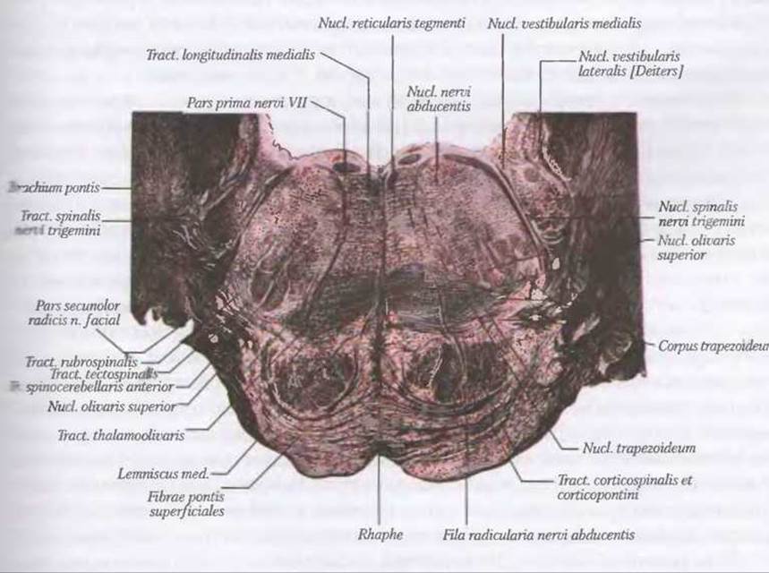
Fig. 138. Internal STRUCTURE OF THE pons (transverse section at the level of the vestibular nuclei) (after Rauber-Kopsch). Cellular structures are shown in red, and fibrous structures in black
The anterior part of the pons consists mainly of nerve fibers, interspersed with numerous clusters of Gray matter known as the pontine nuclei, nuclei pontis.
These nuclei receive the terminating fibers of the corticopontine tract and collaterals from the passing Pyramidal Tracts. The axons of the pontine nuclei cells (which function as Relay stations) project to form the pontocerebellar tract; these fibers cross to the contralateral side as the transverse pontine fibers, fibrae pontis transversae, and enter the middle cerebellar peduncles. Through these fibers, information regarding The Nature of impulses traveling along the corticobulbar and corticospinal tracts to the lower motor Neurons is transmitted to the cerebellar cortex.
The posterior part of the pons (tegmentum of the pons) consists of the Reticular Formation and the brainstem nuclei of cranial nerves (V, VI, VII, VIII). Located in the medial region of the pontine tegmentum is the motor Nucleus of the Trigeminal nerve, nucl. motorius nervi trigeminalis, whose Cell axons form the motor ROOT that projects to and innervates the Muscles of Mastication. Lateral to the motor nucleus lies the principal (pontine) sensory nucleus of the trigeminal nerve, nucl. pontinus nervi trigeminalis, which receives central processes from the sensory Cells of the trigeminal nerve. These processes enter the substance of the pons as the sensory root of the trigeminal nerve at its boundary with the middle cerebellar peduncle.
Within the reticular Formation of the pontine tegmentum lies the motor nucleus of the Facial Nerve, nucl. nervi facialis. The axons of this nucleus form an intramedullary root that follows a complex course. Emerging from the reticular formation, the root loops around the abducens nucleus on the floor of the fourth ventricle, forming the genu of the facial nerve, genu facialis, and then courses toward the cerebellopontine angle. After exiting the skull, the facial nerve fibers reach and innervate the Muscles of facial expression. Posterior to the facial motor nucleus, within the reticular formation, lies the superior salivatory nucleus, nucl. salivarius superior, which is autonomic (secretory) in nature and belongs to the nervus intermedius. It provides parasympathetic innervation to the sublingual, submandibular, and lacrimal glands. Lateral to the superior salivatory nucleus is The Nucleus of the solitary tract, nucl. tractus solitarii, while the nuclei of the Vestibulocochlear nerve are located in the lower lateral part of the pons. In the lateral PARTS OF THE trapezoid body lies the superior olivary nucleus, nucl. olivarius superior. The axons of the anterior and posterior cochlear nuclei, nucl. cochlearis anterior et nucl. cochlearis posterior, and the superior olivary nucleus of the pons contribute to The formation of the lateral lemniscus, lemniscus lateralis, which is a continuation of the trapezoid body. The lateral lemniscus terminates in the cells of the inferior colliculi of the tectal plate and the cells of the medial geniculate body.
Near the midline of the floor of the fourth ventricle lies the abducens nucleus, nucl. n. abducentis, which consists of motor neurons. The root of the Abducens nerve emerges from the pons at the junction between its posterior border and the pyramid of the medulla. Deep to the superior olivary nucleus, above the trapezoid body, lies the medial lemniscus, lemniscus medialis, which is a bundle of proprioceptive sensory fibers.
The WHITE MATTER OF the pons consists of transverse pontine fibers, fibrae pontis transversae, and longitudinal fibers, fibrae pontis longitudinales. The transverse fibers comprise the pontocerebellar fibers, fibrae pontocerebellares, which project from the pontine nuclei proper to the cerebellum, fibers of the lateral lemniscus, and fibers of certain cranial nerves.
The longitudinal fibers are formed by:
- corticospinal, fibrae corticospinales;
- corticonuclear, fibrae corticonucleares;
- corticoreticular, fibrae corticoreticulares;
- corticopontine, fibrae corticopontinae, fibers.
Thus, the pons is of great functional significance: first, it houses the brainstem nuclei of cranial nerves (pairs V, VI, VII, and VIII), the reticular formation, and the pontine nuclei. Second, efferent and afferent pathways pass through the pons, establishing bidirectional communication between the Brain and the spinal cord, which is of vital importance to the Organism.
Last update: 08/08/2026
Editorial and Educational Adaptation: This material has been compiled based on the primary/original source text. The project team performed an editorial review, corrected technical inaccuracies, structured sections, and adapted the content for an educational format.
What was processed:
- elimination of formatting defects (OCR errors, structural breaks, corrupted characters);
- editorial organization of content;
- standardization of terminology in accordance with academic sources;
- verification of factual statements against the original source text.
All mentions of the author, publication year, and origin of the primary text have been preserved in accordance with the source.