Human Anatomy Part 1 - K. A. Dyubenko, A. K. Kolomiytsev, Yu. B. Chaykovsky 2002
Special Part
Reproductive systems, systemata genitalia - Female reproductive system, systema genitale femininum
External female genitalia, organa genitalia feminina externa - Development of external genitalia - Developmental anomalies of the urinary and reproductive systems
Developmental anomalies of the Urinary Organs
Renal developmental anomalies are divided into several groups.
1. Anomalies of renal position (dystopia).
2. Renal relationships and shapes.
3. Anomalies of number.
4. Anomalies of the renal parenchyma.
5. Anomalies of the pelvicalyceal system.
6. Renal vascular anomalies.
Class="center">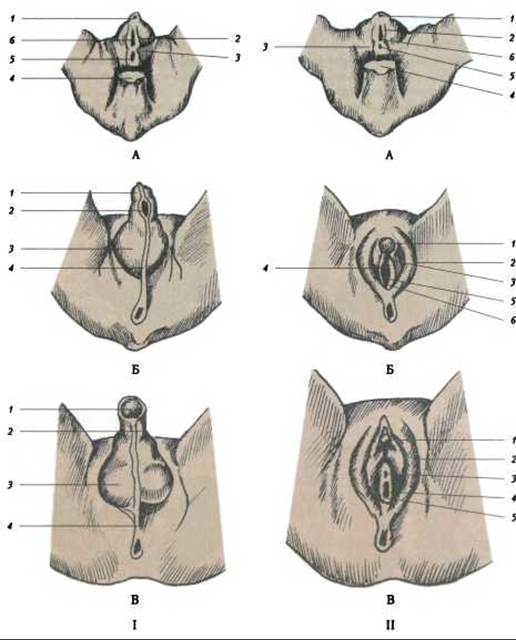
Fig. 340. Diagram of The Development of male and female external genitalia (according to Spaulding): I-A - 7-week embryo: 1 - tuberculum genitale, 2 - plica genitalis; 3 - valla genitalis; 4 - cauda; 5 - anus; 6 - sinus urogenitalis; B - 12-week embryo: 1 - glans Penis; 2 - raphe urethralis;
3 - Scrotum; 4 - raphe scroti; C - 9-month fetus: 1 - glans penis; 2 - raphe urethralis; 3 - scrotum; 4 - raphe scroti; II-B - 12 weeks: 1 - glans clitoridis; 2 - labium majus pudendi; 3 - ostium urethrae externum; 4 - labium minus pudendi; 5 - hymen; 6 - ostium vaginae; C - 9 months: 1 - glans clitoridis; 2 - labium majus pudendi; 3 - ostium urethrae externum; 4 - labium minus; 5 - hymen; 6 - ostium vaginae
1. Anomalies of renal position (dystopia) are associated with impaired ascent and Rotation of the Kidneys during Embryogenesis. Homolateral (thoracic, lumbar, iliac, and pelvic) and heterolateral (crossed) dystopias are distinguished.
2. Anomalies of relationship and shape. Positional and relational anomalies (renal fusion) account for about 13% of all renal anomalies. Horseshoe and dumbbell-shaped kidneys are distinguished, as well as asymmetric fusion forms (S-, L-, and I-shaped kidneys) (Fig. 341).
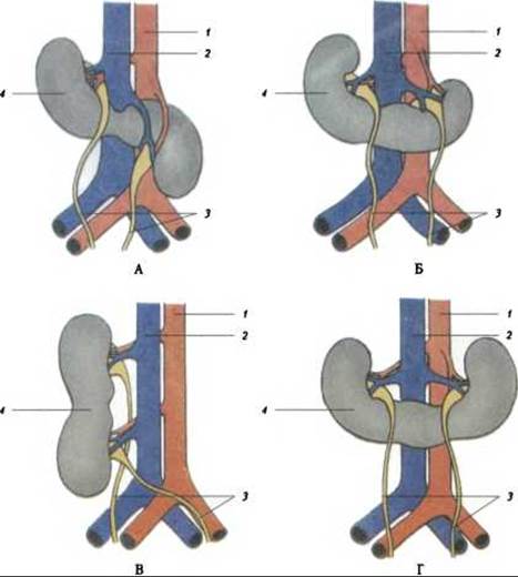
Fig. 341. Schematic representation of kidneys with asymmetric fusion forms (according to N. A. Lopatkin):
A - S-shaped Kidney; B - L-shaped kidney; C - I-shaped kidney; D - Horseshoe kidney; 1 - Abdominal Aorta; 2 - INFERIOR VENA CAVA; 3 - Ureter; 4 - kidney
3. Anomalies of number. Renal duplication is the most common anomaly. Duplication can be complete or incomplete. In incomplete renal duplication, the Blood supply to both halves is provided by a single vascular pedicle.
4. Anomalies of the renal parenchyma. These include: 1) renal agenesis resulting from the failure of the organ bud to form during embryogenesis; 2) renal aplasia, a severe degree of renal parenchymal hypoplasia; 3) hypoplastic kidney, a normally formed kidney reduced in size; 4) supernumerary third kidney, a rare anomaly caused by the Separation of a portion of the metanephric blastema; 5) cystic anomalies of the parenchyma, of which Polycystic Kidney Disease is the most common.
A serious developmental anomaly is the failure of the collecting tubules to connect with the independently formed renal tubules. The accumulation of urine in the latter leads to their transformation into fluid-filled cavities (cysts). These compress the normal tubules, rendering them unable to function. This birth defect is known as congenital cystic kidney.
5. Anomalies of the pelvicalyceal system. These anomalies include: 1) megacalycosis, a Dysplasia of the renal calyces accompanied by their enlargement; 2) calyceal diverticulum, an anomaly characterized by a cystic formation connected to a minor renal calyx via a narrow channel.
6. Anomalies of the renal vessels. These include renal artery atresia, renal artery aplasia, hypoplasia, accessory renal Arteries, congenital stenosis, and renal artery aneurysm.
Ureteral developmental anomalies. According to A. Ya. Pitel and G. M. Chabanyuk (1969), anomalies of the proximal and distal segments of the ureter are distinguished, as well as anomalies of its shape and Structure (Fig. 342).
A duplicated ureter quite frequently occurs on one or both sides. In this case, the metanephric blastema typically divides into two parts, resulting in a duplicated (more accurately, divided) kidney. Other occurrences include megaureter, ureteral stricture, ring-shaped ureter, and retrocaval/retroiliac ureter.
In cases of anal atresia in female newborns, a communication between the rectum and Vagina may develop (rectovaginal fistula), whereas in male newborns, a communication between the rectum and Urinary Bladder may occur (rectovesical fistula). The latter condition requires immediate surgical intervention.
Partial persistence of the urachal lumen can lead to The formation of a fluid-filled cyst, while complete persistence results in an umbilicourinary fistula, which allows urine to leak through the umbilicus.
Sometimes, absence of the urinary bladder (atresia), incomplete duplication of the bladder, bilocular bladder, bladder diverticulum, as well as narrowing and fissure of the bladder are observed.
Last update: 08/08/2026
Editorial and Educational Adaptation: This material has been compiled based on the primary/original source text. The project team performed an editorial review, corrected technical inaccuracies, structured sections, and adapted the content for an educational format.
What was processed:
- elimination of formatting defects (OCR errors, structural breaks, corrupted characters);
- editorial organization of content;
- standardization of terminology in accordance with academic sources;
- verification of factual statements against the original source text.
All mentions of the author, publication year, and origin of the primary text have been preserved in accordance with the source.