Antibiotics (Properties, Application, Interaction) - M.P. Cherenko 1999
Acute purulent infection
Purulent diseases of loose connective tissue
Mastitis. To understand the Pathogenesis, Clinical presentation, and Treatment of mastitis, one must be familiar with certain Anatomical Features of the mammary gland.
The female breast is located on the anterior chest wall at the level of the III–VI Ribs. A pigmented nipple is situated in the center of the breast. The mammary gland consists of 15–20 separate radially arranged lobes, each of which in turn is composed of alveolar glands. The lobes of the gland are surrounded by adipose tissue and Connective Tissue septa. Lactiferous ducts extend radially from the lobes and open at the center of the nipple. The mammary gland is separated by a thin layer of adipose tissue from the retromammary fascia, which covers the pectoralis major Muscle.
The Blood supply to the mammary gland is provided by Branches of the internal thoracic artery, which originates from the Subclavian Artery. Lymph from the breast drains into the axillary, parasternal, as well as sub- and supraclavicular Lymph Nodes.
Non-specific and specific (tuberculosis, Syphilis, actinomycosis) inflammatory processes may develop in the mammary gland.
Acute mastitis (mastitis acuta) is an acute purulent inflammation of the parenchyma (parenchymatous mastitis) or connective tissue (interstitial mastitis) of the mammary gland. It is caused by a staphylococcal infection. Staphylococci penetrate the breast exogenously (through nipple fissures) or endogenously (via hematogenous or lymphogenous routes) from existing inflammatory foci in the body. Less frequently, acute mastitis is caused by streptococci, Escherichia coli, Pseudomonas aeruginosa, Proteus, or anaerobes. Most commonly, acute mastitis develops postpartum during breastfeeding (in 1–5% of nursing mothers). Milk stasis and breast engorgement contribute to The Development of the inflammatory process in the gland. Usually, one breast is affected, less often both.
The inflammatory process develops in several stages. Initially, there is serous infiltration of the mammary gland tissue. Serous exudate containing A large number of leukocytes accumulates in the intercellular space. Later, serous infiltration is replaced by purulent infiltration, resulting in the development of breast Phlegmon. As the process progresses in glandular Organs, inflammation tends to spread (affected tissue undergoes liquefaction, and an abscess forms). In the event of vascular thrombosis, the breast tissue may undergo necrosis, leading to gangrenous mastitis.
Many authors distinguish the following phases of process development: serous, phlegmonous, purulent, and gangrenous.
Acute mastitis, like any inflammatory process in glandular organs, is characterized by a poorly defined tendency toward encapsulation and rapid spread within the tissue. The inflammatory process may be localized exclusively within the milk ducts (galactophoritis) or affect the Glands of the areola (areolitis).
Breast abscesses can be located premammarly (paraareolarly), intramammarly, or retromammarly (Fig. 69).
The clinical picture of acute mastitis depends on the stage of the disease and its localization.
Acute mastitis begins with a small induration in the breast resulting from milk stasis. Initially, it is mildly painful, but over time the pain intensifies, and milk secretion is hindered. The general condition during this period remains undisturbed. With delayed or inadequate treatment, the lump in the breast becomes denser, pain grows intense, and the Skin over the induration turns red. Body Temperature rises to 38–39 °C. Chills, headache, and general weakness may occur. Breastfeeding with the affected breast becomes difficult. Axillary lymph nodes enlarge. Blood counts change (as is typical for an inflammatory process).
During the stage of purulent liquefaction, fluctuation is observed in the center of the induration. Local and general symptoms worsen.
In the gangrenous stage, the patient's general condition is very severe. Body temperature reaches 39–40 °C, the pulse is rapid at 120–140 bpm. Blood pressure may be decreased, the Tongue dry, and the skin pale. The skin over the mammary gland is pale green or bluish-purple. The breast is markedly enlarged in volume. Regional lymph nodes become enlarged and tender. Blood parameters change: high leukocytosis, left shift in the leukocyte formula, and an elevated ESR.
In the abscess formation stage, the Diagnosis of purulent mastitis can be confirmed by breast puncture, which yields pus.
Diagnosing paraareolar and intramammar mastitis is not difficult based on the clinical picture described above. In retromammar mastitis, localized in the tissue posterior to the mammary gland in the area of the pectoralis major muscle, a diffuse painful Swelling is observed without any skin discoloration. The entire affected gland appears elevated or protrudes forward. When the gland is displaced, the patient experiences pain. General manifestations are similar to those of mastitis in other locations. Phenomena of lymphangitis and lymphadenitis are characteristic.
Acute mastitis must be differentiated from so-called milk fever, which occurs in postpartum women on the 4th–5th day after delivery and is caused by milk stasis. Stagnating in the mammary gland and being reabsorbed, the milk exhibits pyrogenic properties.
Treatment of acute mastitis in the early stages is conservative, but strictly under a doctor's supervision. Self-treatment is unacceptable. Therapy should begin with Lactation suppression and systematic milk expression. Breastfeeding is discontinued due to the risk of the infant developing stomatitis, enteritis, or Conjunctivitis. Milk is systematically expressed or extracted using a breast pump. Medications that suppress lactation are used (a combination of estrogens and androgens). The breast is elevated and firmly supported with a bandage. Antibiotics, physiotherapy, and general strengthening agents are prescribed.
Class="center">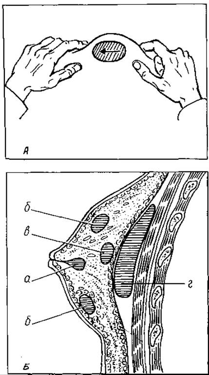
Fig. 69. Technique for detecting the fluctuation sign of an abscess (A). Location of abscesses (B): a — premammar; b, c — intramammar; d — retromammar
Good results are achieved with a retromammar procaine blockade (80 ml of a 0.25% procaine solution + 500,000 IU of monomycin or kanamycin + 10 mg of Trypsin or Chymotrypsin). Concurrently, infusion detoxification therapy is administered (reopolyglucukin, plasma, protein preparations, electrolyte solutions).
If treatment is initiated in the serous or infiltrative stage, a positive effect can be expected: pain decreases, the infiltrate gradually resolves, and body temperature drops.
In the phlegmonous, and especially in the abscessing or gangrenous stage, Surgical treatment is indicated. The operation should be performed under general anesthesia (intravenous or inhalation). This allows for wide incision of the abscess and its pockets, wound exploration, and drainage. If the abscess is located paraareolarly or intramammarly, radial incisions are made in the area of fluctuation or greatest tenderness, directed from the periphery toward the nipple (such incisions do not damage the milk ducts). In cases of retromammar abscess localization, an inframammary curved incision is performed (Bardenheuer's incision). After opening the abscess, its cavity is explored with a finger, breaking down all fibrous strands and septa. Necrotic Tissues are excised. The abscess cavity is washed with an antiseptic solution (furatsilin, chlorhexidine, etc.) and drained using rubber drains and polyvinyl chloride tubes. Postoperative management is identical to that of a purulent wound, continuing general therapy until inflammatory signs completely disappear.
Sometimes, to ensure better pus outflow and drainage, two or three incisions must be made, connecting them together in the depth of the wound.
In recent years, an active surgical method has been introduced into the practice of treating localized forms of Acute Purulent Infection, particularly abscessing mastitis. This method involves excising the abscess, establishing continuous irrigation-suction drainage of the wound cavity, and closing the wound with primary sutures (A.A. Fedorovsky, I.M. Starovoytov, 1967; M.P. Cherenko et al., 1985).
The duration of conservative treatment for acute mastitis averages 12–15 days. In the case of surgical intervention (incision of the abscess and drainage), it extends to 3–4 weeks. Active treatment accelerates wound healing and patient recovery by 2 to 3 times. Furthermore, it provides significantly better functional treatment outcomes.
The Prevention of acute mastitis begins long before childbirth in antenatal clinics (enhancing the body's reactivity, sanitizing existing foci of infection in the woman's body, and educating pregnant women on postpartum self-care). After delivery, mastitis prevention consists, on the one hand, of preventing infection from entering the mammary gland and, on the other hand, of preventing milk stasis. To this end, nipples should be systematically washed with warm Water and soap, wiped with 60–70% ethyl alcohol, and dried with a soft towel. Air baths for 15–20 minutes are beneficial. To prevent nipple fissures, they are lubricated with a neutral fat, underwear is changed frequently, and hands are washed. To prevent milk stasis, the Mammary Glands are fixed with bandages in an elevated position, the baby is breastfed, and milk is thoroughly expressed.
An important role in mastitis prevention is played by maintaining the Sanitary and epidemiological regime in the inpatient facility (wet cleaning of wards, air disinfection with bactericidal lamps, and controlling bacterial carriage).
Sometimes, after the acute process subsides, chronic inflammation of the mammary gland (chronic mastitis) may persist.
Following the acute period, the infection in the inflammatory focus remains latent and can be activated under METABOLISM/18.html">The Influence of any triggering factors. This condition is rare. The Transition of acute mastitis into chronic form is most often caused by spontaneous opening of the purulent focus, small incisions, or errors in treatment and postoperative management.
Chronic mastitis has two clinical forms: fistulous and infiltrative. In the first form, one or more fistulae with scant purulent discharge are present on the mammary gland. A slightly painful infiltrate can be palpated around them. In the second form, there are no fistulae, but indurations of varying sizes remain largely painless within the breast tissue. The skin over them is unchanged. General signs are not pronounced. Axillary lymph nodes may be enlarged, but are painless.
Chronic mastitis responds poorly to Conservative Methods. In cases of a protracted course, a sectoral resection of the mammary gland is performed, accompanied by a histological examination of the removed tissue.
Patients with chronic mastitis must be under dispensary observation due to the potential for malignant transformation of the induration.
Acute paraproctitis (paraproctitis acuta) is acute inflammation of the tissue surrounding the rectum. It is caused by pyogenic infection (Staphylococcus aureus or white staphylococcus, Escherichia coli, anaerobic flora) that penetrates into the pararectal tissue, primarily from fissures and inflamed anal crypts and anal glands of the rectum.
Men develop paraproctitis twice as often as women.
Depending on the spread of infection within the pararectal tissue, the following types of paraproctitis are distinguished: submucosal, subcutaneous, ischiorectal (between the ischial tuberosity and the rectum), pelvirectal (above the levator ani muscle, between the rectum and this muscle), and retrorectal (behind the rectum; Fig. 70). Subcutaneous-submucosal and ischiorectal paraproctitis are the most common.
The Clinical presentation of acute paraproctitis is characterized by severe pain in the rectal area. Walking is difficult for the patient, and acts of defecation and urination are impaired. Superficial abscesses (subcutaneous, submucosal) present with edema, skin erythema, and sharp tenderness upon Palpation. In deep paraproctitis (ischiorectal, pelvirectal), patients experience sharp, throbbing pain in the rectal area, and digital examination of one of the rectal walls reveals marked tenderness. An induration is felt at this site, and fluctuation is present if purulent melting of the infiltrate has occurred.
The patient's general condition progressively deteriorates. Body temperature rises to 38–39 °C, accompanied by chills, loss of appetite, and insomnia. Blood tests show leukocytosis, a left shift in the leukocyte formula, and an increased ESR.
Without adequate treatment, abscesses rupture outward through the skin of the gluteal region (more commonly) or into the rectal lumen. In such cases, acute inflammatory symptoms subside, long-healing fistulae form, and the process becomes chronic (chronic paraproctitis — paraproctitis chronica).
The treatment of acute paraproctitis is surgical. Conservative therapy (UHF therapy, warm microclysters with chamomile infusion, warm sitz baths, antibiotic therapy, normalization of defecation) can be used only for superficial (subcutaneous) localization in the infiltrative stage.
Before surgery, the patient should be given a cleansing enema. Surgery is best performed under general anesthesia. The abscess is incised. The abscess cavity is explored with a finger to locate and treat its internal opening in the rectum.
To prevent premature closure of the wound edges, a triangular skin flap with its apex near the internal opening of the abscess is excised. The wound is loosely drained.
Most pelvirectal abscesses are opened from the rectal side.
It should be noted that a simple incision of a pararectal abscess often fails to cure the disease, merely converting it into a chronic (fistulous) form.
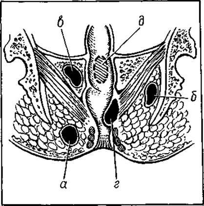
Fig. 70. Location of abscesses in the pararectal tissue: a — subcutaneous abscess; b — ischiorectal; c — pelvirectal; d — submucosal; e — rectorectal
This is due to the fact that already in the acute stage (the abscess stage), paraproctitis is combined with the rectal cavity by an internal fistula that opens into one of the crypts of the posterior (more commonly) or anterior commissure of the anal canal. Consequently, the only radical surgical intervention for acute paraproctitis is one that, along with opening the abscess, is supplemented by the elimination of the internal fistula opening in the rectal wall. In subcutaneous-submucosal and ischiorectal abscesses where they (and the fistulae) are located internal to the anal sphincter, the internal opening in the rectal wall is excised (after opening the abscess) along with the skin and subcutaneous tissue. Triangular flaps with the apex in the area of the internal opening are excised. In acute paraproctitis with an extrasphincteric LOCATION OF THE internal fistula, the latter is removed using more complex methods. One of these methods was proposed by Hippocrates. It involves opening the abscess with the removal of its outer capsule in the form of a triangular flap (this surgical element was proposed by Gabriel) and passing a strong ligature through the internal fistula opening in the rectal wall, which is tied over the anal sphincter and gradually, over a week or even longer, cuts through it. Thanks to the gradual division of the sphincter, the fistula opening is eliminated and sphincter incompetence does not develop.
In the postoperative period, patients receive small doses of antibiotics and tincture of opium (5–6 drops three times a day). Drains are changed every 2–3 days.
Paranephritis is the purulent inflammation of the loose connective and adipose tissue surrounding the Kidney.
The cause of paranephritis is most often the spread of infection from the kidney to the perirenal tissue in Pyonephrosis, tuberculosis, and cortical abscesses of the kidney. Infection can also penetrate the paranephric tissue through stab or gunshot wounds of the kidney, or from purulent foci in abdominal organs (acute appendicitis, cholecystitis, colonic perforation). A metastatic pathway of infection transmission is also possible.
The causative agents of infection are most commonly staphylococci, Escherichia coli, and anaerobic flora.
Paranephritis mainly affects individuals aged 20–40, more frequently men.
The disease begins with serous edema of the perirenal adipose tissue. After some time, it progresses to a phase of inflammatory infiltration, which culminates in purulent melting and sequestration of the fat tissue. Pus may spread downward along the psoas major muscle, forming a so-called psoas abscess.
Against the Background of an underlying condition (most commonly renal), patients suddenly experience severe lower back pain, chills, and a body temperature spike up to 40 °C. The pain intensifies during inhalation and radiates to the shoulder and leg. The sclerae become yellowish, and the patient's general condition deteriorates sharply.
Palpation of the abdomen and lumbar region reveals muscle guarding and tenderness. Pasternatsky's sign (eliciting pain by gently striking the lumbar region with the ulnar edge of the hand) is positive. The subcutaneous tissue in the lumbar region is edematous and slightly indurated. Subsequently, a firm, painful, fixed inflammatory infiltrate begins to be palpable in the hypochondrium or slightly lower. The lumbar region bulges, and the skin becomes hyperemic. Following the descent of pus along the psoas major muscle, the psoas sign is detected on the affected side (inability to straighten the leg flexed at the hip joint).
Blood tests clearly indicate inflammatory changes. Urine is normal. Only in severe cases with a pronounced septic course of the disease may moderate albuminuria and cylindruria be observed.
X-ray Examination reveals a diffuse homogeneous shadowing with the obliteration of the psoas major muscle contours.
Lumbar puncture helps to clarify the diagnosis. Paranephritis can lead to numerous life-threatening complications associated with the rupture of the abscess into the abdominal (Peritonitis) and pleural (empyema) cavities, as well as Sepsis.
The treatment for paranephritis is primarily surgical, involving the incision of the phlegmon and drainage of its cavity.
Last update: 08/08/2026
Editorial and Educational Adaptation: This material has been compiled based on the primary/original source text. The project team performed an editorial review, corrected technical inaccuracies, structured sections, and adapted the content for an educational format.
What was processed:
- elimination of formatting defects (OCR errors, structural breaks, corrupted characters);
- editorial organization of content;
- standardization of terminology in accordance with academic sources;
- verification of factual statements against the original source text.
All mentions of the author, publication year, and origin of the primary text have been preserved in accordance with the source.