HUMAN MEDICAL BIOLOGY, ANATOMY, PHYSIOLOGY AND PATHOLOGY - Y.I. Fedoniuk 2010
BIOLOGY
SECTION 1. BIOLOGICAL BASES OF HUMAN VITAL ACTIVITY
1.2. CELL. TISSUES. CONCEPT OF AN ORGAN, ORGAN SYSTEM, AND ORGANISM
NERVOUS TISSUE
Nervous Tissue is the primary structural component of The Nervous system. Its elements are capable of perceiving stimuli, transforming these stimuli into nerve impulses, rapidly transmitting them, storing information, and producing BIOLOGICALLY ACTIVE SUBSTANCES. Through these Functions, nervous tissue ensures the coordinated activity of the body's Organs and systems, as well as its ADAPTATION TO ENVIRONMENTAL conditions, both internal and external. Nervous tissue is composed of Nerve Cells (Neurons) and supportive elements collectively known as neuroglia.
Neurons (neurocytes) are the core structures of nervous tissue that receive stimuli, generate, and transmit impulses.
A neurocyte consists of a Cell body and processes. The presence of processes is the most characteristic feature of nerve cells. There are two types of processes: the axon and the dendrite.
An axon is a long cell process. There is only one per neuron; it does not branch, conducts nerve impulses away from The Cell body, and terminates with a specialized apparatus on another neuron or an effector organ.
A dendrite is a short, branching process that conducts nerve impulses toward the cell body. The peripheral ends of sensory neuron dendrites bear receptors (sensitive nerve endings). The number of dendrites per cell can vary. Based on the number of processes, nerve cells are classified into unipolar (having a single process, the axon), bipolar (having two processes: an axon and a dendrite), and multipolar (having three or more processes). A variation of bipolar cells is the pseudounipolar neuron (where a single process emerges from the cell body and, at a certain distance, T-junctions into an axon and a dendrite) (Fig. 1.25).
Class="center">
Fig. 1.25. Classification of neurocytes by the number of processes: 1 - unipolar neurocyte; 2 - bipolar neurocyte; 3 - pseudounipolar neurocyte; 4 - multipolar neurocyte; a - axon; b - dendrite; c - neurocyte body.
According to their functional role, nerve cells are divided into receptor (afferent or sensory) neurons, effector (efferent or motor) neurons—which transmit impulses to the contractile or secretory elements of an effector organ—and associative (interneurons), which establish connections between other neurons.
Neuroglia forms the microenvironment in which neurons reside. It has a cellular Structure (Fig. 1.26) and provides trophic, secretory, and protective functions.
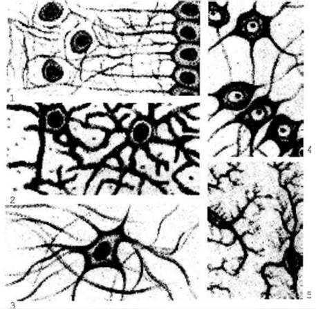
Fig. 1.26. Neuroglia: 1 - ependymal cells; 2 - protoplasmic astrocytes; 3 - fibrous astrocytes; 4 - oligodendrocytes; 5 - microglia.
All neuroglial cells are divided into macroglia and microglia. Macroglia includes ependymocytes, astrocytes, and oligodendrocytes, whereas microglia comprises glial macrophages.
Ependymocytes are cuboidal or columnar cells bearing cilia on their apical surface, which facilitate the movement of CEREBROSPINAL FLUID through the Brain ventricles. Long processes extend from the basal pole of these cells. Ependymocytes line the ventricles of the brain and the central canal of the Spinal Cord, and they participate in The production of cerebrospinal fluid.
Astrocytes are the largest glial cells, found in all Divisions of the nervous system. They possess a stellate shape, with numerous processes radiating from the cell body in all directions. Astrocytes are divided into two groups: protoplasmic and fibrous. Protoplasmic astrocytes are found predominantly in the Gray matter of the Central Nervous System and feature short, thick, highly branched processes.
Fibrous astrocytes are localized primarily in the White matter OF the central nervous system. Their cell bodies give rise to long, straight processes that exhibit little to no branching. Astrocytes form the structural framework (stroma) of the central nervous system.
Oligodendrocytes represent the most numerous group of gliocytes; they are small cells with short processes. They surround the cell bodies of neurons and are incorporated into The structure of nerve fibers and nerve endings.
Microglia consists of small, elongated, stellate cells located mainly along capillaries within the central nervous system. Unlike macroglial cells, microglia develop directly from monocytes. The primary function of microglia is protective (including immune defense).
Microglial cells function as specialized macrophages of the central nervous system. They are highly mobile and become activated during Inflammatory Diseases of the nervous system; upon activation, they retract their processes, assume a rounded shape, and phagocytose cellular debris from dead cells.
Nerve fibers are processes of nerve cells enveloped by specialized sheaths. In different PARTS OF THE nervous system, these sheaths vary significantly in structure; consequently, all nerve fibers are divided into two major groups: myelinated and unmyelinated. Both types consist of a nerve cell process (axon or dendrite) lying at the center—referred to as the axon (or axis cylinder)—and a surrounding sheath formed by oligodendrocytes, which in the Peripheral Nervous System are known as neurolemmocytes (lemmocytes or Schwann cells).
Cytology/practical/65.html">Myelinated nerve fibers are found in both the central and peripheral nervous systems and are characterized by a high conduction velocity of nerve impulses. These are thick fibers containing large-diameter axis cylinders. The axis cylinder is directly surrounded by a specialized myelin sheath, which is enveloped in turn by a thin layer containing the Cytoplasm and Nucleus of the lemmocyte, known as the neurolemma. Externally, the fiber is also covered by a basement membrane.
Unmyelinated nerve fibers belong to the Autonomic nervous system and are characterized by a low conduction velocity of nerve impulses. Their structure is considerably simpler. Unmyelinated fibers consist of an axon (axis cylinder), neurolemma, and basement membrane. The neurolemma is formed by a cord of closely apposed neurolemmocytes (Schwann cells). By indenting the Sheath of the neurolemmocytes, the axon sinks deeply into this cord, while the glial cells surround the process like a sleeve. Externally, the unmyelinated nerve fiber is covered by a basement membrane.
Bundles of myelinated and unmyelinated fibers, enveloped by a Connective Tissue sheath, form nerve trunks (nerves).
All nerve fibers terminate in specialized structures known as nerve endings. According to their functional significance, nerve endings are divided into three groups: 1) effectors, 2) receptors, 3) interneuronal synapses.
Interneuronal synapses are specialized contacts between nerve cells that transmit impulses in a single direction. Based on morphological features, they are classified into: 1) axosomatic synapses (terminal Branches of the axon of the first neuron terminate on the soma of the second); 2) axodendritic synapses (terminal branches of the axon of the first neuron form synaptic connections with the dendrite of the second); 3) axoaxonic synapses (terminal branches of the axon of one neuron terminate on the axon of another).
Based on their morphofunctional characteristics, synapses are divided into chemical (vesicular) and electrical, which are characterized by tight junctions between the Plasmalemma of two neurocytes (primarily their dendrites or cell bodies). Chemical synapses can be either excitatory or inhibitory.
Last update: 08/08/2026
Editorial and Educational Adaptation: This material has been compiled based on the primary/original source text. The project team performed an editorial review, corrected technical inaccuracies, structured sections, and adapted the content for an educational format.
What was processed:
- elimination of formatting defects (OCR errors, structural breaks, corrupted characters);
- editorial organization of content;
- standardization of terminology in accordance with academic sources;
- verification of factual statements against the original source text.
All mentions of the author, publication year, and origin of the primary text have been preserved in accordance with the source.