HUMAN ANATOMY ATLAS - G.L. Bilich - 2014
Muscular System
MUSCLES AND FASCIA OF THE LOWER LIMB
Muscles of the Pelvic Girdle
The pelvic Muscles surround the hip joint on all sides, originating from the pelvic bones, lumbar vertebrae, and sacrum, and inserting into the upper third of the Femur. The pelvic muscles are divided into two groups: internal, located within the pelvic cavity (iliacus, psoas Major and minor, piriformis, obturator internus), and external, located on the lateral surface of the pelvis and the gluteal region (gluteus maximus, medius, and minimus, quadratus femoris, tensor fasciae latae, obturator externus, and the superior and inferior gemelli). The external muscles, arranged in several layers, maintain body balance during standing and walking. In newborns and infants, the gluteal muscles are poorly developed. As children begin to walk, these muscles develop and increase in size.
Class="center">Table 47. Muscles of the Lower Limb
Origin |
Insertion |
Action |
Innervation, Blood Supply |
|
Pelvic muscles. Internal pelvic muscles |
||||
Iliopsoas muscle. Consists of two muscles: Psoas major Iliacus muscle |
Lateral surfaces of the bodies and intervertebral discs of the XII thoracic and I—V lumbar vertebrae, and their transverse processes Iliac fossa of the corresponding bone |
Lesser trochanter of the femur; an iliopectineal bursa is located between the tendon and the lesser trochanter; a bursa of the pectineus muscle lies between the tendon and the pectineus muscle |
Flexes the thigh at the hip joint. When the lower limb is fixed, it tilts the pelvis and trunk forward |
Muscular Branches of the Lumbar plexus (LI-LIV). Aa.: iliolumbar; deep circumflex iliac |
Psoas minor muscle (inconstant) |
Lateral surface of the XII thoracic and I lumbar vertebrae, and the intervertebral disc between them |
By a long tendon to the arcuate line of the ilium and iliopubic eminence; some tendinous fibers continue into the iliac fascia and iliopectineal arch |
Tenses the fascia, flexes the lumbar spine |
Muscular branches of the lumbar plexus (LI-LIV). Aa.: lumbar |
Obturator internus muscle |
Margins of the obturator foramen, internal surface of the obturator membrane, pelvic surface of the ilium above the obturator foramen, obturator fascia |
Medial surface of the greater trochanter |
Laterally rotates the thigh |
Muscular branches of the lumbar and sacral plexuses (LIV-LV, SI—SII). Aa.: inferior gluteal; obturator; internal pudendal |
Piriformis muscle |
Pelvic surface of the sacrum lateral to the pelvic sacral foramina |
Apex of the greater trochanter of the femur |
Laterally rotates the thigh and abducts it slightly |
Muscular branches of the Sacral Plexus (SI—SII). Aa.: superior and inferior gluteal |
External pelvic muscles |
||||
Gluteus maximus muscle |
Gluteal surface of the ilium, dorsal surface of the sacrum and coccyx, sacrotuberous ligament |
Gluteal tuberosity of the femur, iliotibial tract. A trochanteric bursa of the gluteus maximus is situated between the muscle tendon and the greater trochanter |
Extends the thigh at the hip joint and laterally rotates it; with a fixed leg, extends the trunk and maintains balance of the pelvis and trunk. Anterosuperior fibers abduct the thigh, tense the iliotibial tract, and keep the knee joint in an extended position. Posteroinferior fibers adduct the thigh and laterally rotate it |
Inferior gluteal nerve (LV—SII). Aa.: superior and inferior gluteal; medial circumflex femoral |
Gluteus medius muscle |
Gluteal surface of the ilium, between the anterior and posterior gluteal lines, fascia lata |
By a thick flat tendon to the apex and lateral surface of the greater trochanter; a synovial bursa is present |
Abducts the thigh; anterior fibers medially rotate the thigh, posterior fibers laterally rotate it. When the lower limb is fixed, together with the gluteus minimus, it maintains the pelvis and trunk in an upright position |
Superior gluteal nerve (LIV-SI). Aa.: superior gluteal; lateral circumflex femoral |
Gluteus minimus muscle |
External surface of the ilium, between the middle and inferior gluteal lines, margin of the greater sciatic notch |
By a short tendon to the anterolateral surface of the greater trochanter of the femur. A synovial bursa is present between the muscle tendon and the greater trochanter |
Abducts the thigh; anterior fibers medially rotate the thigh, posterior fibers laterally rotate it |
Superior gluteal nerve (LIV-SI). Aa.: superior gluteal; lateral circumflex femoral |
Quadratus femoris muscle |
By a short tendon from the lateral margin of the ischial tuberosity |
Upper part of the intertrochanteric crest of the femur |
Laterally rotates the thigh |
Sciatic nerve (LIV-SI). Aa.: inferior gluteal; obturator; medial circumflex femoral |
Obturator externus muscle |
External surface of the pubic bone and ramus of the ischium near the obturator foramen, obturator membrane |
Trochanteric fossa of the femur |
Laterally rotates the thigh |
Obturator nerve (LII-LIV). Aa.: obturator; lateral circumflex femoral |
Tensor fasciae latae |
Anterior superior iliac spine and adjacent part of the iliac crest |
Continues into the fascia lata (iliotibial tract), lateral condyle of the Tibia |
Tenses the fascia lata, stabilizes the knee joint, and flexes the thigh when the knee is extended |
Superior gluteal nerve (LIV-SI). Aa.: superior gluteal; lateral circumflex femoral |
Superior and inferior gemelli muscles |
Ischial spine, ischial tuberosity |
Medial surface of the greater trochanter of the femur |
Laterally rotate the thigh |
Muscular branches of the sacral plexus (LIV-LV), (SI-SIII). Aa.: inferior gluteal; obturator; internal pudendal |
Muscles of the Free Lower Limb. Thigh muscles. Anterior compartment of the thigh |
||||
Sartorius muscle |
Anterior superior iliac spine |
By a tendinous aponeurosis to the tibial tuberosity and crural fascia. At its insertion, the tendon of the sartorius fuses with the tendons of the gracilis and semitendinosus to form a triangular fibrous plate — the superficial pes anserinus, beneath which lies the anserine bursa |
Flexes the thigh and leg, abducts and laterally rotates the thigh |
Femoral nerve (LII-LIV). Aa.: muscular branches of the femoral; lateral circumflex femoral |
Quadriceps femoris muscle. Consists of four parts: Vastus lateralis muscle |
By tendinous and muscular fibers from the intertrochanteric line, greater trochanter (lower part), lateral lip of the linea aspera of the femur, and lateral intermuscular septum of the thigh |
By a common tendon to the base and lateral margins of the Patella, and the tibial tuberosity. Distally from the apex of the patella, the middle part of the tendon continues as the patellar ligament. Some tendinous fibers continue into the lateral patellar retinaculum. The tendon of the vastus medialis contributes to The formation of the medial patellar retinaculum |
Extends the leg at the knee joint. A suprapatellar bursa, communicating with the knee joint cavity, is located beneath the quadriceps tendon above the patella |
Femoral nerve (LII- LIV). Aa.: femoral; deep artery of the thigh |
Vastus medialis muscle |
Extensive origin: lower half of the intertrochanteric line, medial lip of the linea aspera of the femur, medial intermuscular septum of the thigh |
|||
Vastus intermedius muscle |
By muscular fibers from the anterior and lateral surfaces of the femoral shaft, lower part of the lateral lip of the linea aspera of the thigh, and lateral intermuscular septum of the thigh |
|||
Rectus femoris muscle |
Anterior inferior iliac spine, superior margin of the acetabulum; a synovial bursa is located between the bone and the muscle origin |
Flexes the thigh at the hip joint, extends the leg at the knee joint |
||
Semimembranosus muscle |
Ischial tuberosity |
By three bands to the posterolateral surface of the medial condyle of the tibia. These bands form the deep pes anserinus. One band passes inferiorly and attaches to the tibial collateral ligament; the second passes inferiorly and laterally, blending with the Fascia of the popliteus muscle and ending at the soleal line of the tibia; a robust third band passes laterally to the posterior surface of the lateral condyle of the femur, forming the oblique popliteal ligament. Synovial bursae are located between the tendon and the medial condyle of the femur |
Extends the thigh, flexes the leg, and medially rotates it (when the leg is flexed) |
Muscular branches of the sciatic nerve (LIV-SII). Aa.: medial circumflex femoral; perforating; popliteal |
Medial compartment of the thigh |
||||
Gracilis muscle |
Inferior ramus of the pubis |
Medial surface of the upper third of the tibial shaft, contributing to the Formation of the superficial pes anserinus |
Adducts the thigh, flexes the leg at the knee joint, and medially rotates it |
Obturator and femoral nerves (LII-LIV). Aa.: obturator; external pudendal; femoral |
Pectineus muscle |
Superior ramus and pecten of the pubis |
By a flat, thin tendon to the medial lip of the pectineal line of the femur |
Adducts and flexes the thigh |
Obturator nerves (LII-LIV). Aa.: external pudendal; deep artery of the thigh |
Adductor longus muscle |
Superior ramus of the pubis |
By a broad, flat tendon to the middle third of the medial lip of the linea aspera of the femur |
Adducts the thigh and slightly laterally rotates it |
Obturator nerve (LII-LIV). Aa.: obturator; perforating |
Adductor brevis muscle |
External surface of the body and inferior ramus of the pubis |
By short tendinous fibers to the upper part of the medial lip of the linea aspera of the femur |
Adducts the thigh and slightly laterally rotates it |
Obturator nerve (LII-LIV). Aa.: obturator; perforating |
Adductor magnus muscle |
Ramus of the ischium, ischial tuberosity, and inferior ramus of the pubis |
Medial lip of the linea aspera of the femur. Near its insertion into the adductor tubercle of the femur, the tendon bounds the tendinous hiatus, through which the femoral artery passes from the adductor canal into the popliteal fossa |
Adducts the thigh and laterally rotates it. Medial fibers assist in thigh extension |
Obturator nerve (LII-LIV); sciatic nerve (LIV-SII). Aa.: obturator; perforating |
Muscles of the Leg. Posterior compartment of the leg. Superficial layer |
||||
Triceps surae muscle. Consists of two muscles: Gastrocnemius Lateral |
Popliteal surface of the femur above the lateral condyle. Between the head and the knee Joint Capsule lies the lateral subtendinous bursa of the gastrocnemius |
Common tendon (calcaneal / Achilles tendon) — to the calcaneal tuberosity. A synovial bursa of the calcaneal (Achilles) tendon lies between the tendon and the bone |
Flexes the leg and FOOT (plantar flexion); during standing, Supports the leg, preventing the body from falling forward |
Tibial nerve (LIV—SII). Aa.: posterior tibial |
Medial head Soleus muscle |
Popliteal surface of the femur above the medial condyle. Between the head and the joint capsule lies the medial subtendinous bursa of the gastrocnemius. Posterior surface of the tibia and tendinous arch spanning between the tibia and Fibula |
Common tendon (calcaneal / Achilles tendon) — to the calcaneal tuberosity. A synovial bursa of the calcaneal (Achilles) tendon lies between the tendon and the bone |
Flexes the leg and foot (plantar flexion); during standing, supports the leg, preventing the body from falling forward |
Tibial nerve (LIV—SII). Aa.: posterior tibial |
Plantaris muscle |
Lateral epicondyle of the femur, knee joint capsule |
Blends into the calcaneal (Achilles) tendon |
Flexes the foot (plantar flexion), tenses the knee joint capsule |
Tibial nerve (LIV—SII). Aa.: popliteal |
Deep layer |
||||
Popliteus muscle |
By a thick tendon from the lateral condyle of the femur, knee joint capsule |
Posterior surface of the tibia above the soleal line |
Flexes the leg, medially rotates it, and tenses the knee joint capsule |
Tibial nerve (LIV-SII). Aa.: popliteal |
Flexor digitorum longus |
Posterior surface of the tibia, inferior to the soleal line; crural fascia |
The tendon divides into four tendons that insert into the plantar surfaces of the distal Phalanges of digits II—V, having pierced the tendons of the flexor digitorum brevis |
Flexes the distal phalanges of digits II—V and laterally rotates the foot |
Tibial nerve (LIV-SII). Aa.: posterior tibial |
Tibialis posterior |
Inferior surface of the lateral condyle and upper two-thirds of the posterior surface of the tibial shaft, medial surface of the fibula, interosseous membrane of the leg |
Tuberosity of the navicular bone, plantar surfaces of the cuneiform bones and base of metatarsal bone IV. A bursa of the tibialis posterior is located between the tendon, navicular bone, and the plantar surface of metatarsal bone IV |
Flexes, adducts, and supinates the foot |
Tibial nerve (LIV-SII). Aa.: fibular (peroneal); posterior tibial |
Flexor hallucis longus |
Posterior surface of the fibular shaft (lower two-thirds), interosseous membrane, posterior intermuscular septum of the leg |
Plantar surface of the distal phalanx of digit I |
Flexes digit I of the foot, flexes and adducts the foot, supports its longitudinal arch |
Tibial nerve (LIV-SII). Aa.: fibular (peroneal); posterior tibial |
Anterior compartment of the leg |
||||
Tibialis anterior |
Lateral condyle, upper half of the lateral surface of the tibia, interosseous membrane, and crural fascia |
Plantar surface of the medial cuneiform bone, base of metatarsal bone I. Beneath the tendon at its insertion onto the medial cuneiform and metatarsal I lies the subtendinous bursa of the tibialis anterior |
Extends and supinates the foot, supports the arch of the foot, and when the foot is fixed, tilts the leg forward |
Deep fibular nerve (LIV-SI). Aa.: anterior tibial |
Extensor digitorum longus |
Lateral condyle of the tibia, medial surface of the fibula, interosseous membrane of the leg |
The tendon divides into four tendons that blend into the dorsal expansions of digits II—V |
Extends digits II—V at the metatarsophalangeal joints and the foot at the ankle joint. When the foot is stabilized, holds the leg in a vertical position |
Deep fibular nerve (LIV-SI). Aa.: anterior tibial |
Fibularis tertius (inconstant) |
A small slip arising from the lower part of the extensor digitorum longus |
Tendon inserts into the base of metatarsal bone V |
Elevates the lateral margin of the foot |
Deep fibular nerve (LIV-SI). Aa.: anterior tibial |
Extensor hallucis longus |
Medial surface of the fibula, interosseous membrane of the leg |
Dorsal expansion of digit I. A bursa is located between the tendon and the head of the metatarsal bone |
Extends digit I and the foot |
Deep fibular nerve (LIV—S). Aa.: anterior tibial |
Lateral compartment of the leg |
||||
Fibularis longus |
Head and lateral surface (upper two-thirds) of the fibula, lateral condyle of the tibia, fascia and intermuscular septa of the leg |
Plantar surface of the medial cuneiform bone, bases of Metatarsal Bones I-II |
Flexes the foot, elevates its lateral margin, and supports the arches of the foot |
Superficial fibular nerve (LIV-SI). Aa.: fibular, inferior lateral genicular |
Fibularis brevis |
Lower two-thirds of the lateral surface of the fibula, intermuscular septum of the leg |
Tuberosity of metatarsal bone V |
Flexes the foot and elevates its lateral margin |
Superficial fibular nerve (LIV-SI). Aa.: fibular |
Muscles of the foot. Dorsal muscles |
||||
Extensor digitorum brevis |
Dorsal and lateral surfaces of the calcaneus |
The tendon divides into three tendons that blend into the dorsal digital expansions of digits II—IV |
Extends digits II—IV |
Deep fibular nerve (Liv-SI). Aa.: dorsal artery of the foot |
Extensor hallucis brevis |
Anterior and lateral surfaces of the calcaneus |
Dorsal expansion of digit I |
Extends the great toe |
|
Plantar muscles. Medial compartment |
||||
Abductor hallucis |
Medial process of the calcaneal tuberosity, inferior extensor retinaculum, and plantar aponeurosis |
Medial side of the Base of the proximal phalanx of digit I |
Abducts the great toe in the medial direction |
Medial plantar nerve (Lv-SI). Aa.: medial plantar |
Flexor hallucis brevis |
Plantar surface of the medial cuneiform and cuboid bones |
Proximal phalanx of digit I, sesamoid bone located at the level of the first metatarsophalangeal joint |
Flexes the great toe |
Medial head: medial plantar nerve (Lv-SI). Lateral head: lateral plantar nerve (SI-Sll). Aa.: medial plantar |
Adductor hallucis |
Base of the proximal phalanx of the great toe, lateral sesamoid bone of the great toe |
Adducts and flexes the great toe, and supports the transverse arch of the foot (transverse head) |
Lateral plantar nerve (SI-Sll). Aa.: plantar arch; plantar metatarsal |
|
Oblique head Transverse head |
Cuboid bone, lateral cuneiform bone, bases of metatarsal bones II—V. Tendon of the fibularis longus. Capsule of the II—V metatarsophalangeal joints |
Base of the proximal phalanx of the great toe, lateral sesamoid bone of the great toe |
Adducts and flexes the great toe, and supports the transverse arch of the foot (transverse head) |
Lateral plantar nerve (SI-Sll). Aa.: plantar arch; plantar metatarsal |
Lateral compartment |
||||
Abductor digiti minimi |
By tendinous and muscular fibers from the plantar surface of the calcaneal tuberosity, tuberosity of metatarsal bone V, and plantar aponeurosis |
Lateral side of the proximal phalanx of digit V |
Abducts and flexes the proximal phalanx of digit V |
Lateral plantar nerve (SI-Sll). Aa.: lateral plantar |
Flexor digiti minimi brevis |
Plantar surface of metatarsal bone V |
Base of the proximal phalanx of the little toe |
Flexes digit V |
|
Opponens digiti minimi |
Long plantar ligament and osteofibrous tunnel of the fibularis longus tendon |
Lateral margin of metatarsal bone V |
Opponens digiti minimi muscle: contributes to the support of the lateral longitudinal arch of the foot |
|
Muscles of the central compartment (middle group) |
||||
Flexor digitorum brevis |
Plantar surface of the calcaneal tuberosity, plantar aponeurosis |
Four tendons to the middle phalanges of digits II—V. Each tendon splits at the level of the proximal phalanx into two bands, between which the tendon of the flexor digitorum longus passes. Some fibers blend directly into the fibrous digital sheaths of the foot |
Flexes digits II—V |
Medial plantar nerve (lV—SII). Aa.: lateral and medial plantar |
Quadratus plantae |
Calcaneus |
Lateral margin of the tendons of the flexor digitorum longus (II-V) |
Flexes the toes |
Lateral plantar nerve (SI-Sll). Aa.: lateral and medial plantar |
Lumbricals |
Each of the three laterally placed muscles arises by two heads from the facing surfaces of the tendons of the flexor digitorum longus; the medial muscle arises by a single head from the medial side of the adjacent tendon of the flexor digitorum longus |
Tendon of each muscle — to the medial margin of the proximal phalanx and dorsal aponeurosis of digits II—V |
Flex the proximal phalanges and extend the middle and distal phalanges of digits II—V of the foot, abducting them toward the great toe |
Medial and lateral plantar nerves (lV—SII). Aa.: medial and lateral plantar |
Plantar (3) and dorsal (4) interossei |
Base and medial surface of the shafts of metatarsal bones III—V (plantar); by two heads from the facing surfaces of adjacent metatarsal bones (dorsal) |
Medial surfaces of the bases of the proximal phalanges of digits II—V (plantar); base of the proximal phalanx and tendons of the extensor digitorum longus (dorsal): 1st interosseous muscle — to the medial side of the proximal phalanx of digit II, 2nd—4th — to the lateral side of the proximal phalanges of digits II—IV |
Adduct digits III—V toward digit II, flex the proximal phalanges (plantar). Abduct digits II—IV, flex the proximal phalanges (dorsal) |
Lateral plantar nerve (SI-Sll). Aa.: plantar arch; plantar metatarsal |
Internal pelvic muscles. The iliopsoas muscle (m. iliopsoas) is formed by two muscles — the psoas major and the iliacus, which originate from the lumbar vertebrae and the ilium, uniting to form a single muscle that inserts into the femur. For a considerable extent, the psoas major contributes to the formation of the posterior abdominal wall (Figs. 356, 357).
The psoas major muscle (m. psoas major) is thick, long, and fusiform, located on the posterior abdominal wall and lying against the vertebral bodies. Lateral and posterior to the psoas major lie the quadratus lumborum (superiorly) and the iliacus (inferiorly). The psoas major originates from the lateral surfaces of the bodies, intervertebral discs, and transverse processes of the XII thoracic and I—V lumbar vertebrae. The muscle runs inferiorly, crosses the pelvic brim anteriorly, and unites with the iliacus.
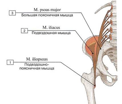
Fig. 356. Origin and insertion sites of the iliopsoas muscle (diagram):
1 — Iliopsoas; 2 — Iliacus; 3 — Psoas major
The iliacus muscle (m. iliacus) is a large, triangular, flat muscle located in the iliac fossa, lying medial to the psoas major. The iliacus originates from the upper two-thirds of the iliac fossa, the internal lip of the iliac crest, the anterior sacroiliac and iliolumbar ligaments. The muscle extends inferiorly and unites with the psoas major to form the iliopsoas muscle (Hoin's muscle). At the junction of the psoas and iliacus muscles lies Bogros' space (syn.: iliac interfascial space, spatium retroinguinale) — a cleft bounded superiorly by the iliac aponeurosis and the fascia propria of the iliacus attached to the iliac crest, and inferiorly by the fusion of these layers (below the middle of the iliac fossa) (Bogros Annet-Jean, 1786—1823 — French anatomist). The triangular area bounded laterally by the medial margin of the psoas major, medially by the lateral margin of THE Vertebral Column, and inferiorly by the iliolumbar ligament, is designated as Marcille's triangle — the site where the obturator nerve originates (Marcille Maurice, 1871 — 1941 — French physician).
The iliopsoas emerges onto the thigh from the pelvic cavity through the muscular lacuna posterior to the inguinal ligament and inserts into the lesser trochanter of the femur. The iliac subtendinea bursa lies between the tendon and the lesser trochanter. The iliopectineal bursa is situated between the tendons of the iliopsoas and pectineus muscles.
Function: the iliopsoas flexes the thigh at the hip joint; when the lower limb is fixed, it tilts the pelvis and trunk forward, and flexes the lumbar spine.
Blood supply: iliolumbar artery, deep circumflex iliac artery.
Innervation: muscular branches of the lumbar plexus (LI-LIV).
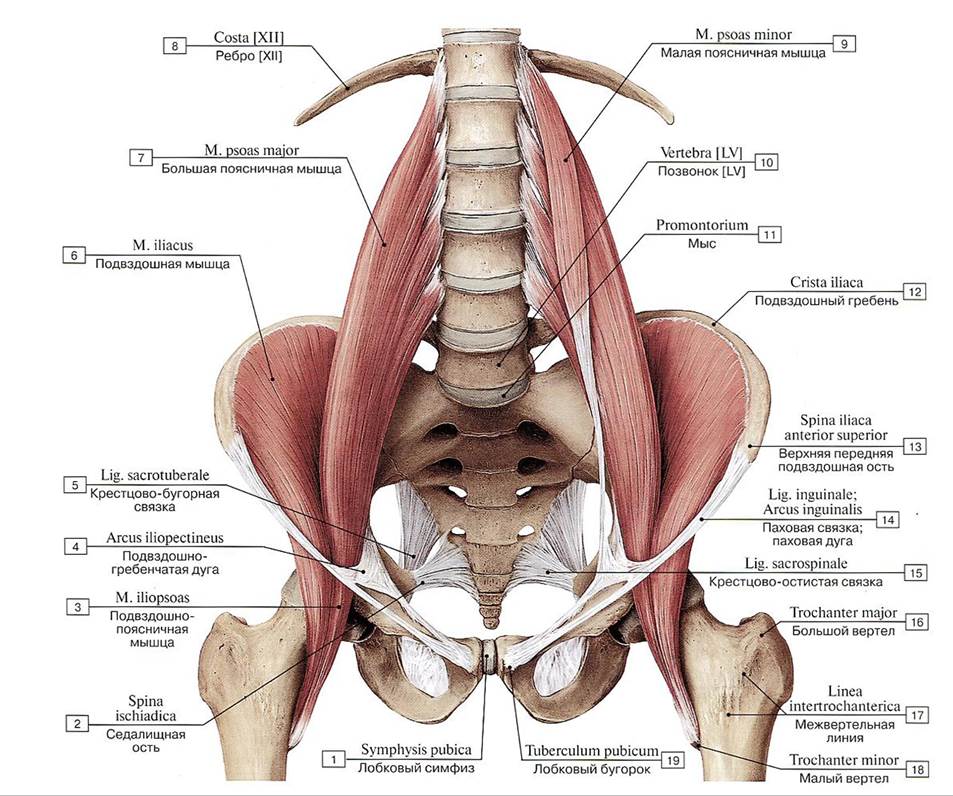
Fig. 357. Iliopsoas muscle, anterior view:
1 — Pubic Symphysis; 2 - Ischial spine; 3 — Iliopsoas; 4 — lliopectineal arch; 5 — Sacrotuberous ligament; 6 — Iliacus; 7 — Psoas major; 8 — Rib [XII]; 9— Psoas minor; 10 — Vertebra [LV]; 11 — Promontory; 12— Iliac crest; 13 — Anterior superior iliac spine; 14 — Inguinal ligament; 15— Sacrospinous ligament; 16 — Greater trochanter; 17— Intertrochanteric line; 18 — Lesser trochanter; 19— Pubic tubercle
The psoas minor muscle (m. psoas minor) is inconstant (present in approximately 60% of individuals), flat, and elongated, located on the anterior surface of the psoas major and fused with its fascia. The psoas minor originates from the lateral surface of the bodies of the XII thoracic and I lumbar vertebrae and the intervertebral disc between them. The short, thin muscle belly transitions into a narrow, long tendon that inserts into the arcuate line of the ilium and the iliopubic eminence. Some tendinous fibers continue into the iliac fascia and the iliopectineal arch.
Function: the psoas minor tenses the iliac fascia, providing support for the iliopsoas muscle, and flexes the lumbar spine.
Blood supply: lumbar Arteries.
Innervation: muscular branches of the lumbar plexus (L—LIV).

Fig. 358. Obturator internus and piriformis muscles (A — internal view from the left, B — posterior view):
1 — Pubic symphysis; 2 — Pubic tubercle; 3 — Obturator canal; 4 — Obturator internus; 5 — Greater sciatic foramen; 6 — Anterior superior iliac spine; 7 —- Iliac crest; 8 — Posterior superior iliac spine; 9 — Piriformis; 10— Sacrum [sacral vertebrae SI—SV]; 11 — Sacrospinous ligament; 12 — Coccyx [coccygeal vertebrae Col—ColV]; 13 — Lesser sciatic foramen; 14 — Sacrotuberous ligament; 15 — Posterior sacral foramina; 16 — Posterior sacro-iliac ligament; 17— Iliofemoral ligament; 18— Ischiofemoral ligament; 19— Greater trochanter; 20 — Obturator internus; Tendon; 21 — Lesser trochanter
The obturator internus muscle (m. obturator internus) is flat and triangular in shape, located on the inner surface of the obturator membrane (Fig. 358). The internal opening of the obturator canal lies between the muscle bundles. The muscle originates from the margins of the obturator foramen (except for the obturator groove), the internal surface of the obturator membrane, the pelvic surface of the ilium, superior to the obturator foramen, and the obturator fascia. Emerging from the lesser pelvis through the lesser sciatic foramen, the muscle transitions into a flat tendon that changes its direction at an acute angle, hooks around the margin of the lesser sciatic notch, and inserts into the trochanteric fossa on the medial surface of the greater trochanter of the femur. A bursa, the trochanteric bursa of the obturator internus muscle (bursa subtendinea musculi obturatorii interni), lies beneath the muscle, positioned between its tendon and the margin of the sciatic notch.
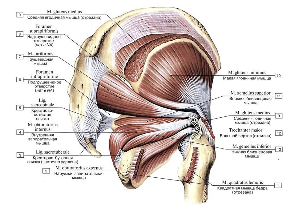
Fig. 359. External muscles of the hip, right view (gluteus maximus, gluteus medius, and quadratus femoris muscles removed):
1 — Quadratus femoris; 2 — Obturator externus; 3 — Sacrotuberous ligament; 4 — Obturator internus; 5 — Sacrospinous ligament; 6 — Infrapiriform foramen; 7— Piriformis; 8— Suprapiriform foramen; 9 — Gluteus medius; 10 — Gluteus minimus; 11 — Gemellus superior; Superior gemellus; 12 — Greater trochanter; 13 — Gemellus inferior; Inferior gemellus
Upon exiting the lesser sciatic foramen, the obturator internus muscle is joined superiorly and inferiorly by two gemellus muscles: the superior and inferior gemelli, which attach to the greater trochanter (its medial surface) alongside the tendon of the obturator internus (Fig. 360-B).
Function: laterally rotates the thigh.
Blood supply: inferior gluteal, obturator, and internal pudendal arteries.
Innervation: muscular branches of the lumbar and sacral plexuses (LIV-LV, SI—SII).
The piriformis muscle (m. piriformis) is cone-shaped; its proximal part is located within the lesser pelvis, while its middle and distal parts lie on the external surface of the pelvis, deep to the gluteus maximus. The piriformis originates from the pelvic surface of the sacrum (II—IV sacral vertebrae) lateral to the anterior sacral foramina. The muscle exits the pelvic cavity through the greater sciatic foramen. Posterior to the femoral neck, the muscle belly transitions into a round tendon that attaches to the apex of the greater trochanter of the femur (see Fig. 358).
Function: the piriformis muscle laterally rotates and slightly abducts the thigh; when standing on one leg, it assists in tilting the pelvis to its own side and forward.
Blood supply: superior and inferior gluteal arteries.
Innervation: muscular branches of the sacral plexus (SI—SIII).
The external pelvic muscles, located in the gluteal region and on the lateral surface of the pelvis, originate from the pelvic bones and the posterior surface of the sacrum, inserting via thick tendons into the femur (Fig. 359, 360). These muscles are arranged in three layers. The superficial layer contains the gluteus maximus and tensor fasciae latae; the middle layer includes the gluteus medius and quadratus femoris. The piriformis, obturator internus, and gemellus muscles also emerge into this middle layer from the pelvic cavity. The deep layer contains the gluteus minimus and obturator externus. All these muscles act upon the hip joint.
The gluteus maximus muscle (m. gluteus maximus) is a thick (up to 3 cm), broad, massive, multipennate quadrilateral muscle that shapes the contour of the gluteal region (Fig. 361, see Fig. 359, 360). This muscle is strongly developed in humans in connection with bipedalism and upright posture. Positioned superficially in the gluteal region, the gluteus maximus originates from the external surface, the tendinous part of the erector spinae, the dorsal surface of the sacrum and coccyx, and the sacrotuberous ligament.
The gluteus maximus extends obliquely downward, laterally, and anteriorly, inserting into the gluteal tuberosity of the femur. Some of its fibers pass superficial to the greater trochanter and blend into the iliotibial tract of the fascia lata. The trochanteric bursa of the gluteus maximus (bursa trochanterica musculi glutei maximi) is situated between the muscle's tendon and the greater trochanter of the femur.

Fig. 360. Origin and insertion sites (A — gluteal and other pelvic muscles, B — internal pelvic muscles) (diagrams):
1 — Gluteus medius; 2 — Gluteus maximus; 3 — Gluteus minimus; 4 — Piriformis; 5 — Tensor fasciae latae; Tensor of fascia lata; 6 — Iliotibial tract; 7 — Obturator internus; 8 —- Gemellus superior; Superior gemellus; Gemellus inferior; Inferior gemellus; 9 — Quadratus femoris
Function: the gluteus maximus extends the thigh at the hip joint and laterally rotates it. The upper fibers of the muscle, acting together with the tensor fasciae latae, assist in extending the leg. Simultaneous contraction of both gluteus maximus muscles with fixed lower limbs tilts the pelvis and trunk backward. These muscles maintain balance, hold the body in an upright position, and provide erect posture. The fulcra for the contraction of both gluteus maximus muscles are the femoral heads.
Blood supply: superior and inferior gluteal arteries, and the medial circumflex femoral artery.
Innervation: inferior gluteal nerve (SI—SII).
The gluteus medius muscle (m. gluteus medius) is thick and triangular; its posterior fibers lie deep to the gluteus maximus (see Fig. 359, 360). The gluteus medius originates from the external (gluteal) surface of the ilium—between the anterior gluteal line anteriorly, the posterior gluteal line inferiorly, and the iliac crest superiorly and posteriorly—as well as from the fascia lata. The muscle fibers converge fan-like downward, transitioning into a thick, flat tendon that inserts into the apex and external surface of the greater trochanter of the femur.
The trochanteric bursa of the gluteus medius (bursa trochanterica musculi glutei medii) is located between the tendon of the gluteus medius and the greater trochanter.
Function: the middle fibers of the muscle abduct the thigh, the anterior fibers rotate it medially, and the posterior fibers rotate it laterally. With a fixed lower extremity, contraction of the anterior fibers rotates the pelvis and consequently the trunk laterally. Contraction of the middle fibers tilts the pelvis toward the same side. The posterior fibers rotate the pelvis medially and tilt it toward the same side.
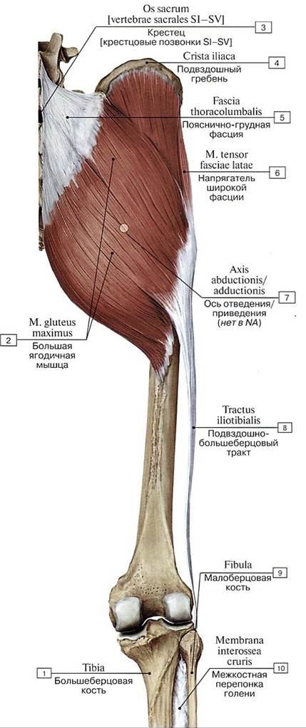
Fig. 361. Gluteus maximus muscle and iliotibial tract, posterior view:
1 — Tibia; 2 — Gluteus maximus; 3 — Sacrum [sacral vertebrae SI—SV]; 4 — Iliac crest; 5 — Thoracolumbar fascia; 6 — Tensor fasciae latae; Tensor of fascia lata; 7 — Axis of abduction/adduction; 8 — Iliotibial tract; 9 — Fibula; 10 — Interosseous membrane of leg
Blood supply: superior gluteal artery, lateral femoral circumflex artery.
Innervation: superior gluteal nerve (LIV—SI).
The gluteus minimus muscle (m. gluteus minimus) is triangular in shape, located beneath the gluteus medius muscle, between the piriformis muscle posteriorly and the tensor fasciae latae muscle anteriorly (see figs. 359, 360). The gluteus minimus originates on the gluteal surface of the ilium between the middle and inferior gluteal lines and at the margin of the greater sciatic notch. The muscle fibers pass downward, converging in a fan-like manner to form a short, flat tendon that inserts into the anterolateral surface of the greater trochanter of the femur, anterior to the insertion site of the gluteus medius. Some fibers of the gluteus minimus blend into the capsule of the hip joint. A trochanteric bursa of the gluteus minimus muscle (bursa trochanterica musculi glutei minimi) is located between the muscle tendon and the greater trochanter.
Function: the gluteus minimus abducts the thigh; its anterior fibers rotate the thigh medially, and its posterior fibers rotate it laterally.
Blood supply: superior gluteal artery; lateral femoral circumflex artery.
Innervation: superior gluteal nerve (LIV—SI).
The quadratus femoris muscle (m. quadratus femoris) is a flat, quadrilateral muscle situated between the gemellus inferior superiorly and the upper margin of the adductor brevis inferiorly. The quadratus femoris originates via a short tendon from the upper part of the lateral margin of the ischial tuberosity. The muscle then runs almost horizontally in a lateral direction and inserts into the upper part of the intertrochanteric crest of the femur (see figs. 359, 360). A synovial bursa is frequently present between the anterior surface of the muscle and the greater trochanter.
Function: the quadratus femoris muscle laterally rotates the thigh.
Blood supply: inferior gluteal artery, medial femoral circumflex artery, obturator artery.
Innervation: sciatic nerve (LIV—SI).
The obturator externus muscle (m. obturatorius externus) is triangular and located on the external surface of the obturator membrane, covered by the pectineus muscle and the proximal part of the adductor longus muscle of the thigh. The obturator externus originates from the medial margin of the obturator foramen, formed by the pubis and ischium, and from the medial two-thirds of the obturator membrane (see fig. 359). The muscle fibers converge and run posteriorly, laterally, and upward, transitioning into a tendon that passes posterior to the hip joint and inserts into the trochanteric fossa on the medial surface of the greater trochanter of the femur, adjacent to the tendon of the obturator internus muscle. Some fibers of the obturator externus blend into the capsule of the hip joint.
Function: the obturator externus muscle laterally rotates the thigh, assists in its flexion, and provides inferior support to the femoral head.
Blood supply: obturator artery, lateral femoral circumflex artery.
Innervation: obturator nerve (LII-LIV).
The tensor fasciae latae (m. tensor fasciae latae) is a flat, elongated, ribbon-like muscle that narrows inferiorly. It originates from the outer lip of the iliac crest, extending forward to the anterior superior iliac spine. The muscle is located in the gluteal region and on the lateral aspect of the thigh, where it lies between the superficial and deep layers of the fascia lata. At the junction of the upper and middle thirds of the thigh, it continues into the iliotibial tract (tractus iliotibialis) of the fascia lata, which extends downward and inserts into the lateral condyle of the tibia (fig. 362). Anteriorly, the muscle borders the sartorius, and posteriorly, it borders the anterior part of the gluteus medius.
Function: the tensor fasciae latae tensions the iliotibial tract, stabilizes the knee joint in full extension, and flexes the thigh.
Blood supply: superior gluteal artery, lateral femoral circumflex artery.
Innervation: superior gluteal nerve (LIV—SI).
The superior gemellus muscle (m. gemellus superior) originates from the ischium.
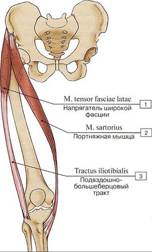
Fig. 362. Tensor fasciae latae muscle and sartorius muscle (schematic):
1 — Tensor fasciae latae; 2 — Sartorius; 3 — Iliotibial tract
The inferior gemellus muscle (m. gemellus inferior) originates from the ischial tuberosity. The distal ends of the flat gemelli muscles attach to the tendon of the obturator internus superiorly and inferiorly, and together with it, insert into the femur on the medial surface of its greater trochanter. Due to this anatomical relationship between the obturator internus and the two gemelli, these three muscles were formerly referred to as the triceps muscle of the hip, which rotates the thigh (see Fig. 360, 361).
Function: The obturator internus, together with the gemelli muscles, laterally rotates the thigh. With a fixed lower limb (when standing on one leg), these muscles help prevent the pelvis from tilting to the opposite side.
Blood supply: Inferior gludetal, obturator, and internal pudendal arteries.
Innervation: Muscular branches of the sacral plexus (LIV—LV, SI— SIII).
Last update: 08/08/2026
Editorial and Educational Adaptation: This material has been compiled based on the primary/original source text. The project team performed an editorial review, corrected technical inaccuracies, structured sections, and adapted the content for an educational format.
What was processed:
- elimination of formatting defects (OCR errors, structural breaks, corrupted characters);
- editorial organization of content;
- standardization of terminology in accordance with academic sources;
- verification of factual statements against the original source text.
All mentions of the author, publication year, and origin of the primary text have been preserved in accordance with the source.