BIOLOGY Volume 1 - A Guide to General Biology - 2004
2. DIVERSITY OF LIFE ON EARTH
2.8. Kingdom Animalia (animals)
2.8.5. Phylum Annelida (annelids, or segmented worms)
The Classification and Characteristic Features of Annelids are given in Table 2.16, and Examples of various annelids are shown in Fig. 2.54. Two of these, Nereis and Lumbricus, are discussed in more detail below. Annelids are coelomate animals.
Class="center">Table 2.16. Classification of the phylum Annelida (annelids, or segmented worms)
Phylum Annelida |
||
Characteristic features Triploblastic body with a true coelom Bilateral Symmetry Metameric segmentation A prostomium—a lip-like outgrowth of the anterior segment—is located in front of the Mouth Well-developed cuticle (outer covering) Body bears setae—Hair-like chitinous structures arranged segmentally (except in leeches)* |
||
|
(polychaetes) |
(oligochaetes, or earthworms) |
Class Hirudinea (leeches) |
Marine animals |
Inhabitants of freshwater environments or damp soil |
Ectoparasites; suckers present at the anterior and posterior ends of the body |
Distinctly demarcated HEAD |
Head poorly demarcated |
Head poorly demarcated |
Numerous setae borne on lateral body outgrowths called parapodia* |
Few setae, arranged singly or in pairs; parapodia absent* |
Few and constant number of body segments; setae and parapodia absent* |
Clitellum absent |
Clitellum present, which secretes cocoon material for egg deposition |
Clitellum absent |
EXAMPLES: Arenicola (lugworm) Nereis (clam worm) |
EXAMPLE: Lumbricus (earthworm) |
EXAMPLE: Hirudo (medicinal leech) |
* Diagnostic features |
||

Fig. 2.54. Diversity of annelids.
Coelomate body plan
As we have seen, in Flatworms the mesoderm completely fills the space between the ectoderm and endoderm layers, forming a dense middle layer. A body plan lacking a coelom is termed acoelomate (Fig. 2.55, A).
In annelids and more advanced animal groups, a body cavity called the coelom develops. It originates as a split within the mesoderm during embryonic development. The resulting cavity is filled with coelomic fluid, dividing the mesoderm into two layers: the somatic layer, facing outward, and the splanchnic layer, facing inward (Fig. 2.55, B). The somatic mesoderm, joining with the ectoderm, forms the body wall. The splanchnic mesoderm, combining with the endoderm, forms the muscular gut wall. Thus, the coelom separates the body wall from the gut wall.
The major part of the mesoderm lining the coelom develops into Muscles; the muscles comprising the body wall provide animal locomotion, while the gut wall muscles drive peristalsis. The Transport of substances between the body wall and the gut wall (and vice versa) is carried out by the vessels of a well-developed Circulatory system. Note that the gut lumen lies within the endoderm. The lining of the coelom is called the Peritoneum. The sheets of peritoneum connecting the gut wall to the body wall are called mesenteries. Any Organs projecting into the coelom, such as reproductive or excretory organs, are supported by the peritoneum (Fig. 2.55, B).
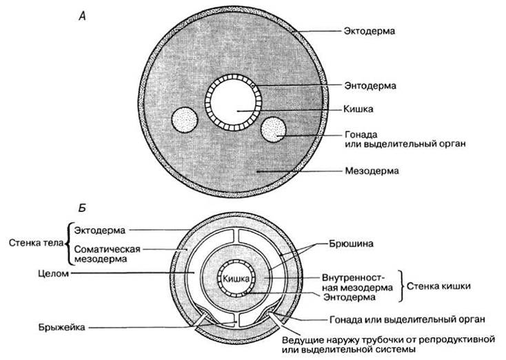
Fig. 2.55. A. Cross-section of a generalized acoelomate animal. B. Cross-section of a generalized coelomate animal.
Biological Significance of the coelom
1. Because the coelom separates the gut from the body wall, the body wall Muscle movements associated with animal locomotion can occur independently of the gut wall movements (peristalsis) that push food through the gut and aid in its mixing. As a result, the animal's locomotory efficiency increases, and different Regions of the gut become specialized to perform diverse Functions. In The Stomach, for example, food mixing takes place, which in turn allows for a more varied diet.
2. The coelom provides a cavity in which various organs can grow, develop, and function independently of one another.
3. The presence of the coelom facilitates an increase in body size and structural complexity, which in turn gives rise to additional challenges related to transport and coordination processes. Food, for instance, is digested in the gut, but the body Cells are separated from the gut by the coelom. Similarly, the gut is located at some distance from the body surface across which gas exchange occurs. As body size increases, these challenges intensify, as previously discussed in Section 2.8.3, necessitating a transport system. Consequently, all coelomate animals have evolved a tubular circulatory system. Blood is a fluid tissue that circulates throughout the animal's body driven by the contraction of muscle fibers located in the walls of Blood Vessels or by the action of a Heart. Due to the presence of Valves in the vessels and The Heart, blood flows in only one direction. Greater complexity also demands more advanced coordination and, consequently, a more sophisticated Nervous system. One of the consequences of this evolutionary trend is cephalization, i.e., The Development of a head (see also below regarding body size and surface area-to-volume ratio).
4. In annelids, the coelom also serves an additional specialized function as a hydrostatic Skeleton, or fluid skeleton. A skeleton performs three primary functions: support, protection, and locomotion. Because coelomic fluid, like any other liquid, is incompressible, muscle contractions can alter the worm's shape without changing its volume. During locomotion, specific body regions alternately become long and thin or short and thick, depending on which muscle group exerts pressure on the coelomic fluid. The protective role of the coelom is ensured by the fluid's ability to rapidly and evenly distribute external pressure in all directions.
5. The coelomic fluid may participate in The transport of nutrients, metabolic wastes, and respiratory gases, although The Vascular System plays the primary role in these functions.
Metamerism
Another evolutionary advantage in the development of coelomate animals is metamerism, or segmentation—an organizational level in which the animal's body is divided by transverse partitions into multiple identical parts, or segments. In other words, the animal's body consists of a long series of repeating segments arranged one after another. Segmentation begins in the mesoderm but typically encompasses both mesodermal and ectodermal regions of the body.
Metamerism is most clearly expressed in segmented worms (annelids), in which this division is readily visible externally (annulations along the entire length of the body). Internal segments are separated from one another by partitions (septa) traversing the coelom. Each segment possesses its own set of muscles, blood vessels, Nerve Cells, and, in some groups, reproductive organs. However, even in annelids, the segments are not entirely independent of one another, as both the nervous and, particularly, the circulatory systems must span the entire length of the animal's body.
Once segmentation and the basic body plan of each segment became evolutionarily established, it paved the way for further evolutionary modifications within individual segments or small groups of segments, as well as for narrower specialization and division of labor among different body regions. This occurs in several ways. Different functions can be performed by different segments; fusion of segments is also possible, as occurred during cephalization when several segments fused to form the head; and even the loss of certain segments can take place, as observed in Arthropods. As we will see later (Section 2.8.6), in arthropods the number of segments has decreased, and segmentation is less obvious externally, such as in the cephalothorax of crustaceans. However, internal segmentation in arthropods remains as distinct as in annelids. In Chordates (Section 2.8.9), external segmentation has been lost, but certain systems still retain clear segmentation, such as myotomes (muscle blocks) in embryos and Spinal Nerves.
Dimensions and Surface-Area-to-Volume Ratio
Organisms with a relatively high surface-area-to-volume ratio can rely on diffusion to meet their transport needs. Oxygen, nutrients, and Metabolic waste products, such as carbon dioxide, can diffuse quickly enough to ensure the Organism's survival without any specialized transport systems. Diffusion, however, can only facilitate substance transport over short distances. Meanwhile, as body size increases, volume increases faster than surface area, resulting in a decreasing surface-area-to-volume ratio. This is easily illustrated using a cube (Fig. 2.56), but the same principle applies to spherical bodies, cells, and entire living organisms. In flat organisms, such as flatworms, the surface area remains high as volume increases. In these organisms as well, diffusion is fully capable of satisfying all transport requirements. Coelomate animals, however, require specialized gas exchange and transport systems.

Fig. 2.56. Effect of increasing body size On the surface-area-to-volume ratio.
Class Polychaeta
The lugworm (Arenicola) inhabits the intertidal zone, burrowing in sandy or soft muddy substrates (Fig. 2.54). Nereis (Fig. 2.57) lives in estuaries, either under stones or burrowing in mud or muddy-sand substrates.
The segmentation of the Nereis body is clearly visible externally. All segments, except for the very anterior and posterior ones, are identical. On the sides of each segment are projections called parapodia. The upper branch of the parapodium is called the notopodium, and the lower branch is the neuropodium (Fig. 2.57). Two groups of hairlike setae extend from both of these structures (notopodium and neuropodium). Each parapodium also bears two additional outgrowths—needle-like structures called the dorsal and ventral cirri. Gas exchange occurs across The surface of the parapodium, which is richly supplied with blood. Nereis crawls using its parapodia like oars. This worm can also swim through the coordinated action of the parapodia and the bending Muscles of the body wall (Skin-muscular sac). The body of Nereis is surrounded by a thin cuticle. The digestive tract extends along the entire body from the mouth to the anus. The Pharynx in Nereis is eversible. After the worm swallows its prey, the pharynx is retracted again.
Nereis possesses a distinct head (Fig. 2.57); such pronounced cephalization is typical of polychaetes, but not of other annelids. The head consists of two sections: anterior and posterior. The anterior section, the prostomium, is formed by the first segment, while the posterior section, the peristomium, is formed by the second segment. Located on the prostomium are a pair of sensory tentacles (on the dorsal side) and two pairs of eyes, while a pair of fleshy, tactile "palps" extends from the ventrolateral regions. The mouth is situated between the two head regions. Four pairs of long, flexible tentacles extend from one of these regions—the peristomium; these are also tactile and bear chemoreceptors that detect various chemical substances, effectively replacing the animal's sense of taste and smell.

Fig. 2.57. Nereis, a polychaete worm.
Class Oligochaeta
The earthworm (Lumbricus terrestris) is an animal with a long, cylindrical body, reaching approximately 12–18 cm in length (Fig. 2.58). Its anterior end is cone-shaped, while the posterior end is flattened dorsoventrally. Although the earthworm lives on land, it has not fully overcome all the challenges associated with a terrestrial lifestyle. To escape desiccation, it lives underground, burrowing in damp soil, and emerges only at night to search for food or a mate.

Fig. 2.58. Anterior end of an earthworm (ventral view).
Differences in the external body Structure of Nereis and the earthworm are due to the latter's ADAPTATION TO A terrestrial lifestyle. The earthworm's body is streamlined and lacks any outgrowths that might hinder its free movement through the soil. The prostomium, located above the mouth, is small and rounded, with no sensory appendages. All segments, except for the first and last, bear four pairs of setae; two of these are located ventrally (on the underside), and two are dorsolateral. The setae emerge from setal sacs located within the body wall. They can be retracted or extended by the action of specialized muscles. The setae facilitate the worm's movement through the soil. They are also used for defense: by hooking them into the walls of its burrow, the worm holds onto it firmly. The longest setae, located on segments 10–15, 26, and 32–37, are used during copulation. Another structure involved in copulation is the clitellum, located on segments 32–37. The epidermis of the clitellum contains numerous glandular cells that form a saddle-shaped thickening on the dorsal and lateral surfaces. The clitellum participates in copulation and cocoon formation.
The mouth and anus are located at opposite ends of the body. The earthworm feeds on detritus (fragments of decaying organic matter), which it ingests along with the soil. The vast majority of the ingested soil passes through the gut and is subsequently deposited on the soil surface in the form of characteristic castings.
Coelomic fluid released through dorsal pores and mucus secreted by epidermal glandular cells constantly moisten the thin cuticle. This prevents the cuticle from drying out, facilitates gas exchange, and provides a kind of lubrication that eases the worm's movement through the soil. Gas exchange occurs through the cuticle via diffusion—a process dependent on a branching network of capillaries within the epidermal layer.
All segments, except for the first three and the last one, bear a pair of nephridia—tubules that perform excretory and osmoregulatory functions. They open to the body surface via pores.
The Reproductive System of the earthworm, as well as its reproductive behavior, is highly complex, which is perhaps explained by its terrestrial lifestyle and the need to protect Gametes and fertilized eggs from desiccation. Lumbricus is a hermaphrodite (i.e., each individual possesses both male and FEMALE REPRODUCTIVE ORGANS). Earthworm encounters with one another are rare, but when they do occur, any two individuals can mate since both are hermaphrodites. As a result, sperm is mutually exchanged, and both worms are fertilized.
The reproductive organs of earthworms are concentrated at the anterior end of the body. External features associated with sexual reproduction are shown in Fig. 2.58. Mating and the subsequent deposition of fertilized eggs is a very complex process. Mating individuals lie alongside each other in opposite directions, such that the head of one partner is directed toward the tail region of the other and vice versa.
Last update: 06/08/2026
Editorial and Educational Adaptation: This material has been compiled based on the primary/original source text. The project team performed an editorial review, corrected technical inaccuracies, structured sections, and adapted the content for an educational format.
What was processed:
- elimination of formatting defects (OCR errors, structural breaks, corrupted characters);
- editorial organization of content;
- standardization of terminology in accordance with academic sources;
- verification of factual statements against the original source text.
All mentions of the author, publication year, and origin of the primary text have been preserved in accordance with the source.