Peptide Self-Regulation of Living Systems (Facts and Hypotheses) - Shataeva L. K. 2003
Peptides in Aqueous Solutions
Tissue-Specific Regulatory Peptides
Atriopeptides
Natriuretic Peptides were first described by De Bold in the early 1980s (De Bold et al., 1981). It was later discovered that these peptides regulate salt excretion in the body. It was shown that atrial peptides (APs) are secreted by the atria when the myocytes of the atrial wall experience stress (stretching). By regulating diuresis and vascular tone, The Heart is able to rid itself of excess circulating fluid volume and regulate Blood pressure (Alekseenko, Orekhovich, 1987). Cardiovascular Homeostasis is thus maintained.
Histological studies have shown that atrial cardiocytes differ significantly from ventricular cardiocytes in that they contain specific granules associated with the Golgi apparatus, which is characteristic of typical endocrine secretory Cells. This indicates the uniqueness of atrial cardiocytes: they combine two physiological Functions—Muscle contraction and endocrine secretion. By performing an endocrine function, The Cell ensures an optimal regime of muscular work for itself (Gunning, Brenner, 1992).
APs originate from a common precursor; Peptides with a molecular weight ranging from 3 to 44 kDa exhibit biological activity. High-molecular-weight forms are converted into more active low-molecular-weight forms by Proteolytic Enzymes. Peptides consisting of 25—33 amino acid residues exhibit the highest activity.
The main action of APs is directed at the Kidneys, where they reduce capillary pressure and increase renal plasma flow. The action of APs begins rapidly, reaches its maximum within 1—2 min, and subsides after 20—30 min.
Regardless of their molecular size, mammalian APs share a common characteristic feature: an intramolecular disulfide bond forms a 17-membered ring Structure. Table IV of the Appendix shows the Amino acid sequences of APs, while Fig. 7 presents the Primary and secondary structures of APs in two different Conformations: cyclic and dimeric. The dimeric form exhibits a prolonged duration of action compared to the cyclic forms. The presence of free N- and C-termini is essential for biological activity, whereas the atrial peptide ring itself is inactive. At the same time, the highly active antiparallel AP dimer features not only a conformation reinforced by cystine bridges, but also a pair of free N- and C-termini.
In addition to the atrial myocardium, APs have been found in the Hypothalamus and Brain Neurons—specifically, brain natriuretic peptide and C-type natriuretic peptide (the last two rows in Table IV of the Appendix), as well as in the ocular chambers of rats (Stone, Glembotski, 1986). Furthermore, highly specific receptors for APs have been discovered not only in the Cells of the renal tubules, Adrenal Glands, aortic smooth muscle, and vascular endothelium, but also in brain cells. Despite some differences in the Amino Acid Composition of the ring, these APs also interact with Antibodies to cardionatrin (Gunning, Brenner, 1992).
Class="center">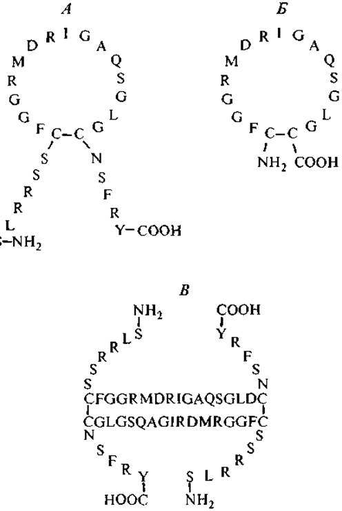
Fig. 7. Amino acid sequences and configurations of human atriopeptids.
A — a-natriuretic peptide; Б — atriopeptin-ring; В — ß-natriuretic peptide — antiparallel dimer.
Thus, this group of peptides demonstrates the same pattern observed in other groups—peptides secreted by somatic tissue cells are found in corresponding brain neurons, which once again confirms the observation that the body's self-regulation is provided by tissue-specific peptides while remaining under the control of the Central Nervous system.
Last update: 06/08/2026
Editorial and Educational Adaptation: This material has been compiled based on the primary/original source text. The project team performed an editorial review, corrected technical inaccuracies, structured sections, and adapted the content for an educational format.
What was processed:
- elimination of formatting defects (OCR errors, structural breaks, corrupted characters);
- editorial organization of content;
- standardization of terminology in accordance with academic sources;
- verification of factual statements against the original source text.
All mentions of the author, publication year, and origin of the primary text have been preserved in accordance with the source.