Biochemistry - The Chemical Reactions of Living Cells, Volume 2 - D. Metzler 1980
Biosynthesis: How New Molecules Are Formed
Regulation of Biosynthetic Processes
Glycogen and Blood Glucose
The Glucose METABOLISM in animals has two major distinctive features [44]. The first of these is the storage of Glycogen, which can be rapidly mobilized as a source of Muscle energy when needed. However, The rate of Glycolysis can be exceptionally high—the entire glycogen reserve in a muscle can be depleted in just 20 s under anaerobic Fermentation or in 3.5 min during oxidative metabolism [45]. Consequently, there must be a mechanism to rapidly switch glycolysis on and off as physiological demands dictate. At the same time, it must be possible to convert lactate back into glucose or glycogen (Gluconeogenesis). Muscle glycogen stores must be replenished by Blood glucose. If The amount of glucose obtained from diet or derived from Liver glycogen proves insufficient, it must be synthesized de novo from Amino Acids.
The second key feature of glucose metabolism is that certain Tissues, including the Brain, Blood Cells, renal medulla, and Testes, derive virtually all of their required energy from The oxidation of glucose. Therefore, blood glucose levels must not be allowed to drop significantly below the normal range (5 mM).
The regulatory mechanisms controlling blood glucose levels are highly complex and not yet fully elucidated, though it is well established that several Hormones are involved.
Insulin (ch. 4, sec. 9.7; ch. 5, sec. B, 5, Supplement 11-B), likely in conjunction with chromium (Supplement 11-C), enhances the rate of glucose uptake by Muscles and other tissues. Glucagon (ch. 6, sec. E, 5) is a peptide hormone consisting of 29 amino acid residues that acts primarily on liver cells. It is secreted by the a-Cells of the pancreatic islets of Langerhans—the same cells that produce insulin. However, the action of glucagon is antagonistic to that of insulin, as it raises blood glucose levels by stimulating hepatic Glycogenolysis. Glucagon also stimulates gluconeogenesis, with both effects being mediated by cyclic AMP [46]. Glucocorticoids (ch. 12, sec. I, 3.6) accelerate gluconeogenesis and hepatic glycogen accumulation through the mechanisms discussed in sec. E, 7.
Supplement 11-B
Diabetes is a well-known and widespread metabolic disorder in humans. Out of one million children aged 8 to 12, about 400 suffer from juvenile-onset diabetes. Approximately 33,000 individuals aged 40 to 50 per million (i.e., over 3%) have diabetes, and among those over 70, the prevalence exceeds 7%a. A predisposition to diabetes is partly inherited; at least two recessive "defective" genes constitute a significant portion of the genetic pool. The severity of the disease varies widely. About half of young patients can manage the condition through diet alone, whereas the other half require insulin injections due to the atrophy of insulin-producing pancreatic ß-cells. Among adult diabetic patients, a common clinical challenge is a reduced sensitivity to administered insulinb. This diminished responsiveness may stem from either an insufficient number of insulin receptors or structural defects within them. A reduced receptor count has also been observed in insulin-resistant obese patients. The Treatment of many diabetic patients with sulfonylurea drugs, specifically 1-butyl-3-p-tolylsulfonylurea:
Class="center">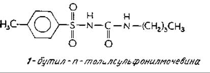
has in some cases led to an increase in the number of insulin receptors. Strict dietary control exerts a similar effect. In certain patients, elevated activities of insulin-degrading Enzymes have been detected. Increased glucagon synthesis is also a common feature of diabetes, a finding that may help resolve some of the unresolved questions regarding the Pathophysiology of the diseaseb.
In recent years, considerable attention has also focused on another peptide hormone, Somatostatin (ch. 16, sec. A, 1), which inhibits the release of both glucagon and insulin from pancreatic cells. The administration of this hormone has been shown to improve the clinical condition of certain diabetic patients.
A hallmark symptom of diabetes is severe hyperglycemia, with blood glucose concentrations reaching 8–60 mMv. Evidently, the failure to utilize glucose properly stems from a breakdown in feedback-loop control. As a result, gluconeogenesis is accelerated, which in turn drives up the Catabolism of Proteins AND AMINO acids. Liver glycogen stores become depleted, and an excess of nitrogen—originating from protein breakdown—appears in the urine. The accumulation of fatty acid degradation products leads to the overproduction of Ketone Bodies (p. 515), while osmotic diuresis caused by elevated urine volume results in tissue dehydration.
Despite extensive research, the strictly chemical mechanisms of insulin action remain unclearg,d. It is generally believed that the hormone acts on the Plasma Membranes of all responsive tissues, inducing marked permeability changes that lead to an increased uptake of glucose, various ions, and other metabolites. Such permeability shifts may account for insulin's profound influence on major biosynthetic pathways, notably the upregulation of glycogen, lipid, and Protein Synthesis. Concurrently, catabolic processes are suppressed, and The activity of catabolic enzymes, such as glucose-6-phosphatase, is reduced. A key to understanding insulin action may lie in identifying The Nature of its "second messenger," analogous in function to cAMP. Although it was once hypothesized that cAMP serves as insulin's second messenger, it is now considered more likely that this role is fulfilled by an ion, possibly K+e.
The total Chemical synthesis of insulin has been achieved in several laboratories, with the most formidable challenge being the correct formation of disulfide bridges within the molecule. To address this, biomimetic approaches have been attempted in which cross-linking is carried out at the proinsulin stage rather than directly on insulin (Fig. 11-9). The Need for synthetic insulin is driven not only by the fact that animal-derived supplies cannot meet the massive clinical demand, but also because chemical synthesis allows for The production of analogs with custom primary structures—particularly sequences distinct from natural animal insulins, which could prove exceptionally valuable for specific groups of diabetic patients.
a Apgar V., Beck I., Is My Baby All Right? Trident Press, New York, 1972.
b Maugh T. H., II, Science, 193, 220–222 and 252 (1976).
c For mild cases of diabetes, the lower of these values is more typical because once glucose concentration exceeds the renal threshold (~8 mM), the excess is excreted in the urine.
d White A., Handler P., Smith E. L., Principles of Biochemistry, 5th ed., pp. 1094–1101, McGraw-Hill, New York, 1973.
e See vol. 1 (pp. 291 and 386) and p. 507 of this volume for discussions on insulin Structure, Cell-surface receptors, and chromium ion "Amplification.".
f Hers H. G., Annu. Rev. Biochem., 45, 167–189 (1976).
g Anonymous, Chem. and Eng. News, 52 (April 29), 19–22 (1974).
Supplement 11-C
Trace Elements: Chromium
In 1959, it was established that animals maintained on chromium-deficient diets exhibit retarded growth and reduced life spans. These animals also display impaired "glucose tolerance," manifested as a blood glucose clearance rate that is half that of normal controlsa,b,c. This metabolic state is essentially indistinguishable from that induced by insulin deficiency. Fractionation of Yeast has yielded a chromium-containing glucose tolerance factor (GTF), which is a complex comprising Cr3+ ions, nicotinic acid, and amino acidsd. There is strong evidence that the chromium within GTF interacts with insulin to potentiate its biological activitye,f. This hypothesis is supported by the finding that normal serum chromium levels, which average approximately 0.03 mM, drop precipitously following a glucose loadg. This indicates that chromium is actively consumed during Carbohydrate Metabolism, likely facilitating the binding of insulin to cell Membrane Receptors. The sharp decline in serum chromium observed during acute infections (despite concurrent elevations in insulin levels) suggests that human chromium metabolism warrants close clinical attention.
Tissue chromium concentrations in animals rarely exceed 2 mM, though significantly higher levels are found in the caudate Nucleus of the brain. Elevated concentrations of Cr3+ ions have also been detected in RNA-Protein Complexes.
Of considerable experimental interest is The Use of chromium in forming Cr(III)–nucleotide complexes, such as with ATPi. Chromium displaces magnesium ions to form exceptionally stable chelate complexes with NUCLEOTIDES. The characteristic light-Absorption Spectra and nuclear paramagnetic properties of these chromium complexes make them powerful probes for investigating the catalytic mechanisms of phosphotransferases.
Recently, The formation of inert ("non-labile") complexes with Cr(III) or Co(III) has been employed as a method for identifying metal-binding sites in proteins and other Biopolymersk,l,m. Divalent ions bound to such sites, such as Mg2+ or Zn2+, are first replaced by Co2+ or Cr2+ ions, after which these ions are oxidized (most commonly using H2O2) to their trivalent state. Such a transformation readily occurs only when the active sites possess an octahedral geometry. Trivalent Co or Cr ions bind tightly to the surrounding ligands, making it possible to cleave away a major portion of the macromolecule while preserving a small fragment that remains firmly bound to the metal.
a Mertz W., Fed. Proc. Fed. Am. Soc. Exp. Biol., 26, 186—193 (1967).
б Mertz W., Cornatzer W. E., Newer Trace Elements in Nutrition, Dekker, New York, 1971.
в Frieden E., Sсi. Am., 227, 52—60 (July 1972).
г Mertz W., Fed. Proc., Fed Am. Soc. Exp Biol., 33, 659 (1974).
д Evans G. W., Roginski E. E., Mertz W., Fed. Proc., 31, 264 (Abstr.), (1972).
e Roginski E. E., Fed. Proc., Fed. Am. Soc. Exp. Biol., 33, 659 (1974).
ж Pekarek R. S., Hauer E. C., Rayfield E. J., Wannemacher R. W., Jr., Beisei W. R., Fed. Proc., Fed. Am. Soc. Exp. Biol., 33, 660 (1974).
з O'Dell B. L., Campbell B. J., Compr. Biochem, 21, 179—266 (1971).
и DePamphills M. L., Cleland W. W., Biochemistry, 12, 3714—3724 (1973).
к Anderson R. A., Vallee B. L., PNAS, 72, 394—397 (1975).
л Wright J. К., Feldman J., Takahashi M., Biochemistry, 15, 3704—3710 (1976).
м Rose S. L., Westhead E. W., Abstr., 172nd ACS Natl. Meet. San Francisco, 1976, BIOL., p. 34.
Last update: 06/08/2026
Editorial and Educational Adaptation: This material has been compiled based on the primary/original source text. The project team performed an editorial review, corrected technical inaccuracies, structured sections, and adapted the content for an educational format.
What was processed:
- elimination of formatting defects (OCR errors, structural breaks, corrupted characters);
- editorial organization of content;
- standardization of terminology in accordance with academic sources;
- verification of factual statements against the original source text.
All mentions of the author, publication year, and origin of the primary text have been preserved in accordance with the source.