Biochemistry: The Chemical Reactions of Living Cells, Volume 1 - D. Metzler 1980
Membranes and Cell Walls
Antigens and Receptors on the Cell Surface
Protein Agglutinins (Lectins)
In recent years, intensive research has focused on Certain plant Proteins that possess a unique ability to cause erythrocyte agglutination. These proteins—Lectins—have attracted attention not only because they serve as specific protein Reagents for sugars and are successfully used to study Cell surfaces, but also because some of them, notably concanavalin A (which accounts for up to 2–3% of the protein in the seeds of the jack bean, Canavalia ensiformis), preferentially agglutinate Cancer Cells. Concanavalin A and several other lectins exhibit mitogenic activity, meaning they stimulate mitotic division and proliferation in resting lymphocytes. The binding sites of Lectins are specific to particular sugars. For instance, concanavalin A binds to a-D-mannopyranose and a-D-glucopyranose residues containing unmodified hydroxyl groups at the C-3, C-4, and C-6 positions [92]. This protein also features specific binding sites for Ca2+ and transition Metal Ions, such as Mn2+. Soybean lectin binds to D-N-acetylgalactosamine and D-galactose residues, whereas wheat germ agglutinin is specific for D-N-acetylglucosamine.
The complete Structure of concanavalin A was determined by X-ray crystallography [93, 94] (Fig. 5-7). This protein is a tetramer composed of subunits with a Molecular Weight of 27,500. At pH ≤ 5.8, it dissociates into dimers. The architecture of concanavalin A is rather unusual in that it lacks a-helical regions. Each monomer consists of 237 residues, 57% of which form a three-layered ß-Structure (Fig. 5-7, B). The protein does not bind CARBOHYDRATES until two metal-binding sites are occupied. It is likely that The formation of the carbohydrate-binding site requires a conformational change in the protein that occurs only upon Metal ion binding.
What role do lectins play in plants and other organisms where they are produced? Do they act as antibody analogs in plants, providing defense against Bacteria and Fungi? Given their ability to accelerate mitosis, can they be assigned a specific role in regulating Cell Division and seed germination? At present, we cannot yet answer these questions. The production of lectin-like proteins by cellular slime Molds [95] and proteins with agglutinating properties by chick embryo fibroblasts [96] suggests that lectins may be components of Cell Recognition systems; it is also possible that they link cell surfaces together by binding polysaccharide groups on two adjacent cells.
Class="center">
FIG. 5-7. Structure of concanavalin A, a lectin from the jack bean (*Canavalia ensiformis*). Schematic representation of the tetrameric Protein Structure [93]. The binding sites for Ca2+ and Mn2+ ions, as well as the carbohydrate (C), are indicated. (Edelman G., Becker I., Reeke O., Jr., Cunningham B., personal communication.)

FIG. 5-7. B. Stereogram of the positions of the a-carbon atoms of the 237 amino acid residues in a single protein subunit [94a]. CA and MN designate the metal-binding sites. The carbohydrate-binding site is labeled CHO, the non-polar binding site is labeled NP, and the N- and C-termini of the polypeptide chain are labeled N and C. C. Stereogram of the Ca2+ and Mn2+ ion-binding sites. Both ions apparently form 6 coordination bonds each. Note that the oxygen atoms of the carboxyl groups of the 10th and 19th residues serve as ligands for both ions. Other ligands include the peptide backbone carbonyl group and an imidazole side chain. The N-3 atom of the latter participates in coordination binding with Mn2+ and apparently forms a Hydrogen bond with the peptide carbonyl group. Small circles bonded to Mn2+ and Ca2+ represent Water molecules; the letters CHO indicate the carbohydrate attachment site. (Courtesy of K. Hardman.)
Supplement 5-E
Among the most intriguing soluble Serum proteins are IMMUNOGLOBULINS (y-globulins), which function as antibodies to protect the body against the threat of invading foreign substancesa,b. Differences in molecular weights and chemical properties allow immunoglobulins to be divided into five classes. The first three—IgG, IgM, and IgA—are quantitatively the most prominent, though the remaining two, IgD and IgE, also play vital roles. For example, individuals prone to allergic reactions exhibit elevated levels of IgE.
The basic structure of all immunoglobulins is a quasisymmetric dimer composed of two light chains and two heavy chains, the lengths of which vary among different immunoglobulin classes. Human antibodies contain two classes of light chains, ϰ and λ. Heavy chains are designated by the Greek letters y, μ, a, δ, and ε (see the accompanying table). IgM and IgA immunoglobulins contain an additional J chainv.
|
Designation |
Mol. Wt. |
Formula |
|
IgG |
150,000 |
ϰ2y2 or λ2y2 |
|
IgM |
950,000 |
(ϰ2μ2)3 or (λ2μ2)5 |
|
IgA |
300,000 or greater |
(ϰ2a2)n or (λ2a2)n |
|
IgD |
160,000 |
ϰ2δ2 or λ2δ2 |
|
IgE |
190,000 |
ϰ2ε2 or λ2ε2 |
Treatment of antibodies with mercaptoethanol cleaves the Disulfide Bonds that link the chains together, yielding preparations of monomeric light and heavy chains. Enzymatic Hydrolysis of immunoglobulin peptide chains produces highly heterogeneous peptide fragments. This result was not unexpected, given the long-standing knowledge that the body contains literally thousands of different antibodies, each possessing a specific binding site for distinct antigenic determinants. While The Mechanism of forming these diverse binding sites was previously unclear, the discovery of Amino Acid Sequence heterogeneity suggested that each antibody possesses a unique amino acid sequence.
Significant progress in understanding the detailed Structure of antibodies was driven by the finding that patients with Lymphatic system tumors (such as Bone Marrow tumors, or multiple myeloma) secrete vast quantities of homogeneous immunoglobulins or their fragments. Soon, similar tumors were discovered in mice, providing a source of experimental material. Bence-Jones proteins secreted in the urine of myeloma patients were found to be immunoglobulin light chains. Amino acid sequencing revealed that while the Bence-Jones protein is homogeneous in any given patient, no two patients were found to secrete the exact same protein. Subsequently, intact homogeneous myeloma globulins and macroglobulins (IgM) were also isolated.
The first report on the complete amino acid sequence of IgG appeared in 1969g. It showed that both heavy chains of the protein contain 446 Amino Acids each, and both light chains contain 214 each, totaling 1,320 amino acids in the IgG molecule. Studies of IgM demonstrated that the longer heavy chains of this protein contain 576 amino acids eachd. In all immunoglobulins, heavy and light chains are interconnected by disulfide bonds. Intrachain disulfide bonds force the chains to fold into loops. IgM is characterized by polymerization driven by additional disulfide bonds between its molecules, leading to the formation of pentamers that are easily visible under an Electron microscope.
Oligosaccharide chains are attached to the heavy chains. As shown in the accompanying figure, IgM contains five such chains.

These consist of mannose and N-acetylglucosamine residues linked to asparagine. Other immunoglobulins (IgA, IgE, and IgG) also contain fucose, galactose, and N-acetylneuraminic acid residuese.
Treatment of the intact IgG molecule with the enzyme Papain cleaves both heavy chains in the region adjacent to the interchain disulfide bridge, within the so-called hinge region. This splits the molecule into three parts: two fragments bearing the antibody-binding sites (Fab fragments), each containing an N-terminal segment of a heavy chain and its associated light chain, and one Fc fragment. The divalent nature of antibodies—their ability to bind two Antigens—was known even before it was established that they could be split into two Fab fragments.
Data on the shape and structure of IgG molecules have been confirmed by Electron Microscopy and X-ray crystallographic analysesg.
Determining The amino acid sequence of immunoglobulins led to unexpected results. Certain regions of molecules from different antibodies exhibit highly variable sequences (variable regions), whereas other regions remain nearly invariant (constant regions). Based on these findings, the antibody molecule can be divided into segments, or domains. The variable regions at the N-termini of light and heavy chains are conventionally designated VL and VH, respectively, while the constant regions are designated CL and CH. Analysis of Cy regions revealed that much of the amino acid sequence repeats approximately every 110 residues. The HEAVY CHAIN CONSTANT region of an IgG molecule consists of three such domains (CH1, CH2, and CH3) with highly similar Amino acid sequences. The IgM molecule contains an additional fourth CH domain. These findings suggest that during the evolutionary development of immunoglobulins, a short Gene encoding a sequence of approximately 110 amino acids underwent successive duplications.
Within the variable regions of immunoglobulin chains lie the so-called hypervariable regions. These are believed to form the antigen-binding sites located at the tips of the Fab fragments, which are formed by both light and heavy chains.
Crystallographic studies indicate a close similarity in the folded structures formed across all domains. Seven extended chain strands form two ß-sheets with a predominantly antiparallel arrangement; hydrophobic side chains are packed in the spaces between the sheets. The overall dimensions of the structural unit are 4.0 × 2.5 × 2.5 nm. At the center of each domain lies an S—S bridge linking the two sheets together.
The structure of the variable domains is somewhat more complex. Individual domains are connected by stretches of extended peptide chains, sometimes referred to as switch regionsh-o.

Recently, it has become possible to determine precisely how Fab fragments bind specific haptens. Haptens are small molecules that can bind to antibodies similarly to antigenic determinants, but are not capable of inducing antibody formation when introduced into an animal Organism. It has been shown that the hypervariable regions of both heavy and light chains take part in binding the hapten phosphorylcholine to one Fab fragment and vitamin K to another.
The pleated sheet structure of a single chain in the constant and variable domains of a Bence-Jones proteinк.
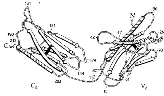
How do immunoglobulins function? One reaction, agglutination, results from the Interaction of a polyvalent antibody with two cells. More frequently, however, the interaction of an antibody with an antigen produces other effects. One such effect is the binding of the C1q protein, a component of Complement (Supplement 5-Ж). It has been established that the C1q protein binds to the CH2 domain of the Fc fragment of IgGп. Binding occurs only after the corresponding antigen (but not a hapten) has bound to the immunoglobulin. In addition, activation of The Complement System requires the presence of an aggregate consisting of two or more IgG molecules (or a single pentameric IgM molecule). It has been suggested that during The process of antigen binding, the immunoglobulin molecule undergoes a conformational change that generates the C1q-binding site. Curiously, however, only polyvalent antigens (i.e., antigens capable of binding to more than one antibody) can induce complement binding to the antibody, whereas haptens are incapable of inducing complement binding and do not even cause noticeable Conformational Changes in the Fab fragment. Thus, A number of important aspects of antibody function remain unclear.
а Edelman G. M., Science, 180, 830—840 (1973).
б Porter R. R., Science, 180, 713—716 (1973).
в Hauptman S. P., Tomasi T. B., JBC, 250, 3891—3896 (1975).
г Edelman G. E., Cunningham B. A., Gall W. E., Gottlieb P. D., Rutishauser U., Waxdal M. PNAS, 63, 78—85 (1969).
д Putnam F. W., Florent G., Paul Q., Shinoda T., Shimizu A., Science, 182, 287—291 (1973).
е Baenziger J., Kornfeld S., Kochwa S., JBC, 249, 1889—1896 (1974).
ж Sarma V. R., Silverton E. W., Davies D. R, Terry W. D., JBC, 246, 3753—3759 (1971).
з Segal D. M., Padlan E. A, Cohen G. H., Rudikoff S., Potter M., Davies D. R., PNAS, 71, 4298—4302 (1974).
и Amzel L. M., Poljak R. J., Saul F., Varga J. M., Richards F. F., PNAS, 71, 1427—1430 (1974).
к Schiffer M., Girling R. L, Ely K. R, Edmundson A. B., Biochemistry, 12, 4620—4631 (1973).
л Epp О., Colman P., Fehlhammer H., Bode W., Schiffer M., Huber R., Palm W., EJB, 45, 513—524 (1974).
м Blake С. C. F., Nature (London), 253, 158 (1975).
н Nisonoff A., Hopper J. E., Spring S. B., The Antibody Molecule, Academic Press, New York, 1975.
о Davies D. R., Padlan E. A., Segal D. M., Annu. Rev. Biochem., 44, 639— 667 (1975).
п Low T. L. К., Liu Y.-S. V., Putnam F. W., Science, 191, 390—392 (1976).
Last update: 06/08/2026
Editorial and Educational Adaptation: This material has been compiled based on the primary/original source text. The project team performed an editorial review, corrected technical inaccuracies, structured sections, and adapted the content for an educational format.
What was processed:
- elimination of formatting defects (OCR errors, structural breaks, corrupted characters);
- editorial organization of content;
- standardization of terminology in accordance with academic sources;
- verification of factual statements against the original source text.
All mentions of the author, publication year, and origin of the primary text have been preserved in accordance with the source.