Principles of Biochemistry, Volume 2 - A. Lehninger 1985
Bioenergetics and Metabolism
The Citric Acid Cycle
The citric acid cycle consists of eight stages
Let us now examine the eight successive stages of The Citric Acid Cycle, paying special attention to the chemical transformations in which the acetyl group of acetyl-CoA is rearranged and ultimately oxidized to CO2 and hydrogen atoms, which are captured in the form of the reduced Coenzymes NADH and FADH2. Figures 16-12 and 16-13 show the balanced equations for the cycle reactions and the structures of the intermediates.
a. Condensation of acetyl-CoA with oxaloacetate yields citrate
The first reaction of the cycle, catalyzed by citrate synthase, is the condensation of acetyl-CoA and oxaloacetate to form citrate (Fig. 16-12). In this reaction, the methyl carbon of the acetyl group of acetyl-CoA binds to the carbonyl carbon of oxaloacetate; simultaneously, the thioester bond is cleaved and coenzyme A is released:
Class="center">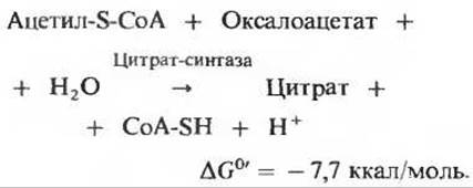
The equilibrium of this reaction within The Cell is largely shifted to the right, as indicated by the large negative Standard Free energy of Hydrolysis that characterizes it. The released CoA-SH can now participate in The oxidative decarboxylation of a new Pyruvate molecule to form a new acetyl-CoA molecule capable of entering the cycle. It is believed that citryl-CoA is an intermediate in the citrate synthase reaction. It is formed in the Active Site of the enzyme and rapidly hydrolyzed, after which free CoA-SH and citrate dissociate from the active site.
Citrate synthase is a regulatory enzyme: in many cell types, the reaction it catalyzes limits the overall rate of The Citric Acid cycle.
b. Citrate is converted to isocitrate via cis-aconitate
The enzyme aconitase catalyzes the reversible conversion of citrate to isocitrate. Tricarboxylic cis-aconitic acid is formed as an intermediate (which normally does not dissociate from the enzyme's active site) (Fig. 16-12). Aconitase catalyzes the reversible addition of H2O to the double bond of cis-aconitate (which remains bound to the enzyme) in two different ways, yielding citrate in one case and isocitrate in the other:
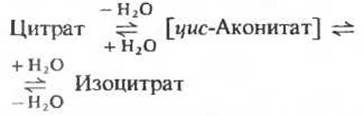
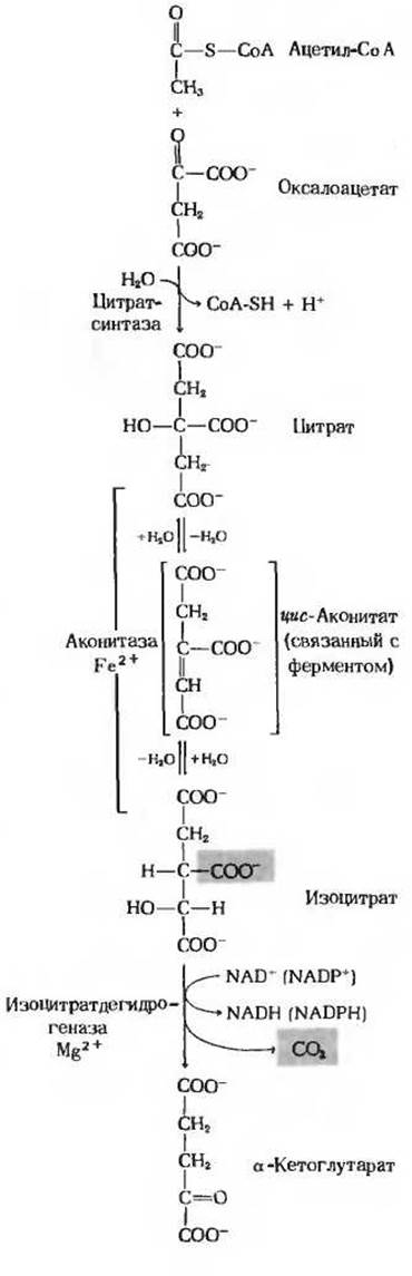
Figure 16-12. The first four Reactions of the citric acid cycle. Overall stoichiometric chemical equations are shown.
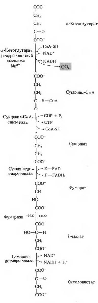
Figure 16-13. The remaining reactions of the citric acid cycle (for preceding stages, see Fig. 16-12).
Although at pH 7.4 and 25 °C the equilibrium mixture contains less than 10% isocitrate, the reaction proceeds forward in the cell because the product, isocitrate, is rapidly consumed in the subsequent steps of the cycle. Aconitase is a rather complex enzyme. It contains iron and acid-labile sulfur atoms grouped into an iron-sulfur center (Section 17.8). The precise function of this iron-sulfur center, which is thought to serve as a prosthetic group of the enzyme, remains unknown.
c. Isocitrate is dehydrogenated to yield a-ketoglutarate and CO2
In the next stage of the cycle, isocitrate is dehydrogenated to form a-ketoglutarate and CO2 (Fig. 16-12) by the action of isocitrate dehydrogenase. There are two types of isocitrate dehydrogenase: one uses NAD+ as an electron acceptor, and the other uses NADP+. Otherwise, the overall Reactions Catalyzed by these Two Types of isocitrate dehydrogenase are identical:
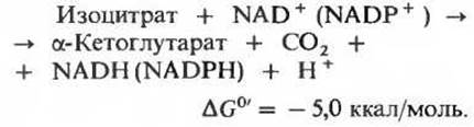
Mitochondria contain both types of isocitrate dehydrogenase, NAD-dependent and NADP-dependent; the first type is found exclusively in mitochondria, whereas the second is present in both mitochondria and the Cytosol. Both mitochondrial Enzymes apparently participate in the citric acid cycle, but the NAD-dependent isocitrate dehydrogenase predominates. Its activity requires Mg2+ or Mn2+ ions as well as its positive modulator ADP, in the absence of which the enzyme is practically inactive. The presence of two types of isocitrate dehydrogenase in mitochondria may be related to the Regulation of the cycle.
d. a-Ketoglutarate is oxidized to succinate and CO2
The next stage of the cycle involves the Oxidative Decarboxylation of a-ketoglutarate to form succinyl-CoA and CO2 (Fig. 16-13), catalyzed by the a-ketoglutarate dehydrogenase complex. The reaction is described by the equation

Note that this reaction is virtually identical to the pyruvate dehydrogenase reaction discussed above; both involve The oxidation of an a-keto acid accompanied by the loss of a carboxyl group as CO2. In both Structure and function, the a-ketoglutarate dehydrogenase complex strongly resembles the pyruvate dehydrogenase complex. It consists of three enzymes analogous to those of the pyruvate dehydrogenase system and also includes enzyme-bound Cofactors: thiamine pyrophosphate, Mg2+, coenzyme A, NAD+, FAD, and Lipoic Acid. There is, however, an important difference: the a-ketoglutarate dehydrogenase system lacks the complex regulatory mechanism characteristic of the pyruvate dehydrogenase complex.
e. Conversion of succinyl-CoA to succinate
Succinyl-CoA, the product of the preceding stage of the cycle, is a high-energy compound. The hydrolysis of its thioester bond, much like that of acetyl-CoA, is characterized by a large negative standard free-energy change.
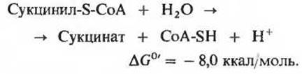
In the cell, however, the Cleavage of the CoA moiety does not occur via simple hydrolysis, as such a pathway would waste free energy. Instead, the breaking of the thioester bond is coupled to a reaction that conserves energy through the phosphorylation of guanosine diphosphate (GDP) to guanosine triphosphate (GTP) (Fig. 16-13):
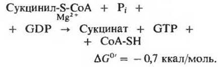
The enzyme catalyzing this reaction, succinyl-CoA synthetase, promotes The formation of free succinate while simultaneously generating a high-energy terminal phosphate group on GTP from GDP and Pi, utilizing the free energy released during succinyl-CoA cleavage. This energy-conserving reaction proceeds via an intermediate step in which the enzyme molecule itself is phosphorylated at a specific Histidine residue in its active site. It is precisely this energy-rich phosphate group, involved in that phosphorylation, which is subsequently transferred to GDP to yield GTP. The coupled formation of GTP driven by The energy released during the Oxidative Phosphorylation of a-ketoglutarate represents yet another example of substrate-level phosphorylation. Recall that we have already encountered another example of this type—namely, the coupled synthesis of ATP driven by the energy released from the oxidation of glyceraldehyde 3-phosphate during Glycolysis (Section 15.7,6). Such reactions are traditionally termed substrate-level phosphorylation because their necessary energy derives directly from the oxidation of an organic substrate. Grouping these reactions under a common heading distinguishes them from oxidative phosphorylation, which is coupled to electron transfer (also referred to as Respiratory Chain phosphorylation; Chapter 17).
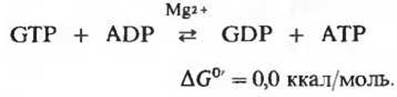
The GTP generated by the action of succinyl-CoA synthetase can subsequently transfer its terminal phosphate group to ADP to yield ATP; this reversible reaction is catalyzed by nucleoside diphosphate kinase (Section 14.18).
e. Dehydrogenation of succinate to fumarate
In the next stage of the cycle (Fig. 16-13), the succinate formed from succinyl-CoA is dehydrogenated to yield fumarate. This reaction is catalyzed by the flavoprotein succinate dehydrogenase, whose molecule contains covalently bound flavin adenine dinucleotide. This redox-active prosthetic group serves as the hydrogen acceptor in the following reaction (E denotes the enzyme protein):
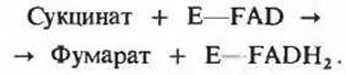
Succinate dehydrogenase is tightly bound to The inner mitochondrial membrane. The Molecular Weight of the enzyme isolated from bovine Heart mitochondria is approximately 100,000. A single molecule of this enzyme contains one covalently bound FAD residue and two iron-sulfur centers: one containing two iron atoms and the other containing four. During the succinate dehydrogenase reaction, these iron atoms undergo valence changes [Fe(II) — Fe(III)], suggesting their direct participation in electron transport (Chapter 17).
Malonate is a competitive inhibitor of succinate dehydrogenase (Section 9.13 and Fig. 16-7); as noted above, investigating its effects played a significant role in elucidating the overall pathway of the citric acid cycle.
f. Hydration of fumarate to malate
The reversible hydration of fumarate to yield L-malate (Fig. 16-13)

is catalyzed by fumarate hydratase. This enzyme, better known as fumarase, has been isolated in crystalline form from pig heart. Fumarase is highly specific: it hydrates only the trans-isomer of the double bond in fumarate, exhibiting no activity toward its cis-isomer or toward the cis- and trans-isomers of monocarboxylic unsaturated acids. In the reverse reaction (L-malate → fumarate), fumarase displays strict optical isomer Specificity, being entirely incapable of catalyzing the dehydration of D-malate. The molecular weight of fumarase is approximately 200,000. The enzyme molecule consists of four subunits (four polypeptide chains). No coenzyme is required for fumarase activity.
g. Dehydrogenation of malate to oxaloacetate
In the final step of the citric acid cycle, the mitochondrial matrix-localized, NAD-dependent L-malate dehydrogenase catalyzes the dehydrogenation of L-malate to form oxaloacetate (Fig. 16-13):

The equilibrium of this reaction under standard conditions (i.e., at 1 M concentrations of all components and pH 7.0) lies strongly to the left. Nevertheless, in intact Cells, the reaction proceeds spontaneously from left to right because the product, oxaloacetate, is rapidly removed (consumed in the citrate synthase reaction), maintaining its steady-state intracellular concentration at an extremely low level, well below 10-6 M.
Last update: 06/08/2026
Editorial and Educational Adaptation: This material has been compiled based on the primary/original source text. The project team performed an editorial review, corrected technical inaccuracies, structured sections, and adapted the content for an educational format.
What was processed:
- elimination of formatting defects (OCR errors, structural breaks, corrupted characters);
- editorial organization of content;
- standardization of terminology in accordance with academic sources;
- verification of factual statements against the original source text.
All mentions of the author, publication year, and origin of the primary text have been preserved in accordance with the source.