Fundamentals of Biochemistry - Filippovich, Y. B. 1999
Biological Oxidation
Free Oxidation
Reactions of free oxidation of Organic compounds in living nature, as well as the enzyme systems that accelerate them, are diverse; many of them have been discussed in previous chapters and in the introductory section of this chapter. This pathway directly oxidizes not only numerous natural and non-natural substrates, but also reduced Coenzymes (NADH, NADPH, FAD ∙ H2, etc.) generated by the action of Primary and secondary dehydrogenases.
Although free oxidation reactions occur in both the Cytosol and the membranes of various subcellular structures, their primary site is the membranes of The Cell's Endoplasmic reticulum. Because the latter yield a microsomal fraction upon cell homogenization and fractionation of subcellular homogenate particles, which can be isolated as a preparation, the Organization and Functions of the microsomal Respiratory Chain are currently being intensively studied. Its first feature is that, despite the presence of Electron Transport Chain Enzymes, no point in this chain is coupled with ADP phosphorylation. The second feature lies in the unique Structure and Functional activity of Cytochromes b5 and P-450 included in its composition. In particular, cytochrome P-450 (M ≈ 50,000, a hemoprotein whose Primary Structure for more than a dozen of its forms has been elucidated) possesses a multitude (hundreds, or perhaps thousands) of forms that emerge in response to the Introduction (or entry) into the Organism of a particular Class of xenobiotics, much like Antibodies are synthesized in response to the presence of Antigens; therefore, cytochrome P-450 is considered a sort of "membrane immunoglobulin."
Finally, the third feature is the high affinity of the terminal oxidase of microsomal chains for oxygen, which allows it to compete for oxygen with mitochondrial cytochrome c oxidase. Consequently, for example, in Liver Cells, the proportion of microsomal oxidation of endogenous substrates is 40%, while mitochondrial oxidation accounts for 60%.
The most unusual reactions of free oxidation, which were virtually untouched upon in previous chapters, are those proceeding with the participation of oxygenases. This subclass of oxidoreductases contains enzymes that accelerate the incorporation of either two (Dioxygenases) or one (Monooxygenases) atoms of molecular oxygen into the substrate being oxidized.
Free oxidation involving dioxygenases. One of the most thoroughly studied dioxygenases is pyrocatechase (catechol 1,2-dioxygenase, or catechol:oxygen 1,2-oxidoreductase (decyclizing)); its M = 85,000. Concentrated solutions of pyrocatechase are red because its active center contains two tightly bound Fe atoms which, according to O. Hayaishi, combine with molecular oxygen to form a complex where oxygen is subsequently activated: Fe2+ + O2 → Fe2+O2 → Fe3 + O2.
Then, after the substrate binds to the active center of pyrocatechase, a ternary complex is formed, the transformation of which leads to the incorporation of molecular oxygen into pyrocatechol:

Analogous action is exhibited by catechol 2,3-dioxygenase (M = 140,000, 1 Fe atom), protocatechuate 3,4-dioxygenase (M = 700,000, 8 x 90,000), which forms β-carboxymuconic acid, Tryptophan 2,3-dioxygenase (M = 123,000), which attaches molecular oxygen across the pyrrole ring of the indole radical, and other dioxygenases. In all cases, molecular oxygen is activated via The addition of an electron lost by the divalent iron of the active center, and the resulting anionic free radical of oxygen (O2-2) attacks and oxygenates the substrate. Elucidating the MECHANISM OF ACTION of dioxygenases has clarified one of the ways in which terminal oxidases activate molecular oxygen.
One of the biologically important dioxygenase reactions is The conversion of β-carotene into vitamin A:
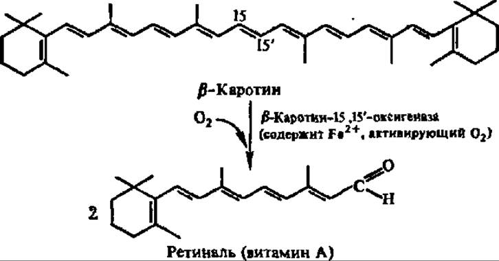
The direct addition of 18O2 across the 15,15'-bond in β-carotene was first proved by B. B. Vartapetyan et al. (1966).
Free oxidation involving monooxygenases. Owing to the high lability of monooxygenases, as well as dioxygenases for that matter, their isolation is fraught with major difficulties. Nevertheless, some of them have been obtained in crystalline form. Monooxygenases are characterized by molecular weights ranging from 65,000 to 200,000, the general absence of heavy Metal Ions in their composition, and the presence of flavin-type coenzymes. In addition, many of them require the participation of a so-called paired donor in the monooxygenation reaction, which supplies hydrogen atoms to one of the molecular oxygen atoms (the second atom is incorporated into the oxidized substrate).
The simplest representative of monooxygenases is phenol hydroxylase (phenol 2-monooxygenase):

Monooxygenases play a major role in The oxidation of Amino Acids (Lysine, Arginine, and tryptophan monooxygenases, Phenylalanine and Tyrosine hydroxylases), hydroxy acids (salicylate hydroxylase), and polyisoprenoid compounds (squalene epoxidase, see p. 404).
The Mechanism of action of monooxygenases remains insufficiently understood. It is hypothesized that the active form of oxygen may be enzyme-bound hydrogen peroxide or its equivalent. Meanwhile, it has been shown that some monooxygenases (fungal phenolase, phenylalanine 4-hydroxylase) contain Cu+ in their composition. In these cases, the following mechanism of oxygen activation is possible:
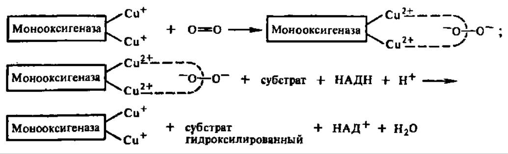
Here, as in the case of dioxygenases, The transfer of electrons from the metal entering into the COMPOSITION OF THE enzyme to molecular oxygen plays a decisive role in its activation. It is quite possible that this pathway of involving molecular oxygen in cellular oxidoreduction processes is universal for oxidases participating in both free and phosphorylation-coupled Biological Oxidation.
Last update: 06/08/2026
Editorial and Educational Adaptation: This material has been compiled based on the primary/original source text. The project team performed an editorial review, corrected technical inaccuracies, structured sections, and adapted the content for an educational format.
What was processed:
- elimination of formatting defects (OCR errors, structural breaks, corrupted characters);
- editorial organization of content;
- standardization of terminology in accordance with academic sources;
- verification of factual statements against the original source text.
All mentions of the author, publication year, and origin of the primary text have been preserved in accordance with the source.