Amino Acids, Peptides and Proteins - Dévényi T., Gergely J. 1976
Gas chromatographic analysis of amino acid derivatives. State of the art. Capabilities and limitations of the method.
Gas chromatography of peptides
Weygand et al. method
In this method, unlike those discussed above, Peptides are first protected and then, with few exceptions, directly subjected to GC. Amino groups are protected with a TFA residue, while the carboxyl group is blocked during Esterification with methanol. The volatility of TFA compounds has been pointed out repeatedly; high-vacuum sublimation has achieved the Separation of the corresponding derivatives of Amino Acids, di-, tri-, and larger peptides [107].
For the GC of these compounds, it was first necessary to solve the Problems associated with Column thermal stability and to select a separation method. Due to their higher polarity compared to amino acids, peptides generally require temperatures above 200°C. Without addressing the issue of phase stability, we note that The Use of polar liquid phases in this case, such as Reoplex or polyphenyl ether, is quite limited. For relatively simple dipeptides, their use yielded good results, but considering that the number of compounds separated was small, it is unlikely that this technique will find wider application in analytical work for amino acid sequencing. Non-polar silicone rubbers SE 30 and SE 52 proved to be satisfactory stationary phases; Apiezon L is less advantageous.
Class="center">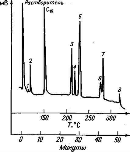
Fig. 75. Gas chromatogram of a partial hydrolysate of Leu-Leu-Val-Val after esterification and trifluoroacetylation.
Column 1.8 m long with 20% SE 30 on Diatoport W; He flow rate 62 ml/min. Initially isothermal mode (150°C); after the elution of C10 — capric acid methyl ester — linear Temperature programming with a gradient of 5 deg/min. 1 — Val; 2 — Leu; 3 — Val-Val; 4 — Leu-Val; 5 — Leu-Leu; 6 — Leu-Val-Val; 7 — Leu-Leu-Val; 8 — Leu-Leu-Val-Val.
When using such a separation method for Amino Acid Sequence analysis, the selectivity of the stationary phases does not play a major role if a relatively small number of compounds are analyzed; the decisive factor in this case will be the number of components detected on a single column. Since tetrapeptides can be detected by gas Chromatography only in exceptional cases, the greatest attention is inevitably focused on the most complete identification of di- and tripeptides. Relevant studies have shown that this can only be achieved with the aforementioned phases.

Fig. 76. Gas chromatogram of a partial acid hydrolysate of Leu-Phe-Val-Val after esterification and trifluoroacetylation.
Column 1.8 m long with 20% SE 30 on Diatoport W; He flow rate 62 ml/min. Initially isothermal mode (150°C); after the elution of C10 — capric acid methyl ester — linear temperature programming with a gradient of 5 deg/min. 1 — Val; 2 — Leu; 3 — Phe; 4 — Val-Val; 5 — Phe-Val; 6 — Leu-Phe; 7 — Phe-Val-Val; 8 — Leu-Phe-Val.
As already noted, since there is A large number of different compounds in the mixture, complete separation of the components cannot be expected, especially since the stationary phases have relatively poor selectivity. In this regard, it is advisable to analyze peptides that are not too long after carrying out their partial Hydrolysis. For example, Cleavage of a decapeptide under the most favorable conditions yields 9 dipeptides, 8 tripeptides, etc. If this peptide consists of 10 different amino acids, all of which are detected by GC, then it is necessary to separate a mixture consisting of 27 compounds, not counting tetrapeptides, which probably can also be detected. Identifying all possible short fragments is, of course, quite difficult. The specified chain length range (from amino acid to tetrapeptide) is undoubtedly the limit of this method's capabilities.
Poor separation, however, is attempted to be compensated for by mass spectrometry. It has been shown that mixtures of simple peptides are fully identified on a mass spectrometer [123], and peak overlapping in no way precludes the identification of peptide derivatives.

Fig. 77. Gas chromatogram of a partial acid hydrolysate of oxytocin after desulfurization and trimethylsilylation.

Column 13 m long with 10% SE 30 on Diatoport P; He flow rate 62 ml/min. Initially isothermal mode (150°C); after the elution of C10 — capric acid methyl ester (standard) — linear temperature programming with a gradient of 5 deg/min. 1 — Ala; 2 — Gly; 3 — Leu; 4 — Asp; 5 — Leu-Gly; 6 — Ala-Pro; 7 — Tyr; 8 — Pro-Leu; 9 — Ile-Glu; (?) Glu-Asp; 10 — Ala-Tyr; 11 — Tyr-Ile.
It is quite understandable that peptides exhibit greater differences in polarity and volatility than amino acids. Peptides elute over a much wider range of retention volumes, and only a very small fraction of the hydrolysate can be separated isothermally. By choosing suitable columns and reaction conditions, good separation could be achieved in this part of the hydrolysate as well—naturally, at a relatively high cost (and loss) of substance. With such a Procedure, a large portion of the hydrolysate taken for analysis is inevitably lost due to poor separation (with more volatile components) or unsatisfactory elution (with less volatile substances). Since the patterns of Isolation and Purification of large peptides are not yet sufficiently understood, any losses should be avoided. The separation of a partial hydrolysate is best carried out in a single analytical run; then, by applying equal amounts of substance, it is much easier to perform a repeat Analysis of the entire mixture during a multi-stage isothermal study, and the Components of the mixture can be reliably identified using appropriate controls.

Fig. 78. Gas chromatogram of a partial acid hydrolysate of Insulin A-chain after esterification and trifluoroacetylation.
Column 1.8 m long with 10% SE 30 on Diatoport P; He flow rate 66 ml/min. Initially isothermal mode (150°C); after the elution of C10 — capric acid methyl ester — linear temperature programming with a gradient of 5 deg/min. 1 — Glu; 2 — Ser-Val; 3 — Ile-Val; 4 — Ser-Leu; 5 — Tyr; 6 — Val-Glu; 7 — Glu-Leu; 8 — Leu-Glu; 9 — Leu-Tyr; 10 — Glu-Glu; C18 — stearic acid methyl ester (standard).
When using temperature-programmed mode, such mixtures are easily separated in a single analytical run, as seen from the chromatograms shown in Figs. 75–80. Individual groups of compounds—amino acids, di-, tri-, and tetrapeptides—elute in different temperature ranges. This allows, despite some overlap (for example, high molecular weight amino acids begin to elute together with dipeptides), the detection of qualitative differences characteristic of different sequences from the appearance of the chromatograms, similar to the "fingerprinting" method [39]. To use GC for Sequence Determination, it is necessary to know which combinations of amino acids in di-, tri-, and longer peptides can be detected. Because the number of such combinations is very large, and the task of synthesizing peptides, especially long ones, is quite complex, one cannot expect to obtain exhaustive data. For the identification of tri- and tetrapeptides, the Selection of reference standards is particularly difficult. However, The behavior of the amino acids themselves during GC can provide significant information, thereby avoiding useless experiments.
The conversion of peptides to N-TFA methyl esters proceeds in a manner completely analogous to amino acids: they are first converted to methyl ester hydrochlorides and then trifluoroacetylated. If individual substances are not to be quantified (since the cleavage of various peptide bonds depends on both the degradation method and the sequence itself), complete conversion is desirable, as this generates fewer cleavage products. During esterification, precautions should be taken, especially when working with enzymatic hydrolysates, to prevent additional cleavage of labile bonds by the acid. A 24-hour incubation in 0.1 N methanolic HCl at room temperature satisfies the necessary conditions [15].
Trifluoroacetylation with an excess of trifluoroacetic acid methyl ester in absolute methanol at room temperature with The addition of triethylamine led to varying yields during amino acid conversions [109]. The risk of diketopiperazine formation, characteristic of dipeptide esters, can be reduced by adding a large excess of the acylating agent [119]. Diketopiperazines can also be subjected to gas chromatography, but they yield highly asymmetric peaks and are therefore unsuitable for sequence analysis [121]. Trifluoroacetylation of peptide hydrochlorides can also be performed by reaction with trifluoroacetic anhydride [119]. Although There is a risk of additional cleavage of peptide bonds, the corresponding products are formed in such low yields that this cannot affect the subsequent analysis [111]. At room temperature, acylation with the anhydride proceeds faster and more completely. As in the Biemann method, N-TFA peptide esters are easily separated from the unreacted portion of the molecules by extraction with acid and Water. During this procedure, O-TFA derivatives formed during the reaction with the anhydride are hydrolyzed. The resulting mixture of protected peptides is dissolved in a suitable solvent, such as tetrahydrofuran, ethyl acetate, or methanol, and subjected to fractionation.

Fig. 79. Gas chromatogram of a partial acid hydrolysate of insulin A-chain after esterification, trifluoroacetylation, and desulfurization.
The separation was carried out on an instrument, the diagram of which is shown in Fig. 72.
C10 — capric acid methyl ester; 1 — Glu; 2 — Ala-Ala; 3 — Val-Ala; 4 — Ser-Val+Ile-Val; 5 — Ala-Asp; 6—Ser-Leu; 7 — Tyr+Glu-Ala; 8 — Val-Glu; 9 — Glu-Leu; 10 — Leu-Glu; 11 — Glu-Asp; 12 — Tyr-Ala; 12 — Glu-Glu; C18 — stearic acid methyl ester.

Fig. 80. Gas chromatogram of a partial acid hydrolysate of insulin A-chain after esterification, trifluoroacetylation, and trimethylsilylation.
The separation was carried out using the apparatus shown schematically in Fig. 72.
C10 — capric acid methyl ester; 1 — Ile-Val; 2 — Ser-Val; 3 — Tyr; 4 — Ser-Leu, (?) Val-Glu; 5 — Glu-Leu; 6 — Leu-Glu; 7 — Glu-Asp (?); 8 — Leu-Tyr; 9 — Glu-Glu; 10 — Tyr-Glu; C18 — stearic acid methyl ester.
GC of di-, tri-, and possibly many tetrapeptides consisting of the amino acids Ala, Val, Leu, Ile, and Pro presents no difficulties. Since Gly has no side chain, the peaks of the corresponding dipeptides often exhibit tailing. This phenomenon is probably caused by adsorption effects, which also likely account for the incomplete elution of N-TFA-Gly-Gly-Gly-OCH3 [19]. Only dipeptides have been studied with Met, and tripeptides as well with Phe. Although, as indicated by the behavior of N-TFA-Ala-Phe-Phe-OCH3, tripeptides composed of high-molecular-weight amino acids do not always elute, satisfactory results can be obtained using short columns with low liquid phase loading.
As for polyfunctional amino acids, dipeptides containing Glu, Lys, and Orn present no difficulties in either derivatization or chromatography. Additional functional groups are protected during routine derivatization. However, for dipeptides containing Lys and Orn, severe peak broadening due to adsorption is observed on columns with less than 5% liquid phase [122]. Isomeric α- and β-peptides containing Glu are separated by GC [114, 121]. In similar studies of N-TFA-derivative methyl esters of Asp-containing peptides, temperature-dependent formation of cyclic imides was detected upon heating, which was more pronounced for the β-peptide [106]. The imides can only be separated from the corresponding α-peptides on capillary columns. Direct on-column injection might prevent such reactions. Tripeptides containing the aforementioned Amino acids have not yet been investigated.
Ser and Thr may yield multiple signals due to thermal β-elimination during high-temperature sample injection. Trimethylsilylation with hexamethyldisilazane proved to be the best method for protecting these amino acids [121]. Tyrosine-containing dipeptides yield sharp, symmetrical peaks As a result of O-trimethylsilylation. These compounds can be prepared by heating for 30 min in an excess of hexamethyldisilazane after esterification and trifluoroacetylation.
To date, dipeptides containing Cys and Cys2 have not been detected. Cys is particularly prone to β-elimination, as already noted in the section on amino acids. Due to the lack of suitable S-protecting groups, desulfurization over Raney nickel had to be employed [121]. According to this method, protected peptide esters are desulfurized over Raney nickel during prolonged heating in 90% aqueous methanol. As a result, Cysteine and cystine compounds yield the corresponding Alanine peptides, which can be distinguished from the alanine peptides present in the original molecule using D2O. Similar to Biemann's method, the SH group is replaced by deuterium, enabling component identification in a mass spectrometer. Methionine compounds are desulfurized simultaneously with Cys, converting into α-aminobutyric acid peptides, which are easily detected by gas chromatography.
Compounds containing Arg, His, and Trp have been very little studied so far. Complex dipeptides containing these Amino acids can hardly be detected. For Arg, there is a possibility of its conversion to Orn using arginase [30]. This can be useful because, after tryptic hydrolysis of large peptides, due to its Specificity, some Arg residues will be located at the C-terminal position of the fragments. Upon treating these peptides with arginase, the corresponding C-terminal amino acid residues are converted to Orn, making the peptides accessible for GC. After Partial Acid Hydrolysis, Trp-containing peptides are virtually not formed, since Trp is destroyed during this procedure.
The sequence of steps in the above analysis can be schematically summarized as follows: 1) partial hydrolysis, 2) esterification, 3) trifluoroacetylation and GC analysis, 4) desulfurization and GC analysis, 5) trimethylsilylation and GC analysis.
By comparing the results of the three GC analyses, THE POSITION OF peptides containing hydroxy amino acids, Cys, and Cys2 can be determined very accurately. After desulfurization, some peaks disappear and others appear; these belong to sulfur-containing peptides. If Met is absent in the starting sample, these must be cysteine (cystine) peptides, which are thus easily identified. The same situation is observed with quantitative trimethylsilylation, which reveals compounds containing Ser, Thr, Hyp, and Tyr. Derivatives of dipeptides containing Hyp and Tyr elute at relatively high temperatures. Thus, definite Conclusions can be drawn even on The basis of the retention volume range.
In Conclusion, GC allows the identification of most Natural Amino Acids in the form of dipeptides. The ability to detect many amino acids even as tripeptides, and in exceptional cases as tetrapeptides, provides researchers with a rapid and relatively simple method for sequencing medium-length oligopeptides. GC is currently used primarily as a fast and efficient separation method. A more complete utilization of GC's advantages is achieved when combined with mass spectrometry [68, 106]; this has already been successfully applied to the analysis of fatty acid mixtures [75]. The use of specially designed ion sources is highly promising, making the attachment of a detector to the gas chromatograph unnecessary [13].
Figs. 75—80 illustrate the GC behavior of N-TFA peptide methyl esters of various lengths subjected to partial acid hydrolysis or methanolysis.
As shown in Figs. 75 and 76, when a tetrapeptide is analyzed, all possible fragments can be detected. In one case, it is even possible to identify the parent peptide. Fig. 77 shows the chromatogram of a partial hydrolysate of oxytocin, which was desulfurized and trimethylsilylated According to the scheme described above. All possible dipeptides were identified, including Asp-containing compounds, which were formed in only small amounts during cleavage. By comparing different gas chromatograms, conclusions can be drawn about the presence of sequences containing Cys (or Cys2) and hydroxy amino acids. Figs. 78, 79, and 80 show gas chromatograms of a partial hydrolysate of the insulin A-chain obtained at different Stages of the transformations discussed above. After desulfurization, as seen by comparing Figs. 78 and 79, new signals 2, 3, and 5 appear, while peaks 7 and 12 become broader. In all these cases, Ala-containing compounds are formed. Similarly, after trimethylsilylation, new peaks 2, 3, 4, 8, and 10 are found in Fig. 80, while signals 2, 4, 5, and 8 present in Fig. 78 disappear. These peaks correspond to A-chain peptides containing Ser and Thr, the latter eluting at relatively high retention temperatures. After silylation, the peaks emerge at higher elution temperatures. As with other peptides, all expected dipeptides except those containing Asp could be detected after acid degradation. Unidentified peaks may belong to tripeptides.
In all chromatograms discussed, the components were identified using reference standards, as mass spectrometric analysis was not feasible.
The information obtained by GC about the parent peptide chain, especially for a relatively large molecule such as the insulin A-chain, is insufficient to determine the complete sequence. For this purpose, various cleavage techniques must additionally be employed. Nevertheless, it is quite obvious that GC, primarily as a separation method, possesses great advantages for research in this field due to its speed, small sample requirements, and high resolution. This chapter on GC cannot be concluded without mentioning further possibilities for its application. For instance, in peptide chemistry, it can provide simple and rapid Answers to A number of questions. As an example, we can mention the opening of internal anhydrides of N-TFA-Glu and N-TFA-Asp by amino esters, which yields isomeric peptides in various ratios [106, 114].
The separation of diastereomeric N-TFA-dipeptide methyl esters has acquired great importance [115]. It was performed quantitatively on capillary columns for a large number of peptides of various sequences [124]. This separation represents the first accurate and reliable METHOD FOR DETERMINING racemization in Peptide Synthesis. Along with various N-acylamino acids, N-acylpeptides can be useful in studying the optimal conditions for different peptide synthesis reactions, provided that the synthesized peptides are subjected to partial hydrolysis prior to the separation of diastereomers [69, 124]. Fig. 81 shows the chromatogram of a partial acid hydrolysate of an incompletely racemized synthetic tetrapeptide. Under these conditions, besides amino acids, only dipeptides belonging to the original sequence were detected. In Fig. 77, the doublet peak of the tyrosine dipeptide also indicates partial racemization of Tyr. This last example demonstrates that, in certain cases, the degree of optical purity of peptides can be monitored and determined rapidly and without difficulty.

Fig. 81. Gas chromatogram of a partial acid hydrolysate of an incompletely racemized Leu-Phe-Ala-Phe tetrapeptide after esterification and trifluoroacetylation.
A 50 m steel capillary column containing polyphenyl ether OS 138; 220°C; N2 flow rate — 2.1 ml/min. 1 — L-Phe-L-Ala; 2 — L-Phe-D-Ala; 3 — L-Ala-L-Phe; 4 — D-Ala-L-Phe; 5 — L-Leu-L-Phe; 6 — L-Leu-D-Phe.
1. Ackman R. G., J. of Gas Chromatogr., 2, 173—179 (1964).
2. Anders К., Gas-Chromatographie, pp. 1—22 (ed. Struppe H. G., Angele H. P., Akademie Verlag, Berlin 1964), 1963.
3. Baraud J., Bull. Soc. Chim. France, 754 (1960).
4. Bayer E., Desty D. H., Gas Chromatography, p. 333, Butterworth, London, 1958.
5. Bayer E., Born F., Reuther K. H., Angew, Chem., 69, 640 (1957).
6. Biemann K., Gapp F., Seibl J., J. Am. Chem. Soc., 81, 2274 (1959).
7. Biemann K., Vetter W., Biochem. Biophys. Res. Commun., 3, 578 (1960).
8. Bier M., Teitelbaum C., Ann. N. Y. Acad. Sci., 72, 641 (1959).
9. Birkofer L., Ritter A., Chem. Ber., 93, 424 (1950).
10. Blau K., Darbre A., Biochem. J., 88, 8P (1963).
11. Blau K., Darbre A., J. Chromatogr., 17, 445 (1965).
12. Boissonnas R. A., Gutimann S., Huguenin R. L., Jaquenoud P. A., Sandrin E., Helv. Chim. Acta, 41, 1867 (1958).
13. Brunnée C., Jenckel L., Kronenberger K., Z. Anal. Chem., 197, 42 (1963).
14. Calvin M., Schallenberg E., J. Am. Chem. Soc., 77, 2779 (1955).
15. Chibnall A. C., Mangan J. L., Rees M. W., Biochem. J., 68, 114 (1958).
16. Gruickshank P. A., Sheehan J. C., Anal. Chem., 36, 1191 (1964).
17. Darbre A., Personal communication.
18. Darbre A., Blau K., Biochem. J., 88, 8P (1963).
19. Darbre A., Blau K., J. Chromatogr., 17, 31 (1965).
20. Dorsey J. A., Hunt R. H., O’Neal M. J., Anal. Chem., 35, 511—515 (1963).
21. Eck R. V., Nature, 193, 241 (1962).
22. Edman P., Acta Chem. Scand., 4, 283 (1950).
23. Engelhardt K., Dissertation, Techn. Hochschule (Technical University), München, 1963.
24. Ettre L. S., Open Tubular Columns in Gas Chromatography, Plenum Press, New York, 1965.
25. Evans R. M., Mfg. Chemists Assoc., 32, 512 (1961).
26. Fischer E., Ber. Deut. Chem. Ges., 34, 433 (1901).
27. Gehrke C. W., Lamkin W. M., Chem. Eng. News, 42, Nr. 37, 62 (1964).
28. Giaccobbo H., Simon W., Pharm. Acta Helv., 39, 162 (1964).
29. Graf J., Wein J. P., Winitz M., Fed. Proc., 22, Part I, 244 (1963).
30. Grassmann W., Hermann H., Janowsky O., Z. Physiol. Chem., 312, 273 (1958).
31. Greenstein J. P., Winitz A4., Chemistry of the Amino Acids, John Wiley and Sons, New York, p. 925, 1961.
32. Hagen P., Black W., Fed. Proc., 23, 371 (1964).
33. Henneberg D., Z. Anal. Chem., 183, 12—23 (1961).
34. Heyns K., Grützmacher H. F., Tetrahedron 1761; Liebings Ann. Chem., 669, 189 (1963).
35. Hoffter D., Dissertation, Technical University, Munich, 1962.
36. Horning E. C., Maddock К. C., Anthony К. V., van den Heuvel W. J. A., Anal. Chem., 35, 526 (1963).
37. Hunter I. R., Dimick К. P., Cobse J. W., Chem. Inds, 294 (1956).
38. Ikekawa N., J. Biochem. (Japan), 54, 279(1963).
39. Ingram V. M., Nature, 178, 792 (1956).
40. Ishii S., Witkop B., J. Am. Chem. Soc., 85, 1832 (1963).
41. Jonak J., Nature, 185, 685 (1960).
42. Johnson D. E., Scott S. J., Meister A., Anal. Chem., 33, 669 (1961).
43. Kaelicke J., Dissertation, Technical University, Berlin, 1958.
44. Raiser R., Gas Chromatography, Vol. II. Capillary Chromatography. Bibliographisches Institut, Mannheim, 2nd ed., 1966.
45. Kaiser R., Gas Chromatography, Vol. III. Tables pp. 78—116, Bibliographisches Institut, Mannheim, 2nd ed., 1968.
46. Kaiser R., Z. Anal. Chem., 205, 284—298 (1964).
47. Kaiser R, Gas Chromatography, Vol. I. Gas Chromatography, pp. 58—59, Bibliographisches Institut, Mannheim, 1965.
48. Kaiser R., Gas Chromatography, Vol. IV. Quantitative Evaluation. Bibliographisches Institut, Mannheim, 1965.
49. Kaiser R., Gas Chromatography, Vol. IV, pp. 26—52, Bibliographisches Institut, Mannheim, 1965.
50. Karrer P., Nicolaus B. J. R., Helv. Chim. Acta, 35, 1581 (1952).
51. Karrer P., Portmann P., Suber A4., Helv. Chim. Acta, 32, 1156 (1949).
52. Koroats E., Wehrli A., Helv. Chim. Acta, 42, 2709 (1959).
53. Lamkin W. W., Gehrke C. IF., Anal. Chem., 37, 385 (1965).
54. Landowne R. A., Lipsky S. R., Nature, 199, 141 (1963).
55. Langheld K., Ber. Deut. Chem. Ges., 42, 2360 (1909).
56. Liberii A., Desty D. H., Gas Chromatography, Butterworth, London, p. 341, 1958.
57. Lorette N. B., Brown J. H., Jr., J. Org. Chem., 24, 261 (1959).
58. Losse G., Losse A., Stock J., Z. Naturforsch., 17b, 785 (1962).
59. Makisumi S., Nicholls C. H., Saroff H. A., J. Chromatogr., 11, 327 (1963).
60. Makisumi S., Saroff H. A., J. Gas Chromatogr., 3 , 21 (1965).
61. McCaldin D. J., Chem. Revs., 60, 39 (1960).
62. Melamed N., Renard R., J. Chromatogr., 4, 339 (1960).
63. Meyer D. M., Jutisz M., Bull. Soc. Chim. France, 1211 (1957).
64. Morita K., Irreverse F., Witkop B., J. Am. Chem. Soc., 85, 2832 (1963).
65. Nicholls C. H., Makisumi S., Saroff H. A., J. Chromatogr., 11, 327 (1963).
66. Pisano J. J., van der Heuvel E. J. A., Horning E. C., Biochem. Biophys. Res. Commun., 7, 82 (1962).
67. Prox A., Dissertation, Technical University, Berlin, 1959.
68. Prox A., Weygand F., in: Peptides, Proc. of the 8th Europ. Peptide-Sympos. Nordwijk, the Netherlands, Sept. 1966, ed. by Beyerman H. C., van de Linde A., van den Brink M., North Holland, p. 158, 1967.
69. Prox A., Weygand F., König W., Schmidhammer L., Proceedings of the 6th Europ. Peptide-Symposium, Athens, 1963, Pergamon Press, Oxford, p. 139, 1966.
70. Rühlmann K., Giesecke W., Angew. Chem., 73, 113 (1961).
71. Rühlmann K., Michael G., Chem. Ber., 94, 1876 (1961).
72. Rühlmann K., Michael G., Gas Chromatography, pp. 221—229, 1963.
73. Rühlmann K., Michael G., J. Gas Chromatogr., I, 18 (1963).
74. Ruttenberg M. A., King T. P., Craig L. C., Biochem. J., 3, 758 (1964).
75. Ryhage R., Anal. Chem., 36, 759 (1964).
76. Sanger F., Biochem. J., 39, 507 (1945).
77. Saroff H. A., Karmen A., Anal. Biochem., 1, 344 (1960).
78. Saroff H. A., Karmen A., Healy J. A., J. Chromatogr., 9, 122 (1962).
79. Shaw P. D., Anal. Chem., 35, 1580 (1963).
80. Shemyakin M. M., Ovchinnikov Yu. A., Ivanov V. T. and Kiryushkin A. A., Tetrahedron Letters, 28, 1927 (1963).
81. Shlyapnikov S. V., Karpeisky M. Ya., Litvin E. F., Biokhimiya, 28, 664; Chem. Abstr., 59, 14278 c (1963).
82. Simmons H. E., Wiley D. W., J. Am. Chem. Soc., 82, 2288 (1960).
83. Stalling D. L., Gille G., Gehrke C. W., Anal. Biochem., 18, 118 (1967).
84. Steglich W., Austel V., Chem. Ber., 100, 547 (1967).
85. Stenhagen E., Z. Anal. Chem., 181, 462 (1961).
86. Stevenson J. M., Luck J. W., J. Biol. Chem., 236, 715 (1961).
87. Tanner H., Dissertation, Techn. Hochschule (Technical University), Munich, 1963.
88. Teuwissen B., Lenain C., Dorlet C., Leonis J., J. Pharm. Belg., 88 (1963).
89. Ulehla J., Sbornik sceskoslov. Akad. Zemedelske ved., Zivocisna vybora, 5 567, (1960); Chem. Abstr., 55, 5542 h. (1961).
90. Van den Heuvel W. J. A., Anal. Chem., 35, 526 (1963).
91. Van den Heuvel W. J. A., Horning E. C., Anal. Chem., 36 (1964).
92. Virtanen A. I., Rautanen N., Suomen Kemistilehti, 19B, 56; Chem. Abstr., 41, 5566 (1946, 1947).
93. Vitt S. V., Soporovskaya M. B., Belikov V. N., Izv. AN Latv. SSR, Ser. Khim., 525 (1963).
94. Voss W., Blanke E., Liebigs Ann. Chem., 485, 258 (1931).
95. Wagner J., Rausch G., Z. Anal. Chem., 194, 350 (1963).
96. Wagner J., Winkler G., Z. Anal. Chem., 183, 1 (1961).
97. Watson J. Th., Biemann K., Anal. Chem., 36, 1135 (1964).
98. Weinstein B., Fenschau A. H., J. Chromatogr., 15, 149 (1964).
99. Weygand F., Structure/133.html">Discussion contribution at the GDCh General Meeting, Berlin, on the occasion of a lecture by F. Bayer on October 9 (1957).
100. Weygand F., Z. Anal. Chem., 205, 406 (1964).
101. Weygand F., Z. Anal. Chem., 205, 413 (1964).
102. Weygand F., Burger K., Engelhardt K., Chem. Ber., 99, 1461 (1966).
103. Weygand F., Csendes E., Angew. Chem., 64, 136 (1952).
104. Weygand F., Eberhardt G., Angew. Chem., 64, 458 (1952).
105. Weygand F., Eichner K., unpublished data.
106. Weygand F., Fritz H., Chem. Ber., 98, 72 (1965).
107. Weygand F., Geiger R., Chem. Ber., 89, 647 (1956).
108. Weygand F., Geiger R., Swodenk W., Angew. Chem., 68, 307 (1956).
109. Weygand F., Geiger R., Chem. Ber., 92, 2099 (1959).
110. Weygand F., Glöckner U., Chem. Ber., 89, 653 (1956).
111. Weygand F., Raelicke J., unpublished data.
112. Weygand F., Klinke P., Eigen I., Chem. Ber., 90, 1896 (1957).
113. Weygand F., Klipping G., Palm D., Chem. Ber., 93, 2619 (1960).
114. Weygand F., Kolb B., Kirchner P., Z. Anal. Chem., 181, 396 (1961).
115. Weygand F., Kolb B., Prox A., Tilak M. A., Tomida I., Hoppe-Seylers, Z. Physiol. Chem., 322, 38 (1960).
116. Weygand F., König W., Prox A., Burger K., Chem. Ber., 99, 1443 (1966),
117. Weygand F., Leising E., Chem. Ber., 87, 248 (1954).
118. Weygand F., Nintz E., unpublished data.
119. Weygand F., Prox А., unpublished data.
120. Weygand F., Prox A., Fessel H.-H., Sun К. K., Z. Naturforsch, 20b, 1169 (1965).
121. Weygand F., Prox A., Jorgensen E. C., Axen R., Kirchner P., Z. Naturforsch., 186, 93 (1963).
122. Weygand F., Prox А., König W., unpublished data.
123. Weygand F., Prox A., König W., Fessel H.-H., Angew. Chem., 75, 724(1963).
124. Weygand F., Prox A., Schnidhammer L., König W., Angew. Chem., 75, 282 (1963).
125. Weygand F., Reiher M., Chem. Ber., 88, 27 (1955).
126. Weygand F., Rinno H., Chem. Ber., 92, 517 (1959).
127. Weygand F., Steglich W., Tanner H., Liebigs Ann. Chem., 658, 128 (1962).
128. Winter L. N., Albro P. W., J. Gas-Chromatogr., 2, 1 (1964).
129. Youngs C. G., Anal. Chem., 31, 1019 (1959).
130. Zlatkis A., Oro J. F., Kimbal A. P., Anal. Chem., 32, 162 (1960).
131. Zomzely G., Marco G., Emery E., Anal. Chem., 34, 1414 (1962).
Last update: 19/08/2026
Editorial and Educational Adaptation: This material has been compiled based on the primary/original source text. The project team performed an editorial review, corrected technical inaccuracies, structured sections, and adapted the content for an educational format.
What was processed:
- elimination of formatting defects (OCR errors, structural breaks, corrupted characters);
- editorial organization of content;
- standardization of terminology in accordance with academic sources;
- verification of factual statements against the original source text.
All mentions of the author, publication year, and origin of the primary text have been preserved in accordance with the source.