Molecular Biology of the Cell - Volume 3 - Alberts, B., Bray, D., Lewis, J., Raff, M., Roberts, K., Watson, J. 1994
From Cells to Multicellular Organisms
Maintenance of Normal Tissue Organization
Fibroblasts and Their Transformations: The Connective Tissue Cell Family
Many differentiated Cells of the adult Organism can be grouped into families, the membership of which is determined by the origin and Properties of the cells. An important example is the connective-tissue Cell family; its members are not only related, but also possess an unusual capacity for interconversion. This family includes fibroblasts, Cartilage cells, and bone cells. All of them are specialized for the secretion of an Extracellular matrix containing Collagen and jointly form the "architectural framework" of the body, along with fat cells and smooth Muscle cells, which apparently share the same origin. Figure 17-40 depicts these cell types and illustrates their possible interconversions. Connective-tissue cells play a central role as structural elements and participants in repair processes in almost every Tissue and organ; the plasticity of their differentiation is of great importance in responses to various types of injury.
Class="center">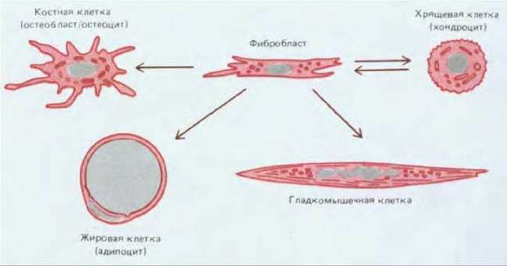
Fig. 17-40. Interconversions apparently occurring within the connective-tissue cell family. For simplicity, fibroblasts are represented as a single cell type, whereas in reality many types of fibroblasts exist; their number, however, remains undetermined, as do their differentiation potentials.
17.7.1. Fibroblasts alter their properties in response to signals from the extracellular matrix [37, 38]
Fibroblasts appear to be the least specialized cells in the connective-tissue family. Within Connective Tissue, they are scattered throughout the body and secrete a soft extracellular matrix rich in type I and/or type III collagen, as described in Chapter 14 (Section 14.6.2). In the event of tissue injury, nearby fibroblasts migrate into the wound, proliferate there, and produce large amounts of collagenous matrix, which helps isolate and repair the damaged tissue. The ability of these cells to survive and function under the unusual conditions of a wound, coupled with their "solitary lifestyle," makes them very easy to culture, rendering them a favored model system for cell biologists (Fig. 17-41).
As shown in Fig. 17-40, fibroblasts appear to be the most plastic of the connective-tissue cells: they exhibit a remarkable ability to differentiate into other members of the same family. However, before delving into details, we must issue a word of caution against hasty Conclusions. There is strong evidence that fibroblasts in different PARTS OF THE body are intrinsically diverse (see the Appendix at the end of this chapter); it is not even certain that all fibroblasts within a single region are equivalent. The simplest assumption is that they are identical, since it has not been proven otherwise. However, it is quite possible that connective tissue contains a mixture of distinct fibroblast lineages, some capable of differentiating into chondrocytes, others into fat cells, and so on; the absence of visible differences does not necessarily mean that only a single cell type with multiple developmental potentials exists. It is also possible that "mature" fibroblasts incapable of transformation exist side by side with "immature" ones (often called mesenchymal cells), which can give rise to various types of mature cells.
Despite these uncertainties, however, in vivo and in vitro studies have clearly demonstrated that The properties of connective-tissue cells can change radically. For example, if a preparation of bone matrix—obtained by grinding bone into a fine powder and eluting the solid mineral component—is implanted into the dermal layer of the Skin, certain cellular elements there (possibly dermal fibroblasts) transform into cartilage cells, and somewhat later other cells transform into bone cells. This results in The formation of a small piece of bone, complete with a Bone Marrow cavity. These findings suggest that extracellular matrix components can profoundly influence the differentiation of connective-tissue cells. As we shall see further on, similar cell transformations play an important role in the healing of bone fractures. It has even been shown that bone matrix contains high concentrations of several growth factors that affect The behavior of connective-tissue cells, such as TGF-ß (transforming growth factor-ß—see Section 13.3.4, Table 13-1), which has been found to induce cartilage differentiation in vitro.
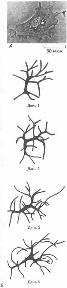
Fig. 17-41. A. Phase-contrast micrograph of a mouse fibroblast in culture. B. Images of a living fibroblast-like cell in the transparent tail of a tadpole, showing changes in its shape and position over several days. Note that although fibroblasts flatten out in culture, in Tissues they may adopt a more complex configuration with numerous processes. (A — courtesy of Guenter Albrecht-Buehler; B — from E. Clark, Am. J. Anat., 13, 351–379, 1912.)
17.7.2. The extracellular matrix can influence the differentiation of connective-tissue cells by altering their shape and attachment [39]
The extracellular matrix can influence the differentiated state of connective-tissue cells both physically and chemically. This has been demonstrated in studies on cartilage cells (chondrocytes) grown in culture. Under appropriate conditions, these cells multiply and maintain their differentiated status, synthesizing over many cell generations large amounts of the highly characteristic cartilage matrix with which they surround themselves. However, if the culture is maintained at a relatively low cell density as a monolayer on a culture dish, a transformation occurs: the cells lose the rounded shape typical of chondrocytes, flatten out on the substrate, and stop producing cartilage matrix. Specifically, they cease the synthesis of type II collagen, characteristic of cartilage, and instead begin to produce type I collagen, characteristic of fibroblasts. Within a month in such culture, almost all cartilage cells switch their collagen Gene Expression and acquire a fibroblast-like appearance. The biochemical changes within The Cell must occur quite rapidly, since very few cells show simultaneous synthesis of both types of collagen.
Various experimental data indicate that these biochemical changes are induced, at least in part, by alterations in cell shape and attachment. For example, chondrocytes that have undergone transformation into fibroblast-like cells can be gently detached from the culture dish surface and transferred to an agarose dish. By forming a gel, agarose keeps the cells in suspension without attachment to a substrate, which forces them to assume a rounded shape. Under these conditions, the cells soon regain their chondrocyte properties and begin to synthesize type II collagen. How cell shape and attachment might influence gene expression is discussed in Section 13.3.6.
For most cells, especially those of connective tissue, attachment opportunities depend on the surrounding matrix, which is normally produced by the cells themselves. Thus, a cell creates its own microenvironment, which in turn acts back on the cell to reinforce its differentiated state. Moreover, the extracellular matrix produced by a cell partially creates the microenvironment for neighboring cells as well, promoting their differentiation along the same pathway. One can observe, for example, how in a developing organism or in a culture dish a group of chondrocytes expands to form a cartilage nodule As a result of adjacent fibroblasts converting into chondrocytes.
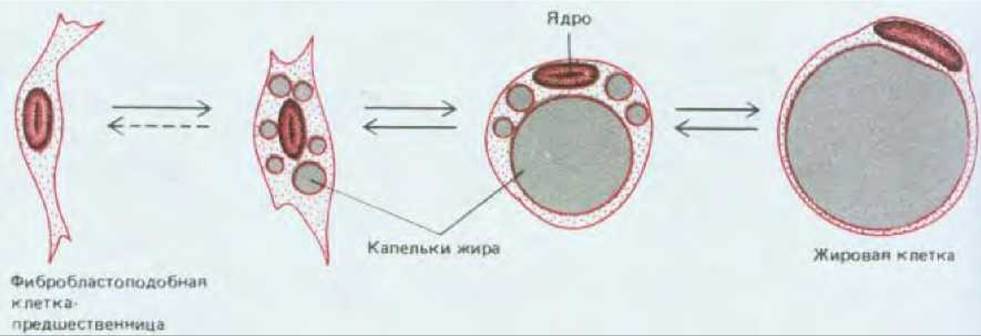
Fig. 17-42. Transformation of a fibroblast-like progenitor cell into a mature fat cell through the accumulation and fusion of lipid droplets. As indicated by the arrows, this process is at least partially reversible. Cells at early or intermediate stages can divide, but a mature fat cell is no longer capable of division.
17.7.3. Various signaling molecules, acting sequentially, regulate the formation of fat cells [40]
It is believed that fat cells, or adipocytes, in mammals also develop from fibroblast-like cells, both during normal development and in various pathologies, such as muscular dystrophy, where muscle cells die and are gradually replaced by fatty connective tissue. The differentiation of a fat cell begins with the Synthesis of specific Enzymes, followed by the accumulation of lipid droplets that subsequently fuse and increase in volume, stretching the cell so severely that only a thin rim of Cytoplasm remains around the fat mass (Fig. 17-42).
The factors influencing this process can be studied in cell culture using established cell lines, such as certain strains of mouse 3T3 cells. It was initially discovered that The Development of fat cells in culture requires the presence of fetal bovine serum, a standard component of culture media. The principal factor in serum that triggers fat Cell Differentiation was later identified as Growth Hormone, a protein normally secreted into the bloodstream by the Pituitary Gland. Evidence indicates that this hormone stimulates the differentiation not only of fat cells but also of chondrocytes, exerting this effect both in vitro and in vivo. However, growth hormone is not the only secreted signaling molecule that regulates fat cell development. Fat cell precursors stimulated by growth hormone become responsive to IGF-1 (Insulin-like growth factor 1), which prompts the differentiating fat cells to proliferate.
Fat cell differentiation, much like chondrocyte differentiation, is influenced by factors that alter cell shape and attachment. For example, the differentiation of 3T3 cells into fat cells is suppressed if they are allowed to spread out On the surface of a culture dish coated with Fibronectin, to which they tightly adhere. However, this suppression can be overcome using cytochalasin, which disrupts Actin filaments and thereby causes the cells to round up.
All these experiments on connective-tissue cells illustrate a general principle: differentiation is regulated by the combined action of soluble signaling molecules and contacts with the extracellular matrix. The Effect of each factor depends on the properties of the responding cell, which in turn depend on the cell's developmental history.
The connective-tissue cell family includes, alongside fibroblasts, cartilage, bone, fat, and smooth muscle cells. Fibroblasts appear capable of transforming into all other cell types of this family, and in some cases this interconversion is reversible. It remains unclear whether this property belongs to a single type of pluripotent fibroblast or represents a mixture of distinct fibroblast populations with narrower potentials. The interconversions of connective-tissue cells are regulated by the COMPOSITION OF THE surrounding extracellular matrix, cell shape, Hormones, and growth factors.
Last update: 12/08/2026
Editorial and Educational Adaptation: This material has been compiled based on the primary/original source text. The project team performed an editorial review, corrected technical inaccuracies, structured sections, and adapted the content for an educational format.
What was processed:
- elimination of formatting defects (OCR errors, structural breaks, corrupted characters);
- editorial organization of content;
- standardization of terminology in accordance with academic sources;
- verification of factual statements against the original source text.
All mentions of the author, publication year, and origin of the primary text have been preserved in accordance with the source.