BIOTECHNOLOGY - V. H. Gerasymenko - 2006
Part I. General Biotechnology
CHAPTER 4. CELLULAR ENGINEERING
4.3. NUCLEAR TRANSPLANTATION BIOTECHNOLOGY
Most studies carried out in recent years on the somatic Cell Hybridization of eukaryotic organisms (primarily mammals) in cell culture have convincingly demonstrated that in a hybrid cell, which possesses a mixed genome due to the heterogeneous Nature of the parental Cells, stable changes occur in the Gene Expression of the synkaryon compared to the expression process in the original parental cells. For example, upon fusing rat hepatoma cells that synthesize and excrete albumin with mouse fibroblasts that do not secrete this protein, hybrid cells were obtained, some clones of which synthesized albumin of only the rat, only the mouse, or albumins of both original parents (rat and mouse).
Based on the results of this and other similar experiments on somatic cell hybridization, the hypothesis was put forward that mammalian cells contain substances capable of directly or indirectly influencing the Cell Nucleus and altering its functional state via positive or negative Regulation of the expression of a specific subset of genes. However, a complex of objective causes inherent to the somatic cell hybridization method (genome heterogeneity, most often with a significant chromosome deficit as well as chromosomal rearrangements that have occurred, all situated within a heterogeneous Cytoplasm) has made it impossible to identify the substances involved in genome expression, or to determine their chemical Structure and MECHANISM OF ACTION. Until now, scientists have only had data indicating that substances contained in animal cytoplasm are involved in regulating the gene expression of The Cell nucleus.
It turned out that in nuclei previously isolated from a differentiated cell and placed into the cytoplasm of an egg cell, processes of cellular differentiation were initiated, and in many cases, a normal Organism developed; that is, the nuclei of specialized cells contained the full scope of information necessary for The Development of a complete organism. Thus, fusing nuclei isolated from tadpole intestinal epithelial cells with an enucleated egg cell in many cases led to the development of a frog. These experiments, performed on amphibians, showed that the cytoplasm regulates nuclear activity; in specialized cells, many nuclear Functions are inhibited by cytoplasmic components. The inhibitory influence coming to The Nucleus from the cytoplasm of the differentiated cell is lifted once it finds itself in a chemically suitable cytoplasmic environment (the egg cell), and processes determining cellular differentiation can resume in the nuclei (Gurdon J.B., 1977). Nuclear transplantation experiments were conducted on amphibians. In the development of mammalian cell nuclear transfer Methods, The Use of cytochalasins—substances synthesized by Fungi—played an essential role.
Class="center">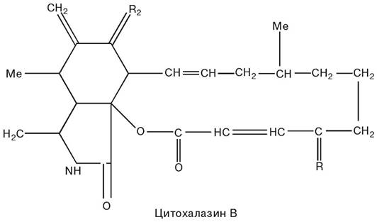
It is suggested that cytochalasin B, by disrupting The structure of microfilaments, promotes a unique positioning of the nucleus wherein it remains connected to the cell by a thin stalk of cytoplasm. Upon centrifugation, the stalk's "umbilical cord" ruptures in most such cells, forming enucleated cells (cytoplasts); the nuclei separated during centrifugation (karyoplasts, or minicells) are surrounded by a thin layer of cytoplasm and Plasma Membrane.
The efficiency of enucleation was monitored by staining (e.g., using the Giemsa method) a cell monolayer located On the surface of one of the dishes or disks.
The next operation aimed at obtaining a cytoplast population is their Separation from the remaining whole cells, because in some cases (the mouse hepatoma cell line Hepa-2), enucleation efficiency does not exceed 50 %. For this purpose, enucleated cells (cytoplasts) and whole cells located on The surface of plastic dishes and Glass disks were detached and treated with Trypsin, resulting in 1 mL of phosphate-buffered saline containing 106 whole cells and cytoplasts. The cell-cytoplast mixture was layered onto 14 mL of a linear 76 % Renografin gradient, the volume concentration of which varied from 15 to 30 %, and centrifuged at 1000 g and a Temperature of 25 oС for 5 min. Clearly demarcated layers of whole cells and cytoplasts were removed from the centrifuge tube and used as intended.
The cytoplasts obtained in the Renografin-76 density gradient were not stained by trypan blue, which served as a control for separation efficiency. They retained The ability to reattach to the surface of the culture dish and could subsequently be used to reconstruct viable cells by fusion with heterologous karyoplasts.
The enucleation methods applied to obtain karyoplasts differ from the Procedures that ensure success in obtaining cytoplasts. Cells intended for karyoplast isolation are seeded 2 days before enucleation onto plastic strips cut from culture flasks, which, prior to placement into a 50 mL centrifuge tube filled with standard nutrient medium, are folded so that their surfaces with attached cells are on the outside. Mouse fibroblast lines A9 were centrifuged at 9.5 thousand g and 35 oС for 15 min. The purpose of this Procedure was to reduce the number of whole cells in the resulting karyoplast pellet. After separating cells weakly attached to the substrate, the remaining strips with monolayer cells were transferred into tubes where the concentration of cytochalasin B reached 10 µg per 1 mL of standard nutrient medium. The tube contents were incubated at 37 oС for 15 min, followed by centrifugation at 35 oС and 7 thousand rpm for 45 min. The resulting supernatant was decanted, and the pellet, consisting mainly of karyoplasts, was resuspended in standard nutrient medium.
Preparations of karyoplasts produced According to the described method contain cytoplasmic fragments, dead karyoplasts, and whole cells. Separation of cytoplasmic fragments is carried out by sedimentation in a 1-6% Ficoll gradient at 1 g and 37 oС for 90 min in a humidified atmosphere containing 5 % СО2. The cytoplasmic fragments located in the top layer are aspirated and discarded, while the karyoplast pellet formed after extraction and dilution with nutrient medium is sedimented again by centrifugation and resuspended in fresh nutrient medium. Whole cells (0.4-4 %) are removed by double 90-minute incubation of the karyoplast suspension in culture dishes.
Incubation for 3 hours makes it possible to sharply reduce the contamination of karyoplasts with whole cells, which is mandatory for all nuclear transplantation experiments.
The next operation is the separation of viable karyoplasts from dead ones. This involves resuspending the pellet in nutrient medium to a concentration of 107 minicells per 1 mL, layering the suspension onto Ficoll-Paque (a solution containing 5 % Ficoll and 9 % sodium diatrizoate) in centrifuge tubes, and carefully adding 1 mL of medium on top so that the added medium and the karyoplast suspension form two distinct layers. Subsequent centrifugation at 800 rpm (130 g) for 75 min at room temperature yields a pellet consisting of 99 % dead karyoplasts; at the interface between the nutrient medium and Ficoll-Paque, There is a layer consisting of 98 % viable karyoplasts.
Other methods are also used to purify karyoplasts, in particular tantalum particles 1-3 µm in size. By the time of enucleation, practically all cells contain more than 12 tantalum particles each. Due to the fact that the density of tantalum exceeds that of the cell by more than 15 times, components containing tantalum particles sediment significantly faster than karyoplasts lacking tantalum. Purification of karyoplasts is sometimes performed using a flow cytometer-sorter.
Properties of cytoplasts and karyoplasts. The ability of cytoplasts to synthesize Proteins, support the Replication of vesicular stomatitis virus and poliovirus, carry out rabies virus-controlled RNA and Protein Synthesis, and release SV40 virus from transformed cells has been established. Cytoplasts contain all types of Organelles characteristic of a normal cell, retain the ability characteristic of whole cells to attach to a substrate and form a folded membrane, move, and perform pinocytosis.
Surrounding the karyoplast is a layer accounting for about 10 % of the cellular cytoplasm, containing components of The Endoplasmic reticulum, a certain number of Mitochondria, and Ribosomes; centrioles are absent in karyoplasts, unlike cytoplasts.
About 10 % of karyoplasts from certain cell lines are able to restore the entire volume of cytoplasm lost during enucleation and transform back into viable cells.
The ability of karyoplasts to regenerate the cytoplasm lost during enucleation and form viable cell colonies depends on The amount of cytoplasm surrounding the nucleus. In the karyoplast fraction where 2-4 % of the amount of cytoplasm contained in an intact cell is concentrated around the nuclei (small karyoplasts), only one in 106 purified elements can form a viable cell colony.
Nuclear transplantation and cell reconstruction.
After cell enucleation, the cytoplast monolayer in 60 mm plastic dishes or glass disks of
14 mm is incubated in nutrient medium at 37 oС for 1-2 hours, and then for 20 min and cooled to 4 oС. At this temperature, the monolayer is washed twice with Earle's solution (pH 8.0), then treated for 20 min with 0.5 mL of chilled Earle's solution containing Sendai virus inactivated by 5 min of irradiation (with an ultraviolet lamp at a distance of 15 cm), after which the cytoplast layer is washed twice more with the same solution, which is then removed.
Karyoplasts suspended in phosphate-buffered saline are added to the cytoplast monolayer culture such that there are 100 karyoplasts per cytoplast. To adsorb karyoplasts onto the virus-coated cytoplasts, incubation is carried out at 4 oС for 45 min; the dishes are gently rocked every 3-5 min. For the fusion of karyoplasts and cytoplasts, the dishes or glass disks are transferred to a thermostat and kept there at 37 oС for 45 min. Upon completion, the monolayer in the dishes is intensively washed several times with Earle's solution or serum-free nutrient medium to remove unfused karyoplasts. Then, nutrient medium suitable for cultivating the type of cells used as karyoplast Donors is poured into the dishes. Cytoplasts that have not fused with nuclei die and detach from the dish surface in approximately 2 days.
To obtain cytoplast-uncontaminated cultures, centrifugation of hybrid cells in a renografin density gradient is recommended.
The use of polyethylene glycol to stimulate the fusion of karyoplasts and cytoplasts during cell hybrid construction is limited due to its toxicity, which significantly exceeds that of Sendai virus.
Mutant cell lines are used to identify hybrid cells, determine the efficiency of nuclear transplantation, and detect the presence of parental cells in a hybrid population.
The efficiency of cell reconstruction via karyoplast and cytoplast fusion depends on numerous factors. For instance, only about 10% of cytoplasts obtained by enucleation of rat HTC line cells fused with karyoplasts isolated from mouse A9 line cells. When hybridization was performed using cytoplasts derived from chick embryo fibroblasts and karyoplasts represented by resting avian erythrocyte nuclei, the reconstruction efficiency exceeded 90%. The high fusion efficiency of isolated, resting avian erythrocyte nuclei with enucleated cytoplasts, combined with experience in activating avian (specifically chicken) erythrocyte nuclei by fusing them with HeLa or other metabolically active cells, has enabled the investigation of nuclear-cytoplasmic interactions in the gene expression of hybrid Eukaryotic cells. It turned out that not all resulting hybrids were viable. Approximately 9% of reconstructed cells can grow and divide (Hightower M., Lucas J., 1985). Morphological changes begin in hybrid cells immediately after fusion. Within a few days, the reconstructed hybrid cells are nearly indistinguishable from the original parental cells used as nuclear donors.
Transplantation of isolated interphase nuclei enclosed in a lipid envelope into somatic cells.
Literature data exist on the Introduction of molecules, including DNA, into cells using erythrocyte "ghosts" (Reyes-Terkelsen M., 1985; *Note: keeping author names natural or standard*) and Liposomes (Straubinger R., Papahadjopoulos D., 1985). Specifically, O.L. Serov (1985) in the Laboratory of Ontogenetic Genetics at the Institute of Cytology and Genetics, Siberian Branch of the USSR Academy of Sciences, conducted experimental work on encapsulating isolated interphase nuclei within Artificial Membranes to protect genetic material from degradation and induce the transformation of isolated nuclei into recipient cells. According to Serov, interphase nuclei isolated from a mink fibroblast culture were suspended in a multicomponent buffer solution containing various concentrations of KCl, NaCl, Tris-HCl, EDTA, sucrose, and spermidine.
To create an artificial membrane around the nuclei, phosphatidylethanolamine at a concentration of 10–20 mg/mL and phosphatidylcholine extracted from chicken egg yolks at a concentration of 150–200 mg/mL of organic solvent were used. The method proposed by O.L. Serov is based on The formation of a phospholipid membrane as isolated nuclei pass through a three-layer system located in a centrifuge tube. This system consists of a bottom layer of 1M sucrose solution, an upper layer of 0.25 M aqueous sucrose solution, and an intermediate layer of organic solvent positioned between the upper and lower sucrose layers in the centrifuge tube, consisting of chloroform, dioxane, and ethyl acetate in a 0.25:0.23:0.51 ratio, along with phosphatidylcholine at a concentration of 150–200 mg/mL of the specified organic solvent.
At the boundary between the three-component organic solvent and the upper sucrose layer, phosphatidylcholine molecules are oriented with their hydrophilic ends toward the aqueous layer; at the interface between the organic solvent and the lower sucrose layer, the phospholipid molecules are similarly oriented with their polar heads
toward the aqueous phase of the denser lower sucrose layer. During centrifugation, as the nuclei pass from the aqueous phase (0.25 M sucrose) into the organic solvent layer, they encounter the polar hydrophilic heads of the phosphatidylcholine molecules at the phase boundary, which form a monolayer lipid membrane on the surface of the isolated interphase nuclei. At the boundary between the organic layer and the denser 1M sucrose layer, the nuclei—already possessing a monolayer lipid membrane on their surface with hydrophobic ends oriented outward—encounter a layer of phosphatidylcholine molecules whose hydrophobic ends are directed toward the organic phase and interact with the hydrophobic ends of the nuclear lipid monolayer, thereby forming a second membrane layer on the surface of the interphase nuclei.
The formed lipid bilayer is firmly retained on the surface of the interphase nuclei and is not washed away during repeated re-sedimentation via centrifugation through a 1M sucrose solution. Using radioautography and Fluorescence Microscopy, it was demonstrated that The Lipid Bilayer is evenly distributed across the nuclear surface, forming an additional membrane or membranes (under these centrifugation conditions, free liposomes cannot enter the nuclear fraction).
Additional experiments demonstrated that the artificially created membrane is imperfect (O.L. Serov). This Conclusion was drawn based on the observed leakage of ATP from nuclei surrounded by a phosphatidylcholine membrane and the penetration of exogenous DNase into these nuclei. Simultaneously, it was established that in both cases, the formed membrane blocked the translocation of both macromolecules (DNase) and relatively small ordinary molecules (ATP). Meanwhile, while natural nuclear membranes are permeable even to polymer molecules, the artificial membrane constructed from phosphatidylcholine is completely impermeable to both large polar molecules and macromolecular structures.
As a result of the experiments conducted, it was suggested that exogenous phosphatidylcholine forms a Membrane Structure around the nucleus, enclosing it within a liposome. This additional membrane imposes certain restrictions on The transport of certain
molecules into and out of the nucleus, though these processes are not completely halted. Upon introducing interphase mink nuclei surrounded by an artificial membrane into a culture of mouse LMTK- cells—which are deficient in the enzyme thymidine kinase (TK)—transfer of relatively large DNA fragments from the mink nuclei into the recipient mouse cells was established, with the majority of studied transformants exhibiting a stable TK+ phenotype.
The proposed method for transferring genetic material using liposome-enclosed interphase nuclei, owing to its high transformation frequency compared to gene transfer via total cellular DNA, indicates a potential protective function of donor Nuclear Proteins shielding DNA from degradation, as well as the involvement of these proteins in the integration of donor and recipient cell Chromatin.
Biotechnology of cybrid production. Cybrids are products formed by the fusion of whole cells from one mutant line with the cytoplasm of enucleated cells from another cell line. To obtain cybrids, cell enucleation was performed using cytochalasin B (adjusted to a concentration of 5 µg/mL of medium), with cells having been pre-labeled with green fluorescent granules added to the nutrient culture medium.
Iongkind J. and Verkerk A. (1985) labeled human fibroblasts with fluorescent Dyes by incubating proliferating cell cultures for 2 days in Ham's F10 medium containing 2×10⁷ dye granules per mL, 100 µg of streptomycin, and 100 units of penicillin, supplemented with 10% fetal bovine serum. During incubation, the cultured cells phagocytosed the granules; granules attached to the outer surface of The cell membrane were washed off with saline solution. As research results showed, labeling with fluorescent granules slows down cell growth.
The enucleated cell suspension (cytoplasts) is also stained with Hoechst fluorescent bisimidazole dyes; fluorescent cytoplasts are isolated by fluorescence-activated cell sorting (FACS). The purity of the cytoplast fraction reached 99.7%, and 90% of the cytoplasts obtained after sorting retained the ability to incorporate 3H-leucine.
Cytoplasts labeled by either method are fused with whole cells that have also been pre-labeled with a fluorescent dye differing in color from the one used to mark the enucleated cells (cytoplasts); The ratio of cytoplasts to whole cells in cybrid production is 3:1. Polyethylene glycol (PEG 1000), dimethyl sulfoxide, or inactivated Sendai virus is used as the agent inducing the fusion of cytoplasts and whole cells. Fusion products (cybrids) exhibit dual-color fluorescence. They are isolated using a FACS II flow cytometer equipped with an argon laser at a wavelength of 488 nm and a constant output of 100 mW. The sorter tubing used for sample delivery is sterilized with 70% ethanol, and the sheath fluid for sterilization is passed through bacterial filters (Millipore, 0.22 µm pores). If the cell suspension contains numerous clumps after the fusion of cytoplasts and whole cells, they are dispersed by passing through the nozzle (70 µm diameter) of the cell sorter.
The yield of the cybrid fraction following sorting is determined by fusing control cytoplasts possessing hypoxanthine-guanine phosphoribosyltransferase (HGPRT+) activity with fibroblasts obtained from Lesch-Nyhan syndrome patients (HGPRT-). Twenty hours after fusion, cells with dual-color fluorescence (cybrids) are sorted onto coverslips, incubated for 20 hours with 3H-hypoxanthine, fixed, and examined by radioautography. The incorporation of hypoxanthine into the cybrid fraction demonstrates that, using Sendai virus, at least 90% of the sorted cells can be classified as true cybrids (HGPRT+).
To determine enzyme activities and substrate concentrations in extremely small sample volumes (5–10 thousand cybrids), micromethods based on fluorimetry were developed, allowing the detection of microgram quantities of substances in minute amounts of lyophilized cells. Ultramicromethods based on microfluorimetry make it possible to measure enzyme activities even in single cells. These techniques have made it possible to study enzyme complementation in cybrids and other cells, as well as to clarify the roles of the nucleus and cytoplasm in the complementation mechanism.
Last update: 11/08/2026
Editorial and Educational Adaptation: This material has been compiled based on the primary/original source text. The project team performed an editorial review, corrected technical inaccuracies, structured sections, and adapted the content for an educational format.
What was processed:
- elimination of formatting defects (OCR errors, structural breaks, corrupted characters);
- editorial organization of content;
- standardization of terminology in accordance with academic sources;
- verification of factual statements against the original source text.
All mentions of the author, publication year, and origin of the primary text have been preserved in accordance with the source.