Review of Medical Physiology - William F. Ganong 2002
Functions of the Nervous System
Reflexes
Monosynaptic reflexes: the stretch reflex
A stretched Skeletal Muscle with intact innervation undergoes contraction. This response is known as the stretch (or myotatic) reflex. The stimulus that triggers this reflex is muscle stretching, and the response is the contraction of the stretched muscle. The sensory receptor performing this function is the muscle spindle. Impulses originating in the muscle spindle travel via fast sensory fibers directly to the Central Nervous system (CNS) and to the motor Neurons innervating that muscle. The neurotransmitter at central synapses is glutamate. Stretch Reflexes are the body's most widely known and best-studied monosynaptic reflexes.
Clinical Examples
Striking the patellar tendon just below the kneecap elicits the knee-jerk reflex, which is a stretch reflex of the quadriceps femoris muscle, since tapping its tendon leads to Muscle contraction. A similar contraction can be observed when stretching the quadriceps manually. Stretch reflexes can also be elicited in most other major skeletal Muscles of the body. Tapping the triceps brachii tendon, for instance, causes elbow extension resulting from the contraction of this muscle; striking the Achilles tendon triggers a stretch reflex driven by the contraction of the gastrocnemius muscle; and a tap on the side of the face elicits a stretch reflex in the masseter muscle. Other stretch reflexes are described in neurology textbooks.
Structure OF THE Muscle Spindle
Each muscle spindle contains approximately 10 muscle fibers enclosed within a Connective Tissue capsule. Structurally, these fibers closely resemble embryonic-type fibers and are less striated than regular muscle fibers. They are referred to as intrafusal fibers, in contrast to extrafusal fibers, which are the standard contractile units of the muscle. Intrafusal fibers run parallel to the other muscle fibers because the ends of the spindle capsule are attached to the tendons at each end of the muscle or to the sides of extrafusal fibers.
Class="center">
Fig. 6-1. Reflex arc. Note that non-propagated graded potentials occur in the receptor and the CNS, which are proportional to the stimulus intensity. The response at the Neuromuscular Junction is also graded, so under normal conditions it is sufficient to trigger a skeletal muscle response. On the other hand, in the impulse-conducting PARTS OF THE reflex arc (afferent and efferent neurons, muscle membrane), Action potentials are generated According to the all-or-none principle.
Mammalian muscle spindles contain Two Types of intrafusal fibers. One fiber type contains numerous nuclei in its expanded central region and is therefore called a nuclear bag fiber (Fig. 6-2). A spindle typically contains two nuclear bag fibers: nuclear bag fiber 1, which has a low Myosin ATPase activity, and nuclear bag fiber 2, which has a high myosin ATPase activity. The other fiber type—nuclear chain fibers—are thinner and shorter, lacking a well-defined nuclear bag. A spindle contains four or more of these fibers, with their ends attached laterally to the nuclear bag fibers. The ends of the intrafusal fibers are contractile, whereas their central regions are presumably non-contractile.
Each spindle contains two types of sensory endings. Primary (annulospiral) endings are the terminations of rapidly conducting group Ia afferent fibers (see Table 2-2). One branch of a Ia fiber innervates nuclear bag fiber 1, while another branch innervates nuclear bag fiber 2 and the nuclear chain fibers. These sensory fibers spiral around the central regions of both nuclear bag and nuclear chain fibers. Secondary (flower-spray) endings are the terminals of group II sensory fibers. They are located near the ends of the intrafusal nuclear chain fibers.
Spindles possess their own motor innervation. These nerves, 3–6 µm in diameter, account for approximately 30% of the fibers in the anterior ROOT and belong to the Erlanger and Gasser Aγ group. Because of their size, they are designated as Lexell's γ-efferents, or the small motor nerve system. These fibers project exclusively to the spindles. Additionally, both intra- and extra-fusal fibers are innervated by larger β-motor neurons. Efferent fiber endings fall into two histological types: motor end-plates (plate endings) on nuclear bag fibers and trail endings (diffuse, net-like terminals) located predominantly on nuclear chain fibers.
Spindles contain two types of sensory fibers—dynamic and static (see below)—while γ- and β-motor neurons provide two functional types of responses. Stimulation of the first type enhances the dynamic response (dynamic fusimotor axons), whereas stimulation of the second type enhances the static response at a constant length (static fusimotor axons).
Central Connections of Afferent Fibers
Experiments demonstrate that Ia fibers originating from the primary endings of a muscle terminate directly on the motor neurons that innervate the extrafusal fibers of the same muscle. The time elapsed between the onset of the stimulus and the response is called the reflex time. In humans, the reflex time of a stretch reflex, such as the knee-jerk reflex, is 19–24 ms. Weak stimulation of the sensory nerve emerging from the muscle, which is known to excite exclusively Ia fibers, elicits a contractile response with the same latency. Since the conduction velocities in afferent and efferent neurons are known, and the distance from the muscle to the Spinal Cord is easily measured, one can calculate the fraction of reaction time required for impulse transmission from the muscle to the spinal cord and back. Subtracting this value from the total reaction time yields a remainder, termed the central delay, which corresponds to the time required for reflex activity within the spinal cord. In humans, the central delay of the knee-jerk reflex is 0.6–0.9 ms. Similar values have also been obtained in animal experiments. Since the minimum synaptic delay is 0.5 ms (see Chapter 4), it is evident that the reflex arc of this reflex type involves only a single synaptic contact.
Excitation from muscle spindles that triggers muscle contraction is propagated via polysynaptic pathways and likely originates from secondary endings. Group II fibers, which form monosynaptic contacts with motor neurons, make only a minor contribution to stretch reflexes.
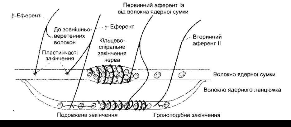
Fig. 6-2. Schematic representation of the Main Components of a mammalian muscle spindle. Each spindle is enclosed by a capsule and contains two nuclear bag fibers and four or more nuclear chain fibers.
Function of Muscle Spindles
When a muscle spindle is stretched, its sensory endings are deformed, generating receptor potentials. This, in turn, induces action potentials in the sensory fibers at a frequency proportional to the degree of stretch. Spindles are arranged in parallel with extrafusal fibers, so when the muscle is passively stretched, the spindles are stretched as well. This results in the contraction of the extrafusal muscle fibers. Conversely, spindle afferents characteristically cease firing if the muscle contracts As a result of electrical stimulation of the nerve fibers supplying the extrafusal fibers, because in that case the muscle shortens while the spindle does not (Fig. 6-3).
Thus, the spindle and its reflex connections function as a feedback mechanism operating in response to an increase in muscle length. When a muscle is stretched, spindle excitation increases, eliciting a reflex muscle contraction. On the other hand, when a muscle contracts without A change in efferent stimulation, spindle excitation decreases, causing the muscle to relax.
When a spindle is stretched, the primary endings of both nuclear bag and nuclear chain fibers are stimulated, although the response pattern differs. Nerves originating from the nuclear bag region mediate a dynamic response—that is, impulse firing occurs at a higher rate while the muscle is actively stretching and decreases during sustained stretch (Fig. 6-4). Nerves originating from the primary endings of nuclear chain fibers produce a static response, meaning they fire at a high frequency throughout the entire duration that the muscle remains stretched. Consequently, primary endings respond to changes in both muscle length and The rate of stretch. This dual response to phase and static phenomena in the muscle is vital, as the rapid phase response helps dampen oscillations caused by conduction delays in the length-regulation feedback loop. Small oscillations in the feedback loop inevitably occur; this physiological tremor has a frequency of approximately 10 Hz. However, this tremor would be much more pronounced were it not for the velocity sensitivity of the spindle.
Effects of Efferent Discharge
Stimulation of the γ-efferent system gives rise to diverse phenomena during extrafusal fiber contraction. It does not directly produce overt muscle shortening because intrafusal fibers lack the bulk and power necessary to drive significant contraction. However, γ-efferents cause the contractile ends of intrafusal fibers to shorten, which stretches the nuclear bag region of the spindle, deforming the annulospiral endings and initiating action potentials in Ia fibers. This, in turn, can lead to reflex muscle contraction. Therefore, a muscle may contract either due to stimulation of α-motor neurons innervating extrafusal fibers or via γ-efferent neurons initiating contraction indirectly through the stretch reflex.
As γ-efferent discharge increases, intrafusal fibers become shorter than extrafusal ones. When the entire muscle is stretched during γ-efferent stimulation, additional action potentials are generated due to the extra stretch placed on the nuclear bag region, further increasing the firing rate of Ia fibers (see Fig. 6-3). Enhanced γ-efferent activity thus heightens spindle sensitivity, and the stretch sensitivity of spindles varies dynamically with the level of γ-efferent input. It is highly probable that γ-efferent discharge increases concurrently with α-motor neuron activation during voluntary movement. Through this co-activation, the spindle shortens alongside the muscle, maintaining its firing throughout the contraction. Consequently, the spindle retains its ability to respond to stretch and reflexively regulate motor neuron excitability continuously during contraction.
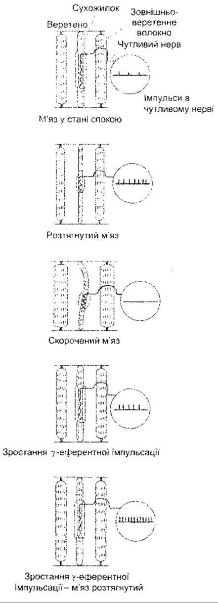
Fig. 6-3. Effect of various conditions on muscle spindle discharge.
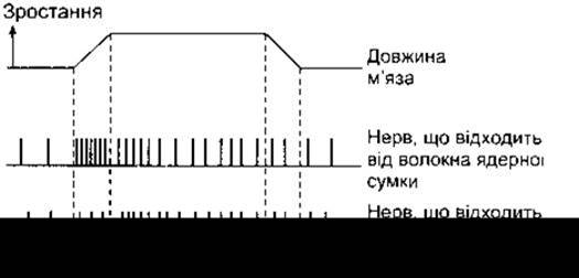
Fig. 6-4. Response of spindle afferents to muscle stretch. The two bottom lines show the discharge frequency in afferent nerve fibers from the primary endings of nuclear bag and nuclear chain fibers when the muscle is stretched and then returned to its initial LENGTH.
Dynamic and static u- and ß-efferents were described previously. Stimulation of dynamic efferents increases the sensitivity of the spindle to the rate of change of muscle length during stretching, whereas static efferents increase spindle sensitivity during steady, sustained stretching. Thus, responses arising in the spindle can be regulated separately in response to phase or static phenomena.
Regulation of u-Efferent Discharge
Motor neurons of the u-efferent system are regulated by descending pathways originating from various Brain regions. These pathways can modulate the sensitivity of muscle spindles and, consequently, the threshold of stretch reflexes across many parts of the body, which is essential for posture (see Chapter 12).
Other factors also influence u-efferent discharge. Anxiety leads to increased firing rates, a fact that likely explains the hyperactive tendon reflexes sometimes observed in anxious patients. Furthermore, unexpected movements are also associated with enhanced efferent discharge. Cutaneous stimulation, particularly by noxious stimuli, leads to increased efferent discharge in the spindles of ipsilateral flexor muscles and decreased discharge in extensor muscle spindles, with opposite effects observed in the contralateral limb. It is well known that attempting to pull the hands apart while the fingers of both hands are interlocked enhances the knee-jerk reflex (Jendrassik maneuver), which may also be mediated by increased u-efferent discharge initiated by afferent impulses originating from the hands.
Reciprocal Innervation
During the stretch reflex, muscles functionally antagonistic to the reflexively contracting muscles—that is, their antagonists—relax. This phenomenon is brought about by reciprocal innervation. Impulses arriving via Ia fibers from the muscle spindles of agonist muscles produce postsynaptic inhibition in the motor neurons supplying the antagonists. The pathway mediating this effect involves two synaptic contacts. A collateral branch of each Ia fiber projects to a spinal cord inhibitory interneuron (the Golgi tendon Cell), which forms a direct synaptic contact with one of the motor neurons innervating the antagonist muscle. An example of this type of postsynaptic inhibition was discussed in Chapter 4, and its pathway is illustrated in Fig. 4-9.
Inverse Stretch Reflex
The greater the degree of strong muscle stretch, the stronger the reflexive contraction. However, when the tension becomes sufficiently high, the contraction suddenly ceases and the muscle relaxes. This relaxation in response to strong stretch is called the inverse stretch reflex, or autogenic inhibition.
The receptor for the inverse stretch reflex is the Golgi tendon organ (Fig. 6-5), a reticulated cluster of encapsulated nerve endings located among the bundles of tendon fibers. A single tendon organ is supplied by 3 to 25 muscle fibers. Nerve fibers emerging from the Golgi tendon organ are myelinated, rapidly conducting sensory fibers of group Ib (see Table 2-2). Stimulation of these fibers produces IPSPs in the motor neurons supplying the muscles from which these sensory fibers originate. Ib fibers terminate in the spinal cord on interneurons, which in turn synapse directly on motor neurons (Fig. 6-6). These fibers also form excitatory contacts with motor neurons innervating the muscle antagonists.
Unlike muscle spindles, which monitor the stretch of a series of muscle fibers, Golgi tendon Organs register the tension of a muscle group; thus, their stimulation results from both passive stretch and active muscle contraction. The excitation threshold of Golgi tendon organs is low. The level of stimulation via passive stretch is minor because most of the elastic muscle fibers absorb the stretching force, meaning that strong stretch leads to relaxation rather than contraction. However, muscle contraction generates continuous impulses, allowing the Golgi tendon organ to function as a transducer in a feedback loop that regulates muscle tension, much like the spindle feedback loop regulates muscle length.
The Importance of primary spindle endings and Golgi tendon organs in regulating the velocity of muscle contraction, muscle length, and muscle force is demonstrated by the fact that sectioning the afferent nerves supplying the arm causes the upper limb to hang limply, resembling hemiparesis. The structure of this described system is shown in Fig. 6-7, and the interaction of spindle discharge, Golgi tendon organ discharge, and reciprocal innervation in determining motor neuron firing rates is illustrated in Fig. 6-8.
Muscle Tone
The resistance of a muscle to stretch is often described as its tone. If the motor nerve to a muscle is cut, the muscle exhibits very little resistance and is said to be flaccid. A hypertonic (spastic) muscle is one in which resistance to stretch is high due to hyperactive stretch reflexes. Somewhere between the relaxed and elastic states lies the poorly defined condition of normal tone. Muscle hypotonicity occurs primarily when u-efferent discharge is low, whereas hypertonicity occurs under the opposite conditions.
Lengthening Reaction
Under conditions of muscle hypertonicity, a distinct sequence is observed: moderate stretch → muscle contraction, strong stretch → muscle relaxation. During passive elbow flexion, for example, an immediate resistance occurs due to the stretch reflex of the triceps brachii muscle. Further stretching activates the inverse stretch reflex. Resistance to flexion suddenly disappears, and the elbow flexion proceeds. Continuing the passive flexion stretches the muscle again, and the sequence repeats. This sequence of resistance as the limb is moved passively is known clinically as the clasp-knife phenomenon. The physiological term for this phenomenon is the lengthening reaction, as it represents the response of a spastic muscle (in the previous example, the triceps brachii) to lengthening.
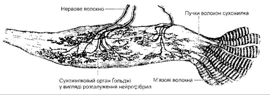
Fig. 6-5. Golgi tendon organ (reproduced with permission from Goss CM [editor] — Gray’s Anatomy of the Human Body, 29th ed. Lea & Febiger, 1973).

Fig. 6-6. Schematic diagram of the pathways mediating the stretch reflex and the inverse stretch reflex. Muscle stretch stimulates the muscle spindle, and impulses propagating along Ia fibers cause excitation in motor neurons. Stretch also stimulates the Golgi tendon organ, and impulses propagating along Ib fibers cause an interneuron to release an inhibitory transmitter, Glycine. Under conditions of strong stretch, the resulting hyperpolarization in the motor neurons is so substantial that firing ceases.
Clonus
Another hallmark of states accompanied by elevated u-efferent discharge is clonus. This neurological sign consists of regular, rhythmic contractions of a muscle subjected to sudden and sustained stretch. A typical example is ankle clonus. It is initiated by a sharp and sustained dorsiflexion of the FOOT, and the response is rhythmic flexion at the ankle joint. The underlying mechanism may be the sequence described above: stretch reflex followed by inverse stretch reflex. However, the response can also occur through synchronized excitations in motor neurons without the participation of impulses from the Golgi tendon organ. The muscle spindles of the tested muscle are hyperactive, and the sudden burst of impulses propagating from them immediately triggers excitation in all motor neurons innervating the muscle. The resulting muscle contraction abruptly halts spindle discharge. Nevertheless, if the stretch is maintained, spindle excitation resumes in the relaxed muscle during renewed stretching.
Last update: 10/08/2026
Editorial and Educational Adaptation: This material has been compiled based on the primary/original source text. The project team performed an editorial review, corrected technical inaccuracies, structured sections, and adapted the content for an educational format.
What was processed:
- elimination of formatting defects (OCR errors, structural breaks, corrupted characters);
- editorial organization of content;
- standardization of terminology in accordance with academic sources;
- verification of factual statements against the original source text.
All mentions of the author, publication year, and origin of the primary text have been preserved in accordance with the source.