Human Anatomy (with Fundamentals of Dynamic and Sports Morphology) - Ivanitsky M. F. 2008
The Study of Internal Secretion Organs
General Part
A characteristic feature of endocrine Organs, or Endocrine glands, is that they lack excretory ducts and release their products, Hormones, directly into the Lymph and Blood (Fig. 141, 142).
Hormones refer to those products of endocrine glands which, upon entering the blood in relatively small quantities, act through it on specific organs and systems, stimulating or, conversely, inhibiting their activity. Some hormones influence the growth of the Organism, the development and formation of individual organs and Organ Systems, others regulate metabolic processes, and still others affect nerve endings or specific structures of the Central Nervous system. Thus, There is a complex relationship between the endocrine organs and The Nervous System. At the same time, The regulation of vital activity in organs is possible not only through neural, but also through humoral pathways (from Latin humor — fluid), i.e., through the effect on organs via the fluid Tissues of the body, particularly the blood. Therefore, we can speak of a unified Neurohumoral regulation of the organism's vital activity and its individual structures.
Class="center">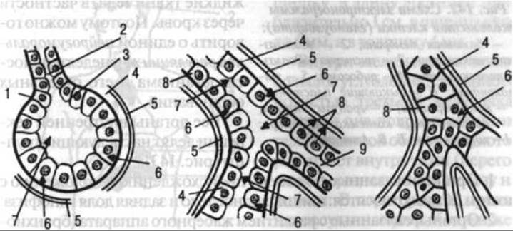
Fig. 141. Diagram of secretion direction in Cells of various glands:
A — gland cells with apically directed secretion (exocrine gland); B — gland cells with bidirectional secretion (Liver trabeculae, the hepatocytes of which secrete Bile in the apical direction and simultaneously release urea and certain other substances into the blood); C — gland cells with basally directed secretion (endocrine gland).
1 — terminal secretory part of the gland; 2 — excretory duct; 3 — junctional plates; 4 — basement membrane of the terminal secretory part of the gland; 5 — blood capillaries; 6 — entry of initial substances into the glandular Cell from the blood; 7 — release of ready secretory substances beyond the lumen of the terminal part (external, or apically directed, secretion; 8 — release of incretions (hormones) into the bloodstream (internal, or basally directed, secretion); 9 — movement of secretion along the excretory duct (after B. V. Aleshin)
All endocrine organs are divided into the following groups (Fig. 143):
1. Organs associated in their origin and Location with the Brain. These include the pineal body and the posterior lobe of the Pituitary Gland.
2. Organs associated with The Development of the gill apparatus (branchiogenic, from Greek branchia — gills). These include the anterior lobe of the pituitary gland, the thyroid, parathyroid, and Thymus glands (discussed in the section "Organs of Immunogenesis", p. 278) (Fig. 144).
3. An organ associated with the Development of the Digestive apparatus. This is the endocrine (internal secretion) part of the Pancreas.
4. Organs developing from the coelomic epithelium (secondary body cavity). These include the adrenal cortex and the epithelium of the Gonads.
5. Organs having Common sources of development with the sympathetic nervous system, i.e., formed As a result of the migration of certain groups of cells from the neural tube. These include The adrenal medulla and Paraganglia (aortic, carotid, etc.). Due to their selective staining with chromium salts (dark brown color), these organs are termed chromaffin. Thus, all three germ layers participate in the development of endocrine organs: the pineal body, the posterior lobe of the pituitary gland, and the adrenal medulla develop from the ectoderm; the adrenal cortex and gonads develop from the mesoderm; and the anterior lobe of the pituitary gland, Thyroid Gland, Parathyroid glands, Thymus gland, and pancreatic islets (endocrine part) develop from the endoderm.

Fig. 142. Diagram of an electron micrograph of a glandular cell (glandulocyte): 1 — basement membrane; 2 — Endoplasmic reticulum; 3 — cistern of The endoplasmic reticulum; 4 — Ribosomes; 5 — Mitochondria; 6 — terminal plates; 7 — microvilli; 8 — secretory granules; 9 — Golgi apparatus (lamellar complex); 10 — Nucleus; 11 — nucleolus (after B. V. Aleshin).
Last update: 08/08/2026
Editorial and Educational Adaptation: This material has been compiled based on the primary/original source text. The project team performed an editorial review, corrected technical inaccuracies, structured sections, and adapted the content for an educational format.
What was processed:
- elimination of formatting defects (OCR errors, structural breaks, corrupted characters);
- editorial organization of content;
- standardization of terminology in accordance with academic sources;
- verification of factual statements against the original source text.
All mentions of the author, publication year, and origin of the primary text have been preserved in accordance with the source.