Human Anatomy - Kotsan I. Y. 2009
Splanchnology
Digestive apparatus
Teeth
Teeth (dentes) are located in the dental alveoli of the upper and lower jaws. In their Chemical Composition and physical properties, they are similar to bones, yet they differ from the latter in origin and Structure. They are not connected to Muscles; instead, their roots are firmly anchored within the jaw sockets, forming a continuous dentoalveolar joint or gomphosis. Teeth serve as Organs for grasping, biting, and grinding food. They also play a role in speech articulation by facilitating the pronunciation of specific sounds.
Class="center">
Fig. 142. Diagram of tooth structure
1 — enamel; 2 — dentin; 3 — dental pulp; 4 — gingiva (gum); 5 — cementum; 6 — periodontium; 7 — bone; I — crown of the tooth; II — neck of the tooth; III — ROOT of the tooth; IV — root canal
Each tooth consists of a crown, a neck, and a root (Fig. 142).
The crown of the tooth (corona dentis) is the external part of the tooth that projects above the Gums. Several surfaces are distinguished on the tooth crown. The surface facing the teeth of the opposite jaw is called the occlusal or masticatory surface (facies occlusalis); the one facing the oral vestibule is the vestibular (facial) surface (facies vestibularis); the one facing the Tongue is the lingual surface (facies lingualis); and the surface facing a neighboring tooth is the contact surface (facies contactus). There are two Contact surfaces: the nearer or mesial surface (facies mesialis) and the farther or distal surface (facies distalis).
The neck of the tooth (collum (cervix) dentis) is a slight constriction between the crown and the root of the tooth, encircled by the gums.
The root of the tooth (radix dentis) is embedded in the dental alveolus. Some teeth have one root, while others have two or three. The root of the tooth terminates in the root apex (apex radicis dentis), which features a small opening known as the apical foramen (foramen apicis dentis).
The bulk of the tooth is composed of dentin (dentinum). In the crown region, dentin is covered by enamel (enamelum), while in the neck and root regions, it is covered by cementum (cementum).
Inside the tooth lies the dental cavity (cavitas dentis), which consists of the pulp cavity (cavitas coronae) and the root canal (canalis radicis dentis). The dental cavity is filled with dental pulp. The dental pulp (pulpa dentis) comprises the coronal pulp (pulpa coronalis) and the radicular pulp (pulpa radicularis). The pulp is formed of loose Connective Tissue penetrated by Blood Vessels and nerves. It is rich in cellular elements (odontocytes, odontoblasts, fibroblasts) and performs a trophic function.
The complex of Tissues that surrounds and fixes the tooth in the jaw socket is called the periodontium (periodontrnm). A major role in tooth stabilization is played by the gums (gingivae), which are the oral mucosa covering the alveolar processes of the upper and lower jaws. The gingival mucosa is very dense and thick, embraces the necks of the teeth, and is tightly fused with the periosteum of the alveolar processes of the jaws and the periodontium.
Based on the shape of their crowns, teeth are divided into incisors, canines, premolars, and molars (Fig. 143).
Incisors (dentes incisivi) are located on either side of the midline (four on each jaw). Medial and lateral incisors are distinguished. Their crowns are chisel-shaped, with a convex labial surface and a concave lingual surface. The crowns of the upper incisors are significantly wider than those of the lower ones. The single root of the incisors is cone-shaped; in the lower incisors, it is slightly flattened laterally.
Canines (dentes canini) are the longest teeth, positioned two above and two below. They are located lateral to the incisors and feature a massive, cone-shaped crown with a strongly convex labial surface and a single long root. Occasionally, the root of the lower canine is bifurcated near the apex.
Premolars (dentes premolares) are situated posterior to the canines. The crown of a premolar has two conical cusps on its occlusal surface: one on the vestibular side (vestibular cusp) and the second on the lingual side (lingual cusp). This shape of the masticatory surface ensures efficient food grinding. Premolars have a single, cone-shaped root. In the first upper premolar, the root may be cleft or double.
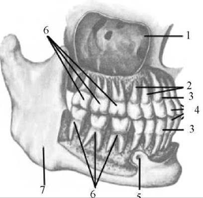
Fig. 143. Permanent teeth of the upper and lower jaws (right view)
1 — maxillary sinus; 2 — premolars; 3 — canines; 4 — incisors; 5 — mental foramen; 6 — molars; 7 — Mandible
Molars (dentes molares) are located posterior to the premolars, three on each side (in both the upper and lower jaws). Their dimensions decrease in a posterior direction. The smallest of them, the last molar, erupts the latest (at 17–30 years of age) and is therefore called the wisdom tooth (dens serotinus). Sometimes the wisdom tooth may be absent. Molars possess a massive, cube-shaped crown. The masticatory surface of the crowns bears 2 to 5 cusps. The presence of this number of cusps on the masticatory surfaces of molars ensures multiple contact points during jaw closure and facilitates the grinding of food. Lower molars have two roots each: an anterior and a posterior. Upper molars have three roots each: one lingual and two buccal (vestibular). The roots of wisdom teeth frequently fuse into a single common root.
The first teeth appear in children at 5–7 months of age, and by the age of 2–2.5 years, their number reaches 20. These are the deciduous teeth (dentes decidui). In children aged 5–7 years, deciduous teeth begin to shed, and new permanent teeth (dentes permanentes) appear in their place. In a normal adult, there are 30–32 permanent teeth in the dental alveoli. Each half of the upper and lower jaws contains two incisors, one canine, two premolars, and three molars.
In anatomy and clinical practice, the number and arrangement of teeth are conventionally designated by a dental formula. The numerator of the formula denotes the upper teeth, and the denominator denotes the lower teeth. Because the right and left halves of the jaws are symmetrical, only one of them is taken into account in the calculation. The first digit indicates the number of incisors, the second represents canines, the third stands for premolars, and the fourth denotes molars. For deciduous teeth, the dental formula looks like  (there are no premolars, and instead of three molars, there are two). For permanent teeth, this formula appears as
Deciduous and permanent teeth erupt during specific age periods (Table 7). The timing of their emergence also depends on the child's Nutrition and other conditions.
Table 7 AVERAGE TIMING OF TOOTH ERUPTION
|
Tooth Name |
Deciduous Teeth |
Permanent Teeth |
|
Medial incisor |
6–8 months |
7–7.5 years |
|
Lateral incisor |
7–9 months |
8–8.5 years |
|
Canine |
15–20 months |
11–11.5 years |
|
First premolar |
— |
10–10.5 years |
|
Second premolar |
— |
11–11.5 years |
|
First molar |
12–15 months |
7–7.5 years |
|
Second molar |
20–24 months |
12–12.5 years |
|
Third molar |
— |
17–25 years and older |
When the mandible is at rest, even if the oral aperture is closed, There is a small free space between the upper and lower teeth because the lower jaw hangs down slightly. The closure of the teeth is termed the bite (mordex). In a normal bite, the teeth of the upper jaw project slightly forward relative to the teeth of the lower jaw due to the fact that the alveolar margin of the upper jaw is somewhat larger than that of the lower. In a normal bite, there is no exact one-to-one correspondence between the upper and lower tooth rows owing to a mismatch in their dimensions. For instance, the upper medial incisor is wider than the lower one and contacts with its edge not only the medial but also the lateral lower incisor. As a result, all subsequent teeth articulate not with one, but with two teeth of the opposing row.
All deviations from a normal bite are classified as malocclusions, which require specialized Treatment to correct. Malocclusions impair the masticatory process and may be either hereditary or caused by certain medical conditions and detrimental habits.
The following types of bite anomalies are distinguished: 1) edge-to-edge (zero) bite, where the upper and lower incisors meet at their cutting edges; 2) open bite, characterized by a gap remaining between all teeth except the molars; 3) crossbite, where some upper teeth overlap the lower ones externally while others do so internally; 4) excessive protrusion of the maxillary teeth; and 5) mandibular prognathism (protrusion of the lower teeth). The latter two variants are caused by the overdevelopment of either the upper or lower jaw.
Dental developmental anomalies are characterized by irregularities in the position, number, and shape of the teeth.
Anomalies of tooth position. Teeth may be located not along the edge of the alveolar processes of the jaws, but rather on their lateral surfaces facing the vestibule or the Oral Cavity proper. Occasionally, teeth erupt into the Nasal cavity, the hard palate, or the maxillary sinus.
Anomalies of tooth number. Cases involving the Congenital absence of the upper lateral incisors and second premolars are sometimes observed.
Anomalies of crown or root shape. Abnormally elongated or shortened roots, as well as roots bent at various angles, can occur. Molars may possess a greater than average number of roots, and the number of cusps on the masticatory surface of the crowns may also vary.
Last update: 08/08/2026
Editorial and Educational Adaptation: This material has been compiled based on the primary/original source text. The project team performed an editorial review, corrected technical inaccuracies, structured sections, and adapted the content for an educational format.
What was processed:
- elimination of formatting defects (OCR errors, structural breaks, corrupted characters);
- editorial organization of content;
- standardization of terminology in accordance with academic sources;
- verification of factual statements against the original source text.
All mentions of the author, publication year, and origin of the primary text have been preserved in accordance with the source.