Human Anatomy Part 2 - K. A. Dyubenko A. K. Kolomiytsev Yu. B. Chaykovsky 2008
Special Part
Nervous system, systema nervosum - Special Part
Cerebral cortex - White matter of the cerebral hemispheres
The White matter, substantia alba, of the cerebral hemispheres is formed by projection and Association Pathways running in various directions. The nerve fibers of these pathways are divided into three groups:
- association nerve fibers;
- commissural nerve fibers;
- projection nerve fibers.
Association nerve fibers, neurofibrae associationes, interconnect cortical fields and areas within the same hemisphere. They are subdivided into short and long fibers (Fig. 161). Short fibers include arcuate fibers, fibrae arcuatae cerebri, which lie directly beneath the cortex and connect two adjacent gyri.
Long fibers lie deeper than the short ones and connect more distant cortical regions. Long association fibers include:
- the cingulum;
- the superior longitudinal fasciculus, fasciculus longitudinalis superior;
- the inferior longitudinal fasciculus, fasciculus longitudinalis inferior;
- the uncinate fasciculus, fasciculus uncinatus.
The cingulum is arch-shaped and located within the cingulate gyrus. It originates from the anterior perforated substance and then extends via the isthmus to the hippocampus and uncus of the temporal lobe. It connects the cortical areas on the Medial surface of the hemispheres and belongs to the rhinencephalon.
The superior longitudinal fasciculus, fasciculus longitudinalis superior, connects the frontal lobe with the parietal and temporal lobes.
Class="center">
Fig. 161. Association PATHWAYS OF THE cerebral hemisphere (after S. P. Dzugayeva).
A: I - central sulcus; II - superior longitudinal fasciculus; III - frontal lobe; IV - fronto-occipital fasciculus; V - inferior fronto-occipital fasciculus; VI - temporal lobe; VII - inferior occipitofrontal fasciculus; VIII - vertical occipital fasciculus. B: I - short association fibers; II - cingulate bundle; III - superior longitudinal fasciculus
The inferior longitudinal fasciculus, fasciculus longitudinalis inferior, connects the occipital lobe with the temporal lobe. It originates from the gyri of the occipital lobe and runs along the outer wall of the posterior and inferior horns of the lateral ventricle.
The uncinate fasciculus, fasciculus uncinatus, connects the inferior part of the frontal lobe, the uncus, and the hippocampus.
Commissural nerve fibers, neurofibrae commissurales, interconnect the cerebral hemispheres and establish communication between them. They form the following commissural bundles:
- the corpus callosum;
- the anterior cerebral commissure, commissura cerebri anterior;
- the hippocampal commissure, commissura fornicis;
- the posterior epithalamic commissure and habenular commissure.
The corpus callosum connects cortical areas of the right and left hemispheres, ensuring The transfer of information from one hemisphere to the other. It consists of 20 million transverse fibers, which give it an oval shape on a sagittal section, as well as a small number of longitudinal fibers that connect cortical areas of different lobes between the right and left hemispheres. It comprises three parts:
- the trunk, truncus corporis callosi;
- the rostrum;
- splenium, splenium corporis callosi;
The upper surface of the corpus callosum is covered with a thin layer of Gray matter known as the indusium griseum, accompanied by longitudinal striae: the medial longitudinal stria, stria longitudinalis medialis, and the lateral longitudinal stria, stria longitudinalis lateralis.
The fibers of the body of the corpus callosum connect the cortex of the precentral gyri, temporal, and parietal lobes, whereas the fibers of the rostrum and genu form the WHITE MATTER OF the frontal lobe cortex, and the splenium consists of fibers from the occipital lobe. The fibers of the genu, rostrum, and splenium curve archwise to form the Major and minor forceps, forceps major et minor.
The anterior commissure, commissura anterior, is horseshoe-shaped and serves as a commissure of the olfactory Brain. Located posterior to the rostrum of the corpus callosum, it consists of two bundles: the first connects the anterior perforated substance, and the second connects the gyrus of the temporal lobe, predominantly the hippocampal gyrus.
The commissure of the fornix, commissura fornicis, has the shape of a triangular lamina. Acting as a commissure of the olfactory brain, it connects the central regions of two arched bundles of nerve fibers that form the fornix beneath the corpus callosum. The fornix comprises the columns of the fornix, columnae fornicis, and the crura of the fornix, crura fornicis. The columns of the fornix reach the subhypothalamic region and terminate in the mammillary bodies.
Projection nerve fibers participate in The formation of complex reflex arcs. They connect the Cerebral Cortex with the nuclei of the Brainstem and Spinal Cord. Projection fibers are divided into two groups: sensory (afferent) and motor (efferent). Sensory fibers project the body surface onto the sensory and motor centers of the cortex. These sensory fibers are processes of Cells from spinal ganglia and cranial nerve ganglia, which function as the first Neurons of ascending pathways.
The axons of the first neurons reach the cellular nuclei of the spinal cord and Medulla Oblongata, where synapses are formed with the second neurons of the sensory pathways, which ascend as part of the medial lemniscus to the ventral nuclei of the thalamus. The thalamic nuclei house the third neurons, whose axons project to the cortical nuclei.
Motor nerve fibers conduct impulses from the motor areas of the cortex to the motor nuclei of the brainstem and spinal cord. Sensory and motor fibers form conduction pathways, and within the substance of the cerebral hemispheres, they constitute The system of radiate bundles. Projection nerve fibers are described in greater detail in the chapter "Conduction Pathways of the Central Nervous system".
Internal capsule, capsula interna (Fig. 162).
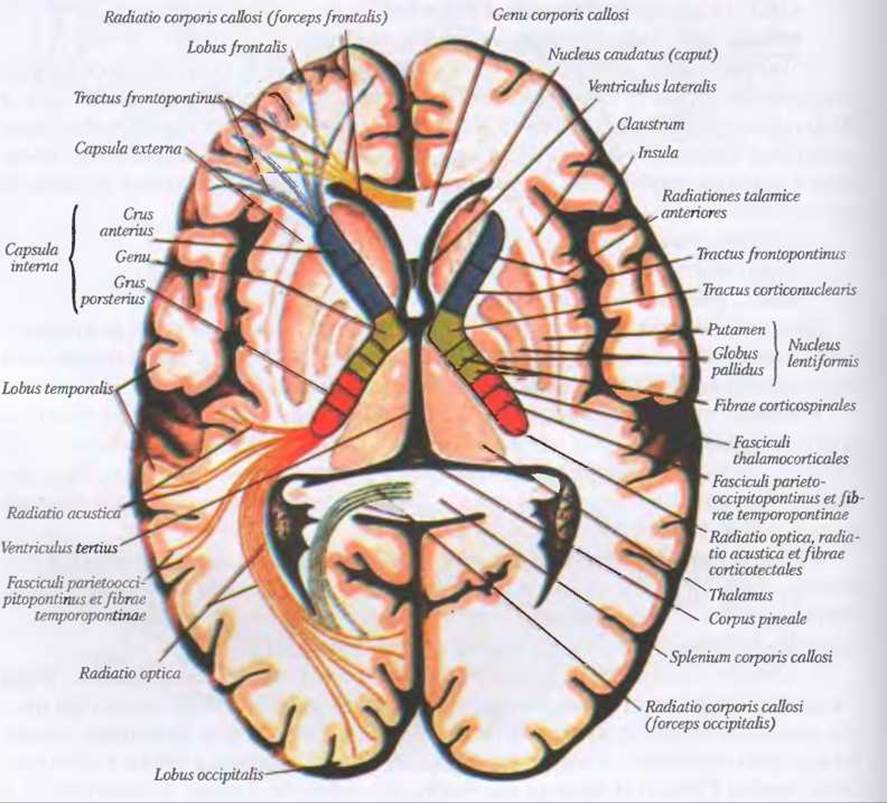
Fig. 162. Internal capsule, capsula interna
The conduction pathways of each hemisphere form a thick, broad band of white matter situated between the thalamus, caudate Nucleus, and lentiform nucleus; this Structure is termed the internal capsule, capsula interna. The internal capsule comprises three parts:
- anterior limb, crus anterior;
- posterior limb, crus posterior;
- genu, genu.
The anterior limb, crus anterior, is located between the caudate and lentiform nuclei. It transmits fibers of the following pathways: tractus frontothalamicus, extending from the frontal lobe to the thalamus, and tractus frontorubralis, extending to the red nucleus, as well as fibers to the pontine nuclei, tractus frontopontinus. Additionally, transverse fibers pass through the anterior limb from the caudate nucleus to the lentiform nucleus.
The posterior limb, crus posterior, is situated between the thalamus and the lentiform nucleus. The anterior two-thirds of the posterior limb are occupied by fibers of the corticospinal tract, tractus corticospinalis, which extend from the Cells of the precentral gyrus to the motor nuclei of the spinal cord. The posterior third of this limb contains sensory fibers—the thalamocortical fascicles, fasciculi thalamocorticales—which connect the thalamic nuclei with the cortex of the postcentral gyrus. Alongside these run feedback fibers from the cortex to the thalamus, forming the corticothalamic fascicles, fasciculi corticothalamici.
The genu, genu, of the internal capsule occupies the angular region between the anterior and posterior limbs. It transmits fibers of the corticonuclear tract, tractus corticonuclearis, running from the Nerve Cells of the precentral gyrus to the cranial nerve nuclei of the Midbrain, Pons, and medulla oblongata. Posteriorly, the posterior limb of the internal capsule continues into the retrolentiform part, pars retrolentiformis capsulae internae. This part is formed by fiber bundles running from the primary visual centers to the occipital lobe cortex. Inferior to the lentiform nucleus lies the sublentiform part, pars sublentiformis capsulae internae, containing fiber bundles passing from the medial geniculate body to the superior temporal gyrus. Lesions of the pathways passing through the internal capsule result in motor and sensory disorders on the opposite side of the body, while complete damage leads to the "three hemis syndrome": *hemiplegia, hemianesthesia, and hemianopia*.
*Hemiplegia, hemiplegia (from Greek: hemi, half + plege, stroke, blow = paralysis of one half of the body).
**Hemianesthesia, hemianesthesia (from Greek: hemi, half + an-... + aisthesis, sensibility = loss of sensation on one half of the body).
***Hemianopia, hemianopsia (from Greek: hemi, half + an-... + opsis, Vision = half-blindness resulting from a lesion of the optic radiation).
Last update: 08/08/2026
Editorial and Educational Adaptation: This material has been compiled based on the primary/original source text. The project team performed an editorial review, corrected technical inaccuracies, structured sections, and adapted the content for an educational format.
What was processed:
- elimination of formatting defects (OCR errors, structural breaks, corrupted characters);
- editorial organization of content;
- standardization of terminology in accordance with academic sources;
- verification of factual statements against the original source text.
All mentions of the author, publication year, and origin of the primary text have been preserved in accordance with the source.