Human Anatomy Part 1 - K. A. Dyubenko, A. K. Kolomiytsev, Yu. B. Chaykovsky 2002
Special Part
Joints of the bones of the lower limb, juncturae membri inferioris — Joints of the free lower limb, articulationes membri inferioris liberi
The hip joint, articulatio coxae1 [iliofemoralis], is the largest ball-and-socket joint, articulatio spheroidea (Fig. 99). It is formed by the lunate surface of the acetabulum of the hip bone and the femoral HEAD. The hip joint plays a vital role in weight-bearing, posture, and locomotion (walking, running, jumping). As a multiaxial joint, movements in the hip occur around three mutually perpendicular axes: frontal (flexion and extension), vertical (supination and pronation), and sagittal (abduction and adduction). In addition, rotational movements (medial and lateral rotation) are possible. These movements are facilitated by the long femoral neck, which offsets the axis of the lower limb from the head, creating a large mechanical lever for the Muscles.
The socket is deepened by the fibrocartilaginous labrum acetabulare, which attaches along the rim of the acetabulum. The strong transverse ligament, lig. transversum acetabuli, spans the acetabular notch. Inside the joint lies the intracapsular ligament of the head of the Femur, lig. capitis femoris, which connects the fovea of the head to the floor of the acetabulum. This ligament transmits a branch of the obturator artery, r. acetabularis, which supplies Blood to the femoral head—a factor that must be considered in ligament ruptures. The hip Joint Capsule originates from the margins of the acetabulum (making the labrum intracapsular) and attaches anteriorly to the intertrochanteric line, while posteriorly it stops short of the intertrochanteric crest. Most of the femoral neck lies within the joint cavity and is covered by the synovial membrane. The joint capsule consists of longitudinal fibers that form a circular zone, the zona orbicularis, around the femoral neck.
The joint capsule is reinforced by extracapsular ligaments (see Fig. 98A):
- the iliofemoral ligament, lig. iliofemorale (lig. Bertini), is the strongest ligament, capable of withstanding loads of up to 350 kg (P. F. Lesgaft); it originates from the anterior inferior iliac spine and attaches to the intertrochanteric line;
- the ischiofemoral ligament, lig. ischiofemorale, originates from the body and tuberosity of the ischium and blends into the capsule;
- the pubofemoral ligament, lig. pubofemorale, originates from the superior ramus of the pubis, blends into the capsule, and attaches just above the lesser trochanter.
Due to the great depth of the acetabulum, strong ligaments, and well-developed surrounding muscles, dislocations of this joint are rare.
Blood supply to the hip joint is provided by the medial and lateral circumflex femoral Arteries (a. circumflexa femoralis medialis et lateralis) and the obturator artery (a. obturatoria). The latter gives off the acetabular branch (r. acetabularis), which travels within the ligament of the head of the femur (lig. capitis femoris) to the femoral head. Additional blood supply comes from inconstant branches originating from the first perforating artery (a. perforantes), the superior and inferior gluteal arteries (aa. gluteae superior et inferior), and the internal pudendal artery (a. pudenda interna), which course toward the femoral neck and the acetabular fossa. Along the outer margin of the fossa, they extensively anastomose to form a closed arterial ring that ensures collateral Circulation in the hip region (T. I. Anikina; P. A. Romanov). Venous drainage occurs via the medial and lateral circumflex femoral Veins into the deep VEINS OF THE thigh and pelvis, namely the deep femoral vein (v. profunda femoris), femoral vein (v. femoralis), and internal iliac vein (v. iliaca interna).
1 Hip joint, Lat. coxa; hence, inflammation of the hip joint is termed coxitis.
Class="center">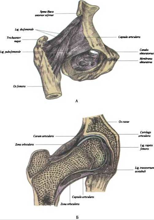
Fig. 99. Hip joint, articulatio coxae: A - anterior surface; B - frontal section
Lymphatic drainage proceeds from the deep and two superficial networks of lymphatic capillaries in the synovial membrane and directs: anteriorly toward the external iliac Lymph Nodes, and posteriorly toward the internal iliac nodes.
Innervation of the joint is supplied by the obturator, femoral, and sciatic nerves (nn. obturatorius, femoralis et ischiadicus), as well as the superior and inferior gluteal nerves (nn. gluteus superior et inferior).
Radiological Anatomy. On anteroposterior radiographs of the hip joint, the upper and medial margins of the acetabulum are visible (Fig. 100). The femoral head is spherical in shape, featuring the fovea of the femoral head, which appears as a depression with uneven edges.
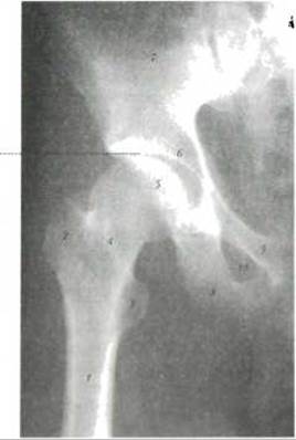
Fig. 100. Overview radiograph of the right hip joint in an anteroposterior projection (according to Ya. I. Kryzhanovsky):
1 - femur, 2 - trochanter major, 3 - trochanter minor; 4 - collum femoris; 5 - caput femoris; 6 - acetabulum; 7 - os ilium; 8 - ramus inferior pubis; 9 - ramus superior os pubis; 10 - foramen obturatum; the joint space is indicated by a dashed line
The radiographic joint space is visualized between the femoral head, which is seated within the acetabulum, and the acetabular roof.
In clinical orthopedic practice, Nélaton's line is used to determine the normal spatial relationship between the femoral head and the acetabulum; it is drawn from the medial contour of the femoral neck to the upper margin of the obturator foramen. Normally, this line is arcuate, but it becomes broken in cases of subluxation.
Developmental anomalies of the Hip Joint. Among developmental defects of the hip, the most common in premature infants (according to V. P. Yurchak, 1994) include: hip Dysplasia (10%), congenital coxa vara, Coxa Valga, dislocations, and subluxations. Hip dysplasia is characterized by underdevelopment of the acetabulum, a reduction in its depth, and a mismatch with the size of the femoral head.
Acquired hip deformities in children also include Hematogenous Osteomyelitis, transient synovitis, and avascular necrosis following femoral neck fractures (O. V. Dolnytskyi, 1992).
Coxa vara is a Varus deformity of the femoral neck characterized by a decreased neck-shaft angle. It clinically manifests as a wide-based stance and a waddling gait.
Coxa valga is a deformity in which the neck-shaft angle is greater than normal. It is characterized by limited hip abduction and may eventually lead to The Development of deforming arthrosis of the hip joint (O. V. Dolnytskyi, 1975).
The knee joint, art. genus1, is the largest and most complex joint structurally, featuring multiple synovial cavities. It is formed by the condyles of the femur, the condyles of the Tibia, and the Patella (a sesamoid bone located within the tendon of the quadriceps femoris Muscle) (Fig. 101A). The articular surfaces of the femoral condyles are convex in both transverse and sagittal directions, representing segments of an ellipsoid (N. D. Dovgyallo).
According to the shape of its articulating surfaces, the knee joint is classified as a bicondylar joint, articulatio bicondylaris. Because the articulating surfaces are incongruent, the joint cavity contains the medial and lateral menisci (menisci medialis et lateralis), which are composed of fibrocartilage (Fig. 101B). The menisci help absorb shocks during movement. The medial meniscus is larger than the lateral one. Their superior surface is concave, while the inferior surface is flat; the outer margin is thickened and fuses with the joint capsule, whereas the inner margin is sharp and projects into the joint cavity. The lateral meniscus is connected to the lateral femoral condyle by the anterior and posterior meniscofemoral ligaments (ligg. meniscofemorale anterius et posterius). Anteriorly, the menisci are joined by the transverse ligament of the knee (lig. transversum genus), located within the joint cavity.
In the middle of the fibrous capsule of the joint are the anterior and posterior cruciate ligaments, lig. cruciatum anterius et posterius (Fig. 102). The anterior ligament originates from the inner surface of the lateral femoral condyle, runs downward and inward, and attaches to the anterior intercondylar fossa. The posterior ligament originates from the outer surface of the medial femoral condyle, runs downward and outward, attaching to the posterior intercondylar fossa of the tibia. The cruciate ligaments divide the joint cavity into anterior and posterior compartments, which prevents the spread of pus from one compartment to the other during inflammation.
1 Knee, Greek *gony*, hence inflammation of the knee joint — *gonitis*.
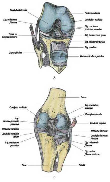
Fig. 101. Knee joint, articulatio genus: A - anterior surface; B - posterior surface

Fig. 102. Menisci and cruciate ligaments of the knee joint
The joint is reinforced by the following ligaments (Fig. 103):
- fibular collateral ligament, lig. collaterale fibulare — runs from the lateral condyle to the head of the Fibula;
- tibial collateral ligament, lig. collaterale tibiale — runs from the medial condyle of the femur to the condyle of the tibia;
- oblique popliteal ligament, lig. popliteum obliquum — runs from the medial condyle of the tibia upward and laterally to the joint capsule;
- arcuate popliteal ligament, lig. popliteum arcuatum — runs from the lateral condyle of the femur as part of the oblique ligament;
- patellar ligament, lig. patellae — runs from the apex of the patella to the tuberositas tibiae. On the sides of this ligament are the medial and lateral patellar retinacula, retinaculum patellae mediale et laterale. The joint capsule of the knee originates 1 cm away from the edge of the Cartilage cover, and fuses with the edge of the menisci along its margins.
The synovial membrane of the knee joint lines the capsule from the inside and has a complex Structure. It covers the cruciate ligaments, enclosing them anteriorly and laterally, and forms folds with layers of adipose tissue. The most developed are the alar folds of the synovial membrane, plicae alares, which are located below the patella. The synovial membrane contains villi, which are numerous around the patella. They contribute to the secretion of synovial fluid. The membrane itself forms 13 recesses (V. V. Kovanov, 1983), which are outpouchings of the synovial membrane extending onto the menisci, ligaments, and bones. There are five recesses on the anterior surface of the joint, four on the posterior surface, and four on the lateral sides.
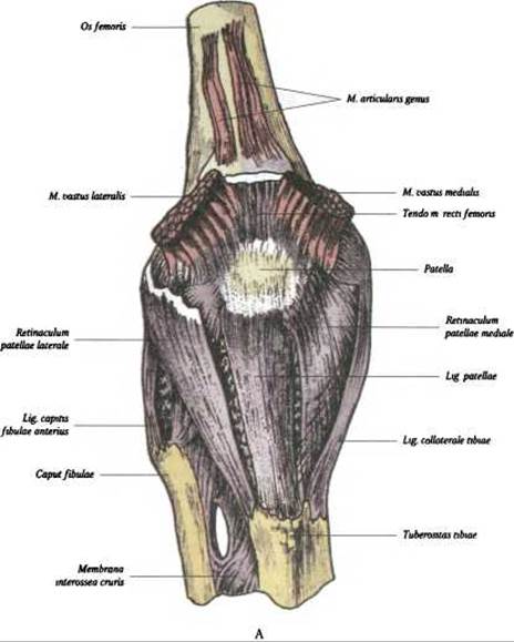
Fig. 103. Ligaments of the knee joint, articulatio genus. A - anterior surface
1 Vladimir Vasilyevich Kovanov — a prominent Russian topographoanatomist and surgeon.
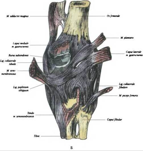
Fig. 103. Ligaments of the knee joint, articulatio genus (Continued): B - posterior surface
Around the knee joint, There is a series of mucous synovial bursae, which are of great importance in the Pathogenesis of paraarticular phlegmons. Some of them communicate with the joint cavity, such as the subcutaneous prepatellar bursa, bursa prepatellaris subcutanea. On the anterior surface of the patella, the following bursae are distinguished: subcutaneous, bursa subcutanea prepatellaris; subfascial, b. prepatellaris subfascialis; and subtendinous, located beneath the aponeurotic expansion of the quadriceps femoris muscle, b. prepatellaris subtendinea. Near the inferior attachment of lig. patellae to the tibia lies the constant deep infrapatellar bursa, b. infrapatellaris profunda, which communicates with the joint cavity. On the posterior surface of the joint, there are seven synovial bursae beneath the attachment sites of almost all muscles (bursa capitis medialis m. gastrocnemii, bursa m. semimembranosi superior, b. semimembranosi inferior, bursa m. sartorii, bursa anserina, bursa superior lig. collateralis tibialis, bursa inferior lig. collateralis tibialis).
The posterolateral group includes four synovial bursae (bursa capitis lateralis m. gastrocnemii, bursa lig. collateralis fibularis, bursa m. poplitei, bursa m. bicipitis).
Among the listed synovial bursae surrounding the knee joint, the most constant and frequently communicating with the joint cavity are the suprapatellar bursa, the bursa of the medial head of m. gastrocnemius, and the bursae of the semimembranosus and popliteus muscles. Under METABOLISM/18.html">The Influence of injury, infection, and other causes, the synovial bursae of the knee joint can become inflamed (bursitis), accompanied by the accumulation of exudate in their cavities.
Movements in the knee joint occur around two axes: the frontal axis (flexion and extension) and the vertical axis when the knee is semi-flexed (a small range of pronation and supination).
Blood supply. The knee joint receives blood from the articular network of the knee, rete articulare genus, which is formed by: aa. genus superiores mediales et laterales, aa. genus inferiores mediales et laterales, a. genus media (from a. poplitea), and a. descendens genus (from a. femoralis). Venous outflow is carried out via companion veins into the deep veins of the lower extremity — vv. tibiales anteriores, v. poplitea, v. femoralis.
Lymphatic drainage from the knee joint is carried out via deep Lymphatic vessels into the popliteal and inguinal lymph nodes, nodi lymphatici poplitei et inguinales.
Innervation of the knee joint is provided by the musculocutaneous Branches of the femoral nerve, which branch out in the upper lateral and upper medial Regions of the knee joint, as well as in the region of the tuberositas tibiae. The anterior and posterior branches of the obturator nerve branch out in the upper medial region of the joint. The saphenous nerve of the lower extremity, n. saphenus, innervates the lower medial region of the knee joint. Branches of the sciatic, tibial, and peroneal nerves reach the posterior wall of the knee joint capsule, as well as the lateral and anterolateral regions of the knee joint.
X-ray anatomy. An anteroposterior radiograph of the knee joint shows the intercondylar eminence of the tibia and the femoral condyles surrounding the intercondylar fossa (Fig. 104 A). The shadow of the patella is projected onto the distal end of the femur. The articular cavity of the knee joint is the widest in the articular system. Lateral radiographs reveal the joint space of the proximal tibiofibular articulation. The lateral view demonstrates THE POSITION OF the patella (Fig. 104 B).
Last update: 08/08/2026
Editorial and Educational Adaptation: This material has been compiled based on the primary/original source text. The project team performed an editorial review, corrected technical inaccuracies, structured sections, and adapted the content for an educational format.
What was processed:
- elimination of formatting defects (OCR errors, structural breaks, corrupted characters);
- editorial organization of content;
- standardization of terminology in accordance with academic sources;
- verification of factual statements against the original source text.
All mentions of the author, publication year, and origin of the primary text have been preserved in accordance with the source.