MEDICAL BIOLOGY, HUMAN ANATOMY, PHYSIOLOGY, AND PATHOLOGY - Y.I. Fedoniuk 2010
BIOLOGY
CHAPTER 2. BIOCOENOTIC LEVEL OF ORGANIZATION OF LIFE AND THE PLACE OF HUMANS IN IT
2.3. MEDICAL PROTOZOOLOGY
Phylum Apicomplexa.
Class Sporozoa
Exclusively parasitic Protozoa adapted to living in Body Cavities or inside the Cells of humans and animals. Due to their parasitic lifestyle, the Organization of sporozoa is extremely simplified: they lack digestive and contractile vacuoles and, as a rule, locomotor Organelles. During reproduction, most species form spores (embryos with a protective coat), hence the name of the Class. Their life cycle is characterized by alternation of hosts, a sequence of asexual reproduction (schizogony, endodyogeny), sexual reproduction, and sporogony (formation of spores and sporozoites). Human parasites belong to the orders Haemosporidia and Coccidia.
Order Haemosporidia. At certain stages of their development, they inhabit the erythrocytes of vertebrates and humans. The spore stage is absent, as haemosporidians are transmitted from one host to another without entering the external environment. Malaria parasites are human pathogens belonging to this group.
Malaria parasites (Plasmodium). Plasmodium species are the causative agents of malaria. Over 100 species of malaria parasites are known, parasitizing reptiles, birds, and mammals. Four species parasitize humans: 1) Plasmodium vivax, the CAUSATIVE AGENT OF tertian malaria; 2) Pl. malariae, the causative agent of quartan malaria; 3) Pl. falciparum, the causative agent of malignant tertian (falciparum) malaria; 4) Pl. ovale, the causative agent of ovale tertian malaria.
Malaria is a vector-borne anthroponotic disease. The source of infection is a sick person. The transmission mechanism is vector-borne. The vector is a female mosquito of the genus Anopheles, which becomes infected while feeding on the Blood of an infected person. Humans are infected through the bite of an infected mosquito. Localization: intracellular parasites; in humans, they reside in erythrocytes and Liver cells. Geographical distribution: Africa, Asia, America, and Europe. Malaria is particularly prevalent in tropical climates, but also occurs in temperate regions. Its distribution is closely linked to the range of the Anopheles mosquito, with ambient Temperature playing a decisive role. The temperature minimum for P. vivax is +16° C. At lower temperatures, The Development of the parasite within infected mosquitoes ceases. Therefore, local cases of malaria do not occur in areas with short and cold summers. In tropical countries, over 300 million people suffer from malaria, of whom one million—predominantly children—die annually. The intensification of international migration in recent years has exacerbated the spread of many infectious diseases, including malaria. Imported cases of malaria are registered in Ukraine. The danger lies in the fact that malaria mosquitoes in Ukraine are susceptible to imported strains of P. vivax.
Life cycle (Fig. 2.9). The intermediate host is a human, and the definitive host is a female Anopheles mosquito. The life cycle of malaria parasites comprises three stages: 1) schizogony, an asexual reproductive phase involving multiple fission, which takes place in The Human Body within liver cells (pre-erythrocytic schizogony) and erythrocytes (erythrocytic schizogony); 2) sexual reproduction, where gamete formation begins in human erythrocytes and is completed within the body of the female Anopheles mosquito, leading to copulation and zygote formation; 3) sporogony, The production of sporozoites (within the mosquito).
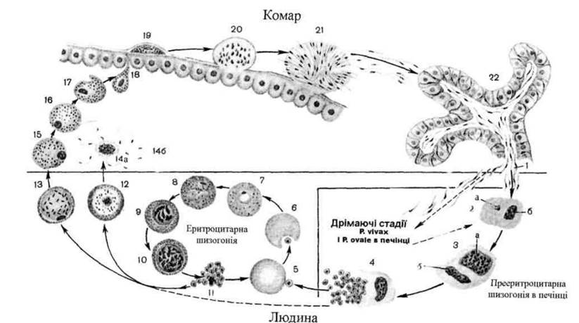
Fig. 2.9. Life Cycle of Plasmodium vivax and P. ovale:
1 - release of a sporozoite from the salivary gland duct and its penetration into a liver Cell; 2 - trophozoite in a liver cell: a - trophozoite, b - liver Cell Nucleus; 3 - schizont in a liver cell: a - schizont, b - cell nucleus; 4 - release of tissue merozoites from the liver cell into Blood Plasma; 5 - attachment of a merozoite to an erythrocyte; 6 - penetration of the merozoite into the erythrocyte; 7 - trophozoite at the ring stage; 8 - young trophozoite in the erythrocyte; 9 - immature erythrocytic schizont; 10 - mature erythrocytic schizont; 11 - erythrocytic merozoites; 12 - male gametocyte (microgametocyte); 13 - female gametocyte (macrogametocyte); 14a - formation of male Gametes; 14b - male gamete; 15 - female gamete; 16-17 - Fertilization; 18 - ookinete; 19-20 - Formation of the oocyst and development of sporozoites; 21 - release of sporozoites from the oocyst into the mosquito's body cavity; 22 - sporozoites in the mosquito's salivary gland.
Pre-erythrocytic schizogony. During a bite, the mosquito injects the malaria parasite in the sporozoite stage (the infective stage for humans) into the human bloodstream along with its saliva. Carried by the blood flow, the sporozoites enter liver cells, where they undergo pre-erythrocytic (tissue) schizogony. Each schizont produces A large number (from 1,000 to 5,000) of tissue merozoites. The pre-erythrocytic cycle occurs only once. It lasts 6 days in Pl. falciparum, 8 days in Pl. vivax, 9 days in Pl. ovale, and 15 days in Pl. malariae. It has been proven that in quartan and malignant tertian malaria, tissue merozoites completely leave The Liver and enter the blood upon completion of tissue schizogony. In tertian malaria, due to sporozoite heterogeneity (tachy- and bradysporozoites), tissue schizogony can occur either directly after the mosquito bite (upon Introduction of tachysporozoites) or 1.5-2 years later (upon introduction of bradysporozoites), which accounts for the prolonged incubation period and delayed relapses caused by the parasite's so-called "dormant" stages. The incubation period is the time elapsed from the pathogen's entry into the human body to the onset of clinical symptoms.
Erythrocytic schizogony. Tissue merozoites enter the bloodstream and invade erythrocytes, marking THE START OF the erythrocytic phase of the malaria parasite's life cycle. Merozoites that have penetrated erythrocytes increase in size, and a vacuole forms near the parasite's nucleus, pushing The Nucleus to the periphery and giving the parasite a ring-like shape (the ring stage). Subsequently, the vacuole decreases in size, and pseudopodia develop (the amoeboid trophozoite stage). Gradually, the plasmodium fills the entire erythrocyte, becomes spherical, and its nucleus shifts to the center (the mature trophozoite stage). During the schizogony stage, the trophozoite contains from 6 to 24 nuclei. Cytoplasmic masses cluster around each nucleus, forming erythrocytic merozoites. The erythrocyte membrane then ruptures, releasing merozoites and toxic Metabolic waste products of the plasmodium into the bloodstream. This event coincides with the onset of malaria paroxysms. Merozoites released into the plasma invade new erythrocytes, and the cycle repeats, generating a new generation of merozoites. This process recurs multiple times. The erythrocytic schizogony cycle lasts 48 hours in Plasmodium vivax, Pl. falciparum, and Pl. ovale, and 72 hours in Pl. malariae. This precise duration determines the intervals between malaria attacks.
Following a series of erythrocytic schizogonic cycles, a fraction of the merozoites invading erythrocytes develops not into schizonts, but into immature sexual forms (gametocytes)—macrogametocytes (immature female forms) and microgametocytes (immature male forms). For further development, these gametocytes must reach The Stomach of the definitive host, a female Anopheles mosquito, where sexual reproduction and sporogony take place. Gametocytes represent the infective stage of the malaria parasite for the mosquito. They enter the stomach of the female mosquito during her blood meal on an infected person (male mosquitoes do not feed on blood).
Sexual reproduction and sporogony. In the stomach of the female Anopheles mosquito, macrogametocytes mature into female gametes (macrogametes). Microgametocytes undergo multiple divisions during maturation to form mature male gametes (microgametes). The micro- and macrogamete fuse (fertilization) to form a zygote. Because it is motile, it is called an ookinete. The ookinete penetrates the epithelial lining of the mosquito's stomach, increases significantly in size, and transforms into an oocyst. Inside the oocyst, multiple Divisions of the nucleus and Cytoplasm occur (sporogony), producing a vast number (up to 10,000) of sporozoites. The oocyst wall ruptures, and sporozoites are released into the hemolymph, from which they migrate to the mosquito's Salivary Glands. During a subsequent bite, sporozoites are injected into human blood along with saliva and subsequently invade liver cells.
Pathogenic effect. Malaria is a severe disease characterized by periodic paroxysms of fever associated with the simultaneous release of a large number of merozoites and their toxic metabolic products from erythrocytes. Each attack includes chills and a temperature spike up to 40° C, lasting up to 6–12 hours. Hepatosplenomegaly (enlargement of the liver and Spleen) and anemia are typical features. Fatal outcomes are possible.
Laboratory Diagnostics. Sample material: blood. Research Methods: Microscopy of blood smears and thick blood films. Blood is collected during a paroxysm or immediately after it, prior to the initiation of specific Treatment. Schizonts and gametocytes are identified.
Prophylaxis. Individual: protection against mosquito bites, taking prophylactic medications. Public: detection and treatment of patients and parasite carriers, eradication of adult mosquitoes and their larvae.
Order Coccidia. Toxoplasma is of the greatest medical significance.
Toxoplasma (Toxoplasma gondii) is the causative agent of Toxoplasmosis.
Geographical distribution: ubiquitous.
Localization: Lymph Nodes, liver, spleen, Lungs, Brain, Uterus, eyes, skeletal Muscles, myocardium.
Morphophysiological characteristics. Toxoplasma is an obligate intracellular parasite. The forms located inside the host cell are called endozoites. An endozoite has the shape of an orange segment or a crescent (hence the name from the Greek toxon - arch). The anterior end of the body is tapered, while the posterior end is expanded and rounded. The length is 5–7 μm, and the width is 2–4 μm. Under an Electron microscope, a conoid can be seen at the anterior end, which performs a supportive function during the parasite's invasion into the host cell (Fig. 2.10).
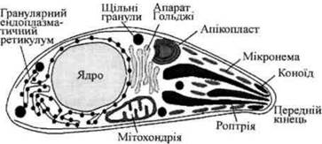
Fig. 2.10. Ultrastructure of Toxoplasma.
The life cycle involves alternating hosts as well as Selection/8.html">Asexual and sexual reproduction. Definitive hosts include domestic cats and other members of the feline family; intermediate hosts include birds and mammals (about 350 species in total), as well as humans. In the intermediate host's body, asexual reproduction occurs via longitudinal division and endodyogeny (internal budding). As a result of multiple divisions, a large number of parasites accumulate within The Cell. An aggregation of these parasites beneath The cell membrane is called a pseudocyst. In chronic toxoplasmosis, true cysts are formed In addition to pseudocysts. A cyst is covered with a thick wall and contains several hundred parasites.
Parasites enter the body of the definitive host (the cat), where sexual reproduction takes place, via the meat of intermediate hosts containing pseudocysts with endozoites. Within the epithelium of the Small Intestine, some endozoites develop first into macrogametocytes and then into macrogametes, while others develop into microgametocytes and subsequently into microgametes. Following copulation, a zygote is formed, which becomes enclosed in a dense protective wall.
This form is known as an oocyst. It is excreted into the external environment with the cat's feces. At temperatures above +20° C, sporogony occurs inside the oocyst, resulting in The formation of two spores with four sporozoites in each (Fig. 2.11).
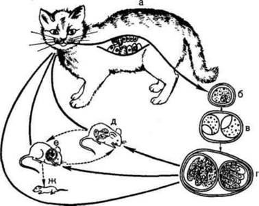
Fig. 2.11. Life cycle of Toxoplasma: a – cat (definitive host); b, c, d – stages of oocyst development in the external environment (a mature oocyst contains two spores with four sporozoites each); e, f – mouse (intermediate host); g – newborn mouse infected transplacentally with the pathogen
Mechanisms of transmission of toxoplasmosis: 1) oral (alimentary) – transmission factors include thermally unprocessed meat, dairy products, eggs containing pseudocysts with endozoites, and hands contaminated with oocysts; 2) percutaneous – through the Skin and mucous membranes (affecting meat Processing plant workers, obstetricians, and laboratory physicians during clinical and laboratory Procedures); 3) transplacental – intrauterine infection of the fetus through the Placenta.
Pathogenic effects. Acquired toxoplasmosis manifests with fever, lymphadenopathy, damage to the Central Nervous system, eyes, myocardium, and skeletal muscles. Congenital toxoplasmosis, resulting from transplacental transmission when a woman is infected during Pregnancy, is characterized by fetal developmental defects (Sabin's triad: chorioretinitis, Hydrocephalus, and cerebral calcifications), CNS damage, fever, jaundice, and rash. Miscarriages or stillbirths are also possible.
Laboratory diagnostics. Test Materials include biopsy tissue fragments, organ and lymph node punctates, CEREBROSPINAL FLUID, and blood. In the acute stage of the disease, the pathogen can be detected in both Internal Organs and blood.
Methods of laboratory diagnostics: 1) serological tests (detection of anti-toxoplasma Antibodies); 2) microscopy of blood smears, cerebrospinal fluid, lymph node punctates, Amniotic Fluid, and fetal membranes; 3) biological assay (intraperitoneal inoculation of white mice with an emulsion of the studied organs); 4) intradermal test with toxoplasmin (an ethereal extract of the peritoneal exudate of white mice infected with toxoplasma).
Prevention. Individual measures include consuming only thoroughly cooked or thermally processed meat and washing hands before eating. Public health measures involve educational outreach, primarily targeting pregnant women.
Last update: 08/08/2026
Editorial and Educational Adaptation: This material has been compiled based on the primary/original source text. The project team performed an editorial review, corrected technical inaccuracies, structured sections, and adapted the content for an educational format.
What was processed:
- elimination of formatting defects (OCR errors, structural breaks, corrupted characters);
- editorial organization of content;
- standardization of terminology in accordance with academic sources;
- verification of factual statements against the original source text.
All mentions of the author, publication year, and origin of the primary text have been preserved in accordance with the source.