HUMAN ANATOMY ATLAS - G.L. Bilich - 2014
Muscular System
MUSCLES AND FASCIAE OF THE ABDOMEN
Muscles of the Anterior and Lateral Abdominal Walls
The Muscle fascicles of the lateral abdominal walls run in various directions. The fibers of the external and internal oblique Muscles intersect at nearly a right angle, while the fascicles of the transverse abdominal muscle run almost horizontally. The anterior PARTS OF THE lateral abdominal wall muscles continue into broad, flat tendons known as aponeuroses, which enclose the rectus abdominis muscle anteriorly and posteriorly, forming its sheath. Along the midline, extending from the xiphoid process of the Sternum to the Pubic Symphysis, the linea alba is formed by the interlocking of the aponeurotic fibers from the oblique and transverse Abdominal muscles of both sides. The umbilical ring is located near its midpoint. Collectively, the Muscles of the anterolateral abdominal wall are referred to as Haller's girdle.
The external oblique muscle (m. obliquus externus abdominis) is the broadest of the abdominal muscles. Located on the anterior and lateral aspects of the abdomen and partially the Thorax (see Fig. 281), it originates by seven to eight large digitations from the external surface of Ribs V–XII. The upper five digitations interdigitate with those of the serratus anterior muscle, and the lower three interdigitate with the latissimus dorsi. The upper fascicles of the external oblique originate on the ribs near their cartilages and run almost horizontally. The lower fascicles course obliquely downward and medially. The fibers of the lowermost part of the muscle are oriented almost vertically downward (Fig. 278). Due to this diverse orientation of muscle fascicles, the tendinous fibers of the external oblique aponeurosis have a broad attachment span. Featuring a broad aponeurosis, the muscle attaches via its tendinous fibers to the outer lip of the iliac crest and the pubic symphysis, and on the anterior abdominal wall, it contributes to The formation of the anterior wall of the rectus sheath.
Along the anterior midline, the aponeurotic fibers meet those of the corresponding muscle of the opposite side, intertwining to form the linea alba.
The thickened lower margin of the aponeurosis of the external oblique muscle, stretched like a trough-like fold between the anterior superior iliac spine and the pubic tubercle, forms the inguinal ligament (ligamentum inguinale, s. arcus inguinalis)—also known as Poupart's ligament, Fallopius's ligament, or Vesalius's ligament, varying in length from 12 to 16 cm. This ligament serves as the inferior wall of the Inguinal Canal (François Poupart [1616–1708], French anatomist and surgeon).
Class="center">
Fig. 278. External oblique muscle, all other muscles removed, origins and insertions (schematic):
1 — External oblique
At its attachment to the pubic bone, the aponeurosis splits into two crura: medial and lateral. The medial crus (crus mediale) attaches to the anterior surface of the pubic symphysis, and the lateral crus (crus laterale) attaches to the pubic tubercle. Intercrural fibers (fibrae intercrurales), belonging to the proper Fascia of the external oblique muscle, run transversely between the crura. Medially, the intercrural cleft is bounded by a Connective Tissue plate known as the reflected ligament (ligamentum reflexum). The intercrural cleft forms the superficial (subcutaneous) ring (opening) of the inguinal canal, through which the Spermatic Cord (in males) or the round ligament of the Uterus (in females) exits the canal beneath the Skin. The subcutaneous inguinal ring is larger in males than in females.
A triangular space—the lumbar triangle—is located between the posterior margin of the external oblique muscle laterally, the iliac crest inferiorly, and the anteroinferior margin of the latissimus dorsi medially. The floor of this triangle is formed by the fibers of the Water/144.html">Origin of the internal oblique muscle (above the iliac crest).
Function: When contracting bilaterally, the external oblique lowers the ribs, flexes THE Vertebral Column, and—in the supine position—raises the pelvis; when contracting unilaterally, it rotates the trunk to the opposite side. The muscle is a component of the abdominal press.
Blood supply: Posterior intercostal, lateral thoracic, and superficial circumflex iliac Arteries.
Innervation: Lower intercostal nerves (ThV–ThXI), iliohypogastric nerve (ThXII–LI), and ilioinguinal nerve (LI).
The internal oblique muscle (m. obliquus internus abdominis) is a flat, broad musculotendinous sheet located directly deep to the external oblique, forming the second
muscle layer of the abdominal wall. The muscle originates via muscular fascicles from the intermediate line of the iliac crest, the thoracolumbar fascia, and the lateral half of the inguinal ligament. Its muscular fascicles fan out to attach to the outer surface of the lower rib cartilages and via a broad tendon insert into the linea alba along its entire length. The posterosuperior fascicles of the internal oblique run anterosuperiorly and attach to the lower rib cartilages, sharing the same direction as the fibers of the internal intercostal muscles. The fascicles of the internal oblique course medially and transition into a broad aponeurosis (along a line connecting the Cartilage of rib X to the pubic bone, remaining 1–3 cm lateral to the lateral margin of the rectus abdominis) (Fig. 279). Superior to the umbilicus, the aponeurosis of the internal oblique splits into two laminae: anterior and posterior. The anterior (external) lamina passes superficial to the rectus abdominis, while the posterior (internal) lamina passes deep to it. 2–5 cm below the umbilicus, the entire aponeurosis of the internal oblique passes anterior to the rectus abdominis, contributing to the Formation of the anterior wall of its tendinous sheath.
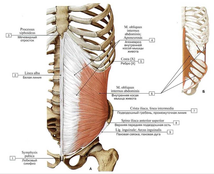
Fig. 279. Internal oblique muscle, all other muscles removed (A — general anterior view, B — origins and insertions (schematic)):
1 — Pubic symphysis; 2 — Linea alba; 3 — Xiphoid process; 4 — Internal oblique; Aponeurosis; 5 — Rib [XII]; 6 — Internal oblique; 7— Iliac crest, intermediate zone; 8 — Anterior superior iliac spine; 9 — Inguinal ligament
The fascicles of the lower part of the internal oblique, together with fibers originating from the transverse abdominal muscle, become part of the spermatic cord and form the cremaster muscle (m. cremaster).
Function: When contracting unilaterally, the internal oblique rotates the trunk toward its own side (along with the contralateral external oblique). When contracting bilaterally, these muscles pull The thoracic cage downward, flex the vertebral column, and—with a fixed thorax—elevate the pelvis.
Blood supply: Posterior intercostal, superior and inferior epigastric, and musculophrenic arteries.
Innervation: Intercostal nerves (ThVI–ThXII), iliohypogastric nerve (ThXII–LI), and ilioinguinal nerve (LI).
The transversus abdominis muscle (m. transversus abdominis), located deep to the internal oblique, is a thin musculotendinous sheet with transversely directed muscle fibers. It forms the deep (third) layer of the lateral abdominal wall muscles. The muscle fibers run horizontally from back to front and medially. They originate via flat musculotendinous slips from the inner surface of ribs VII–XII, interleaving with the slips of the costal part of the Diaphragm, from the Deep Layer of the thoracolumbar fascia, the anterior half of the inner lip of the iliac crest, and the lateral third of the inguinal ligament (Fig. 280). Near the lateral margin of the rectus abdominis muscle, the muscle fibers of the transversus abdominis transition into a broad aponeurosis along a medially concave line known as the semilunar line (linea semilunaris)—or Spigelian line, which extends downward from the costal margin to the inguinal ligament (Adriaan van den Spiegel [Spigelius Adrianus], 1578–1625, a Belgian anatomist and surgeon).
The upper two-thirds of the aponeurosis of the transversus abdominis fuse with the posterior lamina of the internal oblique aponeurosis to form the posterior wall of the rectus sheath. The lower third of the transversus abdominis aponeurosis fuses with the aponeuroses of the oblique muscles and contributes to the anterior wall of the rectus sheath. Together with the aponeuroses of the external and internal obliques, the aponeurosis of the transversus abdominis participates in the formation of the linea alba. The inner surface of the transversus abdominis is lined by a part of the extraperitoneal fascia known as the Spigelian fascia (syn.: transversalis fascia, fascia transversalis).
Function: As an essential component of the abdominal press, the transversus abdominis decreases the volume of the Abdominal cavity AND pulls the ribs forward toward the midline. Unilateral contraction bends the trunk to the same side.
Blood supply: Posterior intercostal, superior and inferior epigastric, and musculophrenic arteries.
Innervation: Intercostal nerves (ThVI—ThX||), iliohypogastric nerve (ThXII—LI), and ilioinguinal nerve (LI).
Muscles of the anterior abdominal wall. The rectus abdominis muscle (m. rectus abdominis) is a flat, long, straplike muscle that is wide superiorly and tapers inferiorly, positioned lateral to the midline. Both rectus muscles are separated from each other by the linea alba. The muscle originates by two heads from the pubic crest and pubic symphysis. At its attachment to the pubic bone, the fibers decussate, forming Bourgery's anterior cruciate ligament of the rectus abdominis (Jean-Marc Bourgery, 1797–1869, a French surgeon and anatomist). Ascending from inferior to superior, the muscle broadens and attaches to the anterior surface of the xiphoid process and the external surface of the cartilages of ribs V–VII via muscular slips (Figs. 281, 282).
Along its course, the muscle features three to four transversely oriented tendinous intersections (intersectiones tendineae) that are fused with the anterior layer of the rectus sheath. These tendinous intersections indicate the segmental origin of the muscle from multiple myotomes, serving as remnants of the connective tissue septa (myosepta) between the myotomes from which the muscle developed. The first (superior) tendinous intersection is located at the level of the eighth rib cartilage, the second midway between the first intersection and the umbilicus, the third at the level of the umbilicus, and the fourth (inconstant and poorly defined) at the level of the arcuate line of the posterior wall of the rectus sheath.

Fig. 280. Transversus abdominis muscle, all other muscles removed (A — general view from the front, B — origins and insertions (diagram)):
1 — Pubic symphysis; 2 - Rectus sheath. anterior layer; 3 — Arcuate line; 4 — Linea alba; 5 — Xiphoid process; 6 — Body of sternum; 7 — Rectus sheath, posterior layer; 8 — Transversus abdominis; Transverse abdominal; 9 — Transversus abdominis; Transverse abdominal; Aponeurosis; 10 — Iliae crest, inner lip; 11 — Anterior superior iliacspine; 12— Inguinal ligament
Function: When the spine and Pelvic Girdle are fixed, the rectus abdominis depresses the ribs, pulls the thorax (sternum and ribs) downward, and flexes the spine; when the thorax is fixed, it elevates the pelvis.
Blood supply: Superior and inferior epigastric, and posterior intercostal arteries.
Innervation: Intercostal nerves (ThV—ThXII), and iliohypogastric nerve (ThXII—LI).
The pyramidalis muscle (m. pyramidalis), triangular in shape with its apex directed upward, is located anterior to the lower part of the rectus abdominis within its fibrous sheath, originating from the pubic crest. Its fibers run upward and blend into the linea alba. The pyramidalis muscle sometimes extends as far as the umbilicus or may be absent. Two to three tendinous intersections have been described in this muscle (see Fig. 281).
Function: The pyramidalis muscle tenses the linea alba.
Blood supply: Inferior epigastric and cremasteric arteries.

Fig. 281. Abdominal muscles, anterior view:
1 — Pyramidalis; 2 — Media! crus; 3 — Lateral crus; 4=2 + 3 — Superficial inguinal ring; 5 — lntercrural ftbres; 6 — Anterior superior iliac spine; 7— Linea alba; 8 — Umbilical ring; 9 —External oblique; 10 — Rectus abdominis; 11 — Rectus abdominis, tendinous intersection; 12 — Rectus sheath, anterior layer; 13 — Serratus anterior; 14 — Costoxiphoid ligaments; 15 — Pectoralis major, stemocostal HEAD; 16 — Pectoralis major, abdominal part; 17 — Intemal intercostal muscle; 18 — Internal oblique; Aponeurosis; 19 — Internal oblique; 20 —- External oblique; Aponeurosis; 21 — Spermatic cord; Cremaster; 22 — Retlected ligament
Innervation: Ilioinguinal nerve, iliohypogastric nerve (ThXII—LI).
It should be emphasized that the Description of the Functions of each individual muscle of the anterior and lateral abdominal walls is somewhat conventional, as they contract simultaneously. Muscle and tendinous fiber bundles running in four different directions (transverse, longitudinal, superior-inferior and medial, inferior-superior and medial) form a complex structural framework within the thickness of the anterior and lateral abdominal walls (Fig. 283). Consequently, upon simultaneous contraction of all abdominal muscles, the abdominal wall constricts in all directions, thereby reducing the volume of the abdominal cavity and displacing the Internal Organs upward (Fig. 284).

Fig. 282. Rectus abdominis, pyramidalis, iliopsoas, and quadratus lumborum muscles, anterior view (all other muscles removed):
1 — Pubic symphysis; 2 — Lesser trochanter; 3 — Pubic lubercle; 4 — Iliopsoas; 5 — Iliacus; 6 — Iliae crest; 7 — Psoas major; 8 — Quadratus lumborum; 9— Xiphoid process; 10 — Linea alba; 11 — Tendinous intersections; 12 — Iliae fossa; 13 — Rectus abdominis; 14— Inguinal ligament; 15 — Pyramidalis
Muscles attaching to the BONES OF THE thorax pull it downward, which facilitates expiration. Simultaneously, these muscles act upon the spine, flexing it and the trunk forward. When the thorax is fixed (such as hanging by the arms from athletic equipment) and the lower extremities are free, the abdominal muscles flex the lumbar spine along with the pelvis, elevating the anterior pelvis. Unilateral contraction of the oblique abdominal muscles lateral flexes the thorax to the side and slightly forward (see Fig. 284).
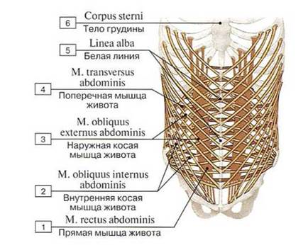
Fig. 283. Direction of muscle fibers of the abdominal wall (diagram):
1 — Rectus abdominis; 2 — Internal oblique; 3 — External oblique; 4 — Transversus abdominis; Transverse abdominal; 5 — Linea alba; 6 — Body of sternum
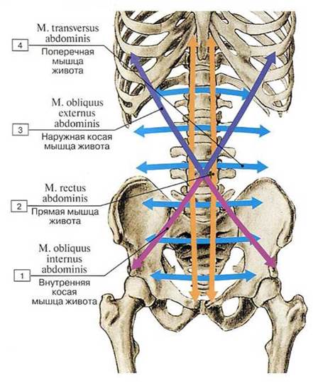
Fig. 284. Direction of contraction of the abdominal muscles (diagram):
1 — Internal oblique; 2 — Rectus abdominis; 3 — External oblique; 4 — Transversus abdominis; Transverse abdominal
Last update: 08/08/2026
Editorial and Educational Adaptation: This material has been compiled based on the primary/original source text. The project team performed an editorial review, corrected technical inaccuracies, structured sections, and adapted the content for an educational format.
What was processed:
- elimination of formatting defects (OCR errors, structural breaks, corrupted characters);
- editorial organization of content;
- standardization of terminology in accordance with academic sources;
- verification of factual statements against the original source text.
All mentions of the author, publication year, and origin of the primary text have been preserved in accordance with the source.