Algology - Kostikov I.Yu. - 2009-2013
Chapter 18. Dictyochophytes – Dictyochophyta
Dictyochophytes are a specific group of chromists that until recently were included in the division Chrysophyta, treated either as a separate and independent order, Dictyochales, within the Class Prymnesiophyceae, or as several distinct genera within the class Chrysophyceae. Dictyochophytes were elevated to an independent chromist taxon with a rank of at least a class, and likely a division, As a result of intensive electron microscopic studies in the 1980s and 1990s. The very first molecular phylogenetic studies of the Gene encoding 18S rRNA in dictyochophytes confirmed the validity of this approach and, moreover, demonstrated that dictyochophytes represent an independent phylum within chromists, comparable to diatoms orraphidophytes—meaning that from a phylogenetic perspective, their rank corresponds to a division. Within the dictyochophytes, two distinct classes are clearly outlined: Dictyochophyceae (silicoflagellates) and Pedinellophyceae (Fig. 18.1).
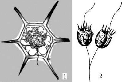
Fig. 18.1. Dictyochophytes representing the two main evolutionary lineages of the division: 1 - Dictyocha, 2 - Pedinella (after Matvienko, 1965).
Modern dictyochophytes comprise about 40 species of microscopic unicellular Algae with a monadoid or amoeboid Organization. A significant number of fossil dictyochophytes are also known, with the oldest representatives dating back about 120 million years. Dictyochophytes are widely distributed, primarily in the plankton of marine and freshwater habitats.
Taxonomic CHARACTERISTICS OF THE Division
Pigments and Storage Compounds
The Biochemical characteristics of the division include the presence of chlorophylls "a" and "c", which are masked by accessory pigments, primarily fucoxanthin. Consequently, the METABOLISM/14.html">Chloroplasts of dictyochophytes are yellow or light brown in color. Other xanthophylls detected include diatoxanthin, diadinoxanthin, and lutein. Among carotenes, only β-carotene has been found. The reserve storage products are oil and chrysolaminarin.
Cytological Characteristics
Cell coverings occur in two main variants: in silicoflagellates, it is a Plasmalemma overlain by a siliceous Skeleton enclosed in a mucous capsule. In pedinellophyte algae, cell coverings are represented by a plasmalemma typically covered with submicroscopic scales.
Nuclear apparatus. Dictyochophytes are uninucleate. The Nucleus is of the eukaryotic type, separated from the Cytoplasm by a double-membrane envelope containing one or more nucleoli. Two basal bodies of the flagella attach to the nuclear envelope on the apical side. The Golgi apparatus also directly abuts the nuclear envelope; at this site, the outer nuclear membrane is capable of pinching off vesicles that are subsequently incorporated into the cisternae of the Golgi complex. In addition, dictyochophytes possess specialized cytoplasmic microtubule structures that originate from the nuclear surface and form additional supporting elements in the cytoplasm, while in amoeboid forms, they extend into rhizopodia and participate in their movement.
Mitochondria. On ultrathin sections, mitochondrial profiles feature tubular cristae.
Photosynthetic apparatus. The vast majority of dictyochophytes are photoautotrophs or mixotrophs, although four obligately heterotrophic genera are known among the pedinellacean algae. In photosynthetic representatives, The Cell typically contains several parietal chloroplasts. Each chloroplast is enclosed by a four-membrane envelope. The two inner membranes belong to the chloroplast proper, while the two outer membranes form the chloroplast Endoplasmic reticulum. Between the chloroplast-proper membranes and the membranes of the chloroplast endoplasmic reticulum lies a fairly large periplastidal space containing a periplastidal reticulum and 80S Ribosomes.
A girdle lamella runs beneath the chloroplast envelope, followed by regular thylakoids, which are mostly grouped in threes. The chloroplast stroma also contains 70S ribosomes and chloroplast DNA (Fig. 18.2).
Flagellar apparatus exhibits typical features of chromists on the one hand, and A number of specific traits that clearly set dictyochophytes apart from other chromist divisions on the other. Typical features include a heterokont, heteromorphic, and heterodynamic two-flagellate structural plan. Typically, the short flagellum is completely reduced, leaving only its basal body. An exception is found only in the zoospores of dictyochophyte algae. The long flagellum bears two rows of tripartite mastigonemes typical of chromists (Fig. 18.2).
Unique Features of the dictyochophyte flagellar apparatus include the complete absence of microtubular flagellar roots and the direct attachment of flagellar basal bodies to the nuclear membrane. A feature linking the flagellar apparatus of dictyochophytes to golden, yellow-green, and eustigmatophyte algae is the presence of an intermediate helical Structure located in the transition zone of the flagellum—between the axoneme and the basal body.
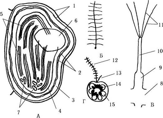
Fig. 18.2. Diagram of The structure of a chloroplast (A), flagellum (B), mastigoneme (C), and zoospore (D) in dictyochophyte algae (using Dictyocha as an example): 1 - outer chloroplast membranes; 2 - inner chloroplast membranes; 3 - periplastidal space; 4 - girdle lamella; 5 - lamellae consisting of thylakoids grouped in threes; 6 - semi-embedded pyrenoid; 7 - chloroplast DNA consisting of several closed-ring isolated segments; 8 - flagellar axoneme; 9 - 11 - mastigoneme (9 - expanded region, 10 - intermediate region, 11 - terminal hairs); 12 - tinsel flagellum of the zoospore; 13 - smooth flagellum; 14 - chloroplasts; 15 - vacuole (after Moestrup, Thompson, 1990; Van Valkenburg, 1971).
Reproduction
Reproduction occurs either by vegetative Cell Division into two or via zoospores. Cysts are also known in pedinellacean algae; outwardly they resemble the cysts of golden algae, but they are not impregnated with silica.
System of the Division
The division includes two classes, which are separated based on the Structural Features of their Cells: Dictyochophyceae (silicoflagellates) and Pedinellophyceae. The main differences between these classes lie in the general plan of cell organization, the Structure of Cell coverings, the number and arrangement of the Golgi apparatus, details of the photosynthetic apparatus organization, the mode of reproduction, and the presence of cysts.
Class Dictyochophyceae
Plan of cell organization. The cells of dictyochophytes are represented by uniflagellate monads. The cell protoplast is differentiated into two zones—a central perinuclear zone and a peripheral zone. The peripheral zone consists of numerous radially arranged cytoplasmic strands that anastomose with one another and expand at the apex. Chloroplasts and mitochondria are located within these expansions (Fig. 18.3). The perinuclear zone contains the nucleus and Golgi complexes; the flagellum and sometimes a pseudopodium, referred to as a tentacle, extend from the central zone.
Cell coverings are formed by a mucous capsule, inside of which lies the cell protoplast, separated from the mucus solely by the plasmalemma. In the peripheral part of the mucous capsule, yet external to the plasmalemma, lies a silica skeleton. The skeleton is shaped like a basket formed by interconnected hollow silica tubes (Fig. 18.4).
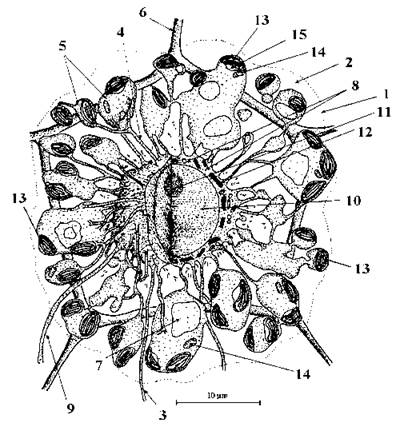
Fig. 18.3. Cell Structure of dictyochophyte algae (using the genus Dictyocha as an example, top view): 1 - boundary of the mucous capsule; 2 - amorphous mucus; 3 - pseudopodium; 4 - cytoplasmic strand connecting the peripheral and perinuclear Zones of the cell; 5 - peripheral zone of the protoplast; 6 - element of the silica skeleton; 7 - vacuole; 8 - perinuclear zone of the protoplast; 9 - flagellum; 10 - nucleus; 11 - nucleolus; 12 - Golgi apparatus; 13 - chloroplast; 14 - mitochondrion; 15 - pyrenoid (after Van Valkenburg, 1971).
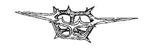
Fig. 18.4. Silica skeleton of Dictyocha, side view (after Deflandre, 1952).
The nucleus contains a single large nucleolus; several (sometimes up to several dozen) Golgi complexes are located around the nuclear envelope.
The photosynthetic apparatus of silicoflagellates exhibits three distinct features compared to pedinellophytes: first, the chloroplasts are numerous (up to several dozen); second, the chloroplast envelope is not continuous with the nuclear envelope; and third, the chloroplast DNA is scattered throughout the stroma as several fragments.
Flagellar stages are represented by vegetative cells and zoospores.
Reproduction in known Representatives of the class occurs via zoospores. Zoospores of dictyochophyte algae are naked, lacking a silica skeleton, and unlike vegetative cells, they bear two heterokont and heterodynamic flagella emerging from an apical depression (see Fig. 18.2). Each zoospore develops into a regular vegetative cell. Cysts have not been observed in dictyochophytes.
System of the class. The class is represented by a single order, Dictyochales, and a single family, Dictyochaceae. In the modern flora, two genera are known: Dictyocha and Distephanus (Fig. 18.5).
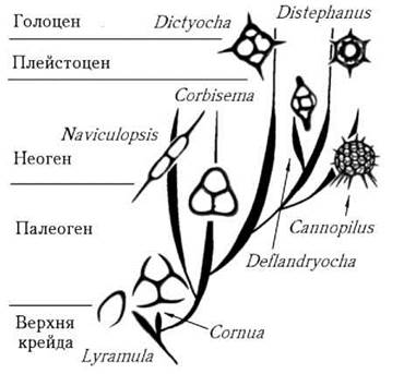
Fig. 18.5. Diagram of the evolutionary Development of the main genera of dictyochophyte algae (after Glezer, 1977).
Additionally, based on certain ultrastructural features and the degree of similarity of the gene encoding 18S rRNA, the marine picoplanktonic algae Pelagococcus and Pelagomonas are closely related to dictyochophytes. Some authors treat them either as a separate order of dictyochophytes, a separate class of dictyochophytes, or even as a distinct phylum of chromists related to Dictyochophyceae.
Distribution, ecological features, and significance. Despite their low diversity, dictyochophyte algae are extremely widespread in the seas and oceans of high latitudes. The Abundance of modern silicoflagellates in the plankton is generally low, inferior to that of diatoms, dinophytes, and prymnesiophytes. However, in the late 1980s, alarming data emerged regarding a new toxic "bloom" caused by the silicoflagellate Distephanus speculum.
One of the striking Water/58.html">Ecological features of dictyochophytes is their pronounced psychrophily (cold preference). The Temperature optimum for vegetation in members of Dictyochophyceae ranges from 5 to 12 °C. In this trait, silicoflagellates are similar to diatoms.
The oldest fossil remains of dictyochophyte algae date back about 120 million years, which may indicate the relative "youth" of these organisms. It is believed that the evolution of silicoflagellates was associated with the gradual complexification of the silica skeleton (Fig. 18.5). Because the skeletons of silicoflagellates are well preserved in the fossil record, these algae are used in geological practice to determine the age of sedimentary rocks and to reconstruct the temperature conditions of the World Ocean during recent geological epochs.
Class Pedinellophyceae
Cell organization plan. Cells of pedinellophyte algae are monadoid and uniflagellate. The flagellum emerges from an apical depression. Typically, the cell can form pseudopodia at the anterior end and a stalk at the posterior end, which is capable of contraction in some species. The protoplast is not differentiated into peripheral and perinuclear zones (Fig. 18.6).
Cell coverings are represented by a plasmalemma, upon The surface of which submicroscopic scales are deposited. These scales are formed by the Golgi apparatus and extruded outward at the posterior end.
The nucleus is directly connected to the outer membrane of the chloroplast, the basal bodies of the flagella, and the tritubular cytoskeletal structures. A single Golgi complex adjoins the basal side of the nucleus. From this side, the outer nuclear membrane is capable of budding off vesicles that are subsequently incorporated into the Golgi complex.
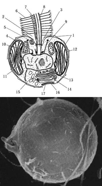
Fig. 18.6. STRUCTURE OF THE cell and cyst of pedinellophyte algae (using Mesopedinella arctica as an example): 1 - scales, 2 - subapical flagellar depression, 3 - basal body of the long flagellum, 4 - basal body of the reduced short flagellum, 5 - intermediate spiral structure, 6 - tripartite mastigoneme, 7 - paraxial strand, 8 - flagellar axoneme, 9 - tritubular cytoskeletal structure, 10 - mitochondrial profile with tubular cristae, 11 - nucleus, whose outer membrane is connected to the outer chloroplast membrane, 12 - chloroplast, 13 - nuclear-membrane vesicles, 14 - cisternae of the Golgi apparatus, 15 - vacuole, 16 - Golgi vesicle with a cell covering plate, 17 - zone of stalk formation and extrusion of cell covering plates (after Daugbjerg, 1996).
The photosynthetic apparatus is reduced in some representatives. In photoautotrophic pedinellophytes, it most commonly comprises six parietal chloroplasts. The outer chloroplast membrane, at least in pedinellalean algae, is directly continuous with the outer nuclear membrane. Chloroplast DNA is not scattered throughout the stroma, but concentrated as a single closed-ring molecule.
Monadoid cells are invariably uniflagellate. The Cytoskeleton is based on a supporting microtubular system composed of trimicrotubular structures. These trimicrotubular structures form a system of struts and rings. One end of the strut is attached to the nuclear membrane, while the opposite end connects to six to twelve microtubule triplets that close into a ring around the subapical depression.
Reproduction occurs by binary fission of cells in the motile state. In stalked species, longitudinal splitting of the stalk is observed simultaneously with cell division. Pedinellophytes survive unfavorable conditions in the form of cysts (Fig. 18.6). Cysts are more or less spherical, un-silicified, possess an opening closed by a plug, and are enclosed in a three-layered wall surrounded by a layer of scales.
Class system. Within the class, two orders are represented—Pedinellales and Rhizochromulinales. These orders are differentiated primarily by ultrastructural features. For instance, Rhizochromulinales share certain similarities with dictyochophyte algae: the flagellum lacks a paraxial rod, the cell is not covered with submicroscopic scales, the outer chloroplast membrane is not continuous with the nuclear envelope, and a stalk is absent. In other characteristics, this order aligns with the features of the class Pedinellophyceae (including the absence of a siliceous skeleton). Molecular phylogenetic studies based on 18S rRNA have confirmed the phylogenetic affinity between Pedinellales and Rhizochromulinales. Two representatives of the class, Pedinella and Mesopedinella, are shown in Fig. 18.1 and 18.6.
Distribution and ecological features. Pedinellophyte algae are distributed predominantly in cold marine waters, and are less commonly found in freshwater habitats. One species, Pedinella hexocostata, has been recorded in Ukraine within the lakes of the Donetsk region.
Thus, the division Dictyochophyta exhibits a unique type of Cellular Organization that sets it apart from other chromist divisions—chiefly, the absence of flagellar roots, the direct connection of basal bodies with the nuclear envelope, and a specific type of cytoskeleton. Ultrastructural data concerning the distinctiveness and phylogenetic independence of dictyochophytes are in good agreement with the results of molecular phylogenetic reconstructions of chromist relationships.
Last update: 07/08/2026
Editorial and Educational Adaptation: This material has been compiled based on the primary/original source text. The project team performed an editorial review, corrected technical inaccuracies, structured sections, and adapted the content for an educational format.
What was processed:
- elimination of formatting defects (OCR errors, structural breaks, corrupted characters);
- editorial organization of content;
- standardization of terminology in accordance with academic sources;
- verification of factual statements against the original source text.
All mentions of the author, publication year, and origin of the primary text have been preserved in accordance with the source.