BIOLOGY Volume 3 - A Guide to General Biology - 2004
21. REPRODUCTION
21.7. Human Reproductive Systems
21.7.4. Spermatogenesis - Sperm Development
Sperm are produced at a rate of approximately 120 million per day. The formation of a single sperm takes about 70 days. The outer layer of the seminiferous tubule wall consists of germinal epithelium Cells, which undergo repeated divisions to form the remaining six Cell layers (Figs. 21.36 and 21.37, A and B). These layers correspond to successive STAGES OF SPERM development. The initial Divisions of the germinal epithelium cells give rise to numerous spermatogonia, which increase in size to form primary spermatocytes. Following the first meiotic division, these spermatocytes turn into haploid secondary spermatocytes, and after the second meiotic division, into spermatids. Large Sertoli cells, extending from the outer layer of the tubule to its lumen, are interspersed between these rows of developing cells.
Class="center">
Fig. 21.36. Micrograph of a Testis cross-section showing seminiferous tubules and interstitial cells (Leydig cells).

Fig. 21.37, A. Group of seminiferous tubules in cross-section. B. Diagram of the wall Structure of a seminiferous tubule, showing cells at various stages of Spermatogenesis.
21.6. Sertoli cells are rich in agranular Endoplasmic reticulum elements, contain an extensive Golgi apparatus, and possess numerous Mitochondria and Lysosomes. What can be deduced about the Functions of these cells based on such a structure?
Spermatocytes are located in numerous invaginations on the lateral surfaces of Sertoli cells; here they develop into spermatids before moving to the apices of the Sertoli cells facing the lumen of the seminiferous tubule, where they finally mature into spermatozoa. Sertoli cells facilitate the transformation of spermatids into spermatozoa. All supplies of nutrients and oxygen to the developing sperm, as well as the removal of Metabolic waste products, occur through the Sertoli cells. They also secrete the fluid in which sperm travel through the tubules. The entire developmental process from spermatogonium to spermatozoon takes about two months.
Spermatozoa
Spermatozoa are very small cells, only 2.5 µm in diameter (compared to an average animal cell size of 20 µm) and approximately 50 µm in length. The structure of a sperm cell is shown in Fig. 21.38. Its HEAD contains a Nucleus with a haploid number of Chromosomes (23 in humans). The Nucleus also houses the acrosome—a large lysosome containing hydrolytic Enzymes that subsequently assist the sperm in penetrating the layers of cells surrounding the oocyte immediately before Fertilization.
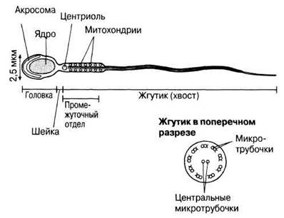
Fig. 21.38. Diagram of the structure of a mature human spermatozoon.
The short neck of the sperm contains a pair of centrioles positioned at right angles to each other. During sperm maturation, the microtubules of one centriole elongate to form the axial filament (axoneme) of the flagellum, or tail, which runs along the entire remaining length of the sperm.
The midpiece of the sperm marks the beginning of the flagellum; it is enlarged due to numerous mitochondria arranged in a spiral around the axial filament. The mitochondria drive aerobic Respiration and produce ATP as an energy source. This energy powers the flagellar movements that propel the sperm at a speed of approximately 1–4 mm/min. A cross-section of the flagellum reveals the typical eukaryotic flagellar arrangement of nine peripheral pairs of microtubules surrounding a central pair.
Activation of the flagellum occurs once it enters the Vagina. Flagellar movement alone is insufficient to cover the distance from the vagina to the site of fertilization. The primary locomotory task of spermatozoa is to gather around the oocyte and orient themselves properly before penetrating the oocyte membranes.
Hormonal Regulation of Spermatogenesis
Spermatogenesis is regulated by Hormones from the Hypothalamus and the anterior Pituitary Gland acting in concert. The hypothalamus is a region of the Brain (Fig. 17.24), and the pituitary gland is located directly beneath it. The hypothalamus secretes gonadotropin-releasing hormone (GnRH), which travels from the hypothalamus to the pituitary via a small portal vein. In turn, GnRH stimulates the anterior pituitary to secrete two hormones known as gonadotropins. (A gonadotropin is a hormone that stimulates gonad activity, in this case, the testis.) These two gonadotropins are follicle-stimulating hormone (FSH) and luteinizing hormone (LH) (Fig. 21.39). The same two hormones are secreted in females (see Section 21.7.6). Chemically, they are Glycoproteins. FSH stimulates spermatogenesis by prompting Sertoli cells to support the final maturation of sperm from spermatids. LH stimulates the Leydig cells (interstitial cells) of the testis to synthesize the hormone testosterone, also known in males as interstitial cell-stimulating hormone. Testosterone is a steroid hormone derived from Cholesterol. It stimulates the GROWTH AND DEVELOPMENT of sperm from germinal epithelium cells and, together with FSH, exerts a stimulatory effect on Sertoli cells. A negative feedback mechanism operates such that rising levels of testosterone lead to a reduction in GnRH secretion by the hypothalamus, as illustrated in Fig. 21.39. This, in turn, leads to a decrease in LH and FSH levels. It is also possible that testosterone acts directly on the anterior pituitary to suppress LH secretion, although this effect is less pronounced.
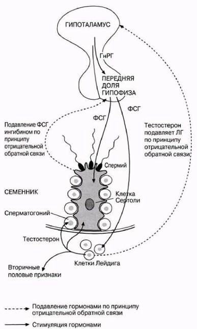
Fig. 21.39. Hormonal regulation of spermatogenesis. GnRH produced by the hypothalamus stimulates the pituitary to secrete FSH and LH. LH stimulates testosterone secretion, which in turn promotes sperm production while also acting as a negative feedback inhibitor on the hypothalamus. FSH stimulates Sertoli cells to produce inhibin. Inhibin provides a second negative feedback loop that regulates FSH production by the anterior pituitary gland.
Role of Inhibin
Sertoli cells produce another glycoprotein hormone, inhibin, which regulates sperm production via a negative feedback loop. If spermatogenesis proceeds too rapidly, inhibin synthesis increases, which acts on the anterior pituitary to suppress FSH secretion (Fig. 21.39). Inhibin also acts on the hypothalamus to reduce GnRH secretion. When The rate of spermatogenesis is low, inhibin secretion ceases, allowing FSH to stimulate spermatogenesis.
Role of Cyclic ATP
Both FSH and LH trigger the release of cyclic AMP (cyclic adenosine monophosphate, cAMP) within the cells they stimulate; cAMP acts as the 'second messenger' discussed in Section 17.6.1. It is released into the Cytoplasm and then enters the nucleus, where it stimulates enzyme synthesis. In the case of LH, for example, these are the enzymes involved in the synthesis of testosterone from cholesterol.
Secondary sexual characteristics
Testosterone is the primary male sex hormone, influencing The Development of both Primary and secondary sexual characteristics. Primary characteristics are those present from birth, whereas secondary characteristics develop by the onset of Puberty. Successful sperm production requires both testosterone and FSH, whereas the development of secondary sexual characteristics and their maintenance throughout adult life are controlled solely by testosterone. Secondary sexual characteristics include:
1) development and enlargement of the Testes, Penis, and reproductive tract glands;
2) enlargement of the Larynx, resulting in a deeper voice;
3) enhanced muscular development;
4) growth of pubic Hair, along with increased hair growth in the armpits and on the chest;
5) behavioral patterns associated with courtship, mating, sexual activity, and parental care.
Last update: 06/08/2026
Editorial and Educational Adaptation: This material has been compiled based on the primary/original source text. The project team performed an editorial review, corrected technical inaccuracies, structured sections, and adapted the content for an educational format.
What was processed:
- elimination of formatting defects (OCR errors, structural breaks, corrupted characters);
- editorial organization of content;
- standardization of terminology in accordance with academic sources;
- verification of factual statements against the original source text.
All mentions of the author, publication year, and origin of the primary text have been preserved in accordance with the source.