BIOLOGY Volume 3 - A Guide to General Biology - 2004
21. REPRODUCTION
21.7. Human Reproductive Systems
21.7.5. Oogenesis - The Development of Egg Cells
Unlike Spermatogenesis, which in males begins only at Puberty, oogenesis in females starts even before birth. The stages of oogenesis are outlined in Fig. 21.35. During fetal development, numerous oogonia are formed from primordial Germ Cells. Through mitotic divisions, oogonia differentiate into primary oocytes, which remain arrested in the prophase stage of Meiosis I practically until ovulation. Primary oocytes are surrounded by a single layer of granulosa cells, forming structures known as primordial follicles (Figs. 21.40–21.42). By birth, a female fetus contains approximately 2 million follicles, yet only about 450 of these will ever mature into secondary oocytes and be released from the Ovary during the reproductive years. Throughout the fertile period, one primordial follicle matures each month into a mature follicle called a Graafian follicle. This process is driven by FSH (follicle-stimulating hormone). Within each developing follicle, the primary oocyte begins to develop into an ovum. Initially, the primordial follicle becomes a primary follicle as the granulosa cells proliferate, forming multiple layers around the primary oocyte (Figs. 21.40–21.42). Additionally, further layers of cells derived from the ovarian stroma form around these cells, collectively known as the theca. The outer layer of the theca contains Blood Vessels and merges with the stroma, while the inner layer, much like the granulosa cells, secretes Female Sex Hormones.
Class="center">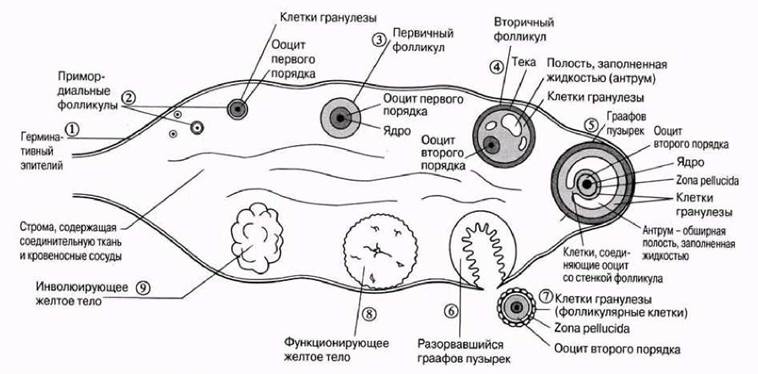
Fig. 21.40. Cross-section of a human ovary, illustrating the sequential stages (numbered) of Graafian follicle development, ovulation, corpus luteum formation, and its regression. In reality, all these stages cannot be observed simultaneously.
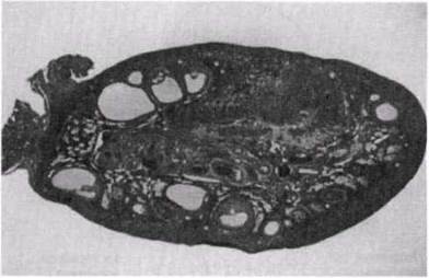
Fig. 21.41. Cytology/practical/54.html">Longitudinal section of a human ovary showing developing follicles; x7.
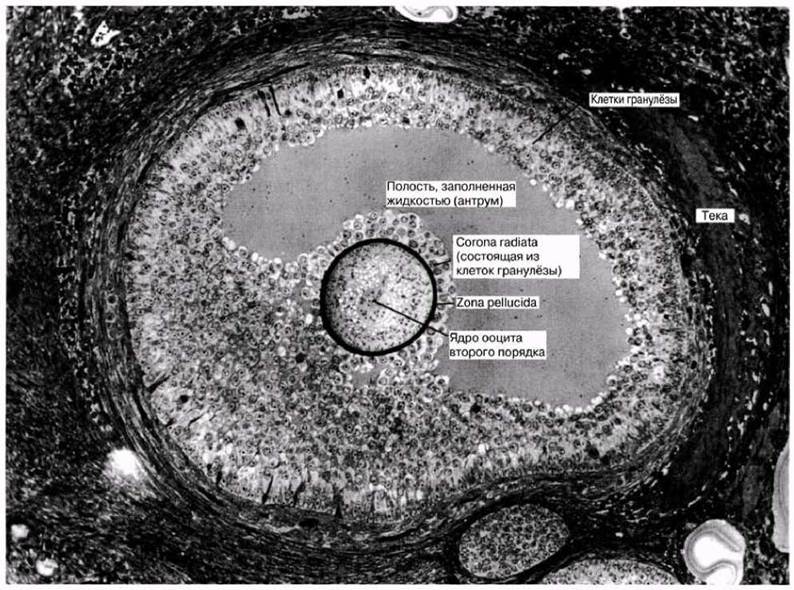
Fig. 21.42. A mature human Graafian follicle.
As the primary follicle develops, the granulosa cells secrete a fluid containing estrogen, one of the female sex hormones. A fluid-filled cavity, the antrum, forms within the follicle, which is now termed a secondary follicle. Estrogen stimulates follicle growth, ultimately transforming it into a mature follicle known as a Graafian follicle. This follicle reaches up to 1 cm in diameter and contains a secondary oocyte and the first polar body, produced During the first meiotic division of the primary oocyte (Fig. 21.35). The secondary oocyte possesses a haploid number of Chromosomes. The second meiotic division reaches metaphase but arrests there until the oocyte fuses with a sperm. Upon Fertilization, the secondary oocyte completes the second meiotic division, yielding a large ovum and a second polar body. All polar bodies are small cells that do not participate in oogenesis and eventually degenerate. The Structure of a secondary oocyte (ovum) is illustrated in Fig. 21.43.

Fig. 21.43. Structure of a secondary oocyte. An ovum shares the same structure, with the sole difference that it possesses two polar bodies. The second polar body is formed by the division of the secondary oocyte, which takes place upon fertilization. The zona pellucida is surrounded by granulosa cells (follicular cells), though they are omitted from this diagram.
21.7. (a) In what key aspects is the structure of an ovum similar to that of a sperm?
(b) Briefly list the major differences between sperm and egg cells.
Last update: 06/08/2026
Editorial and Educational Adaptation: This material has been compiled based on the primary/original source text. The project team performed an editorial review, corrected technical inaccuracies, structured sections, and adapted the content for an educational format.
What was processed:
- elimination of formatting defects (OCR errors, structural breaks, corrupted characters);
- editorial organization of content;
- standardization of terminology in accordance with academic sources;
- verification of factual statements against the original source text.
All mentions of the author, publication year, and origin of the primary text have been preserved in accordance with the source.