Human Biochemistry, Volume 2 - Murray R. 1993
Biochemistry of Intracellular and Intercellular Communication
Sex Hormones
Regulation and Physiological Action of Ovarian Hormones
Maturation and Maintenance of FEMALE Reproductive System Function
The primary function of Ovarian Hormones is to prepare the Structural components of the female reproductive system (see below) for reproduction. This preparation involves: 1) the maturation of primordial Germ Cells; 2) The Development of Tissues required for blastocyst implantation; 3) the hormonal control of the timing of ovulation; 4) the establishment, via placental hormones, of an environment necessary to maintain Pregnancy; and 5) the Hormonal Regulation of parturition and Lactation.
Estrogens stimulate the development of tissues involved in reproduction. As a rule, these hormones increase The rate of synthesis of protein, rRNA, tRNA, mRNA, and DNA, leading to an increase in the size and number of cells in the target tissues. Estrogenic stimulation causes proliferation and Differentiation of the vaginal epithelium, proliferation of the endometrium, hypertrophy with elongation of its glands, the appearance of intrinsic rhythmic motility in the myometrium, and proliferation of the mammary ducts. Estradiol also exerts an anabolic effect on bone and Cartilage, thereby promoting linear growth. By acting on peripheral Blood Vessels, estrogens generally cause vasodilation and enhance heat dissipation.
The manifestation of progestin activity typically requires the prior or simultaneous action of estrogens. Thus, these two classes of hormones often function synergistically, although they can also act as antagonists. Progestins diminish the stimulating effect of estrogens on vaginal epithelial proliferation and facilitate the transition of the uterine epithelium from the proliferative to the secretory phase (characterized by enlargement and enhanced function of the secretory glands and an increased Glycogen content), thereby preparing it for the implantation of a fertilized ovum. These hormones enhance the Development of the mammary acinar tissue following estrogen-induced ductal development. Progestins decrease peripheral blood flow, thereby reducing heat loss. Consequently, during the luteal phase of the Menstrual cycle—when this hormone is produced—body Temperature rises. Such temperature spikes, typically around 0.5°C, are used as an indicator of ovulation.
The maximum number of oogonia in the human fetal Ovary reaches 6–7 million by approximately the fifth month of embryonic development. By birth, this number drops to about 2 million, and by the onset of menarche, it is 100,000 to 200,000. Approximately 400 to 500 of these develop into mature oocytes. The rest gradually disappear through a yet-unidentified process, though it is known that ovarian androgens are involved. Follicular maturation begins in infancy; throughout the prepubertal period, the Ovaries increase in size due to follicular enlargement driven by the growth of granulosa cells, the accumulation of atretic follicular tissue, and an increase in medullary stromal mass containing interstitial and theca cells capable of producing Steroid Hormones.
During childhood, sex hormone concentrations are low, although exogenous gonadotropins can increase their production. Consequently, immature ovaries retain the capacity to synthesize estrogen. It has been hypothesized that in prepubertal girls, the low levels of sex Steroids present suppress gonadotropin production, whereas at Puberty, the hypothalamic-pituitary axis becomes less sensitive to the inhibitory action of these hormones. Puberty is marked by the onset of pulsatile GnRH (gonadotropin-releasing hormone) secretion; under METABOLISM/18.html">The Influence of LH, ovarian hormone production increases sharply, while FSH—the primary stimulator of estrogen secretion—drives follicular maturation and triggers ovulation.
The Menstrual Cycle
The frequency of ovulation and readiness for copulation are determined by hormones. In monoestrous species, ovulation and mating occur once a year; in polyestrous species, ovulation recurs several times a year. In primates, which are characterized by menstrual cycles with endometrial shedding at the end of each cycle, Sexual Behavior is not tightly coupled to ovulation. In humans, the menstrual cycle is governed by a complex interplay among the Hypothalamus, Pituitary Gland, and ovaries. Under normal conditions, the duration of the menstrual cycle ranges from 25 to 35 days (averaging 28 days). It can be divided into the follicular phase, the luteal phase, and menstruation (Fig. 50.8).
A. Follicular Phase. For reasons that remain unclear, under the influence of FSH, only a single follicle begins to enlarge. During the first week of the follicular phase, the E2 concentration remains low, but then begins to rise progressively as the follicle grows. Twenty-four hours prior to the LH (FSH) surge, the E2 level peaks and sensitizes the pituitary gland to the action of gonadotropin-releasing hormone (GnRH).
Class="center">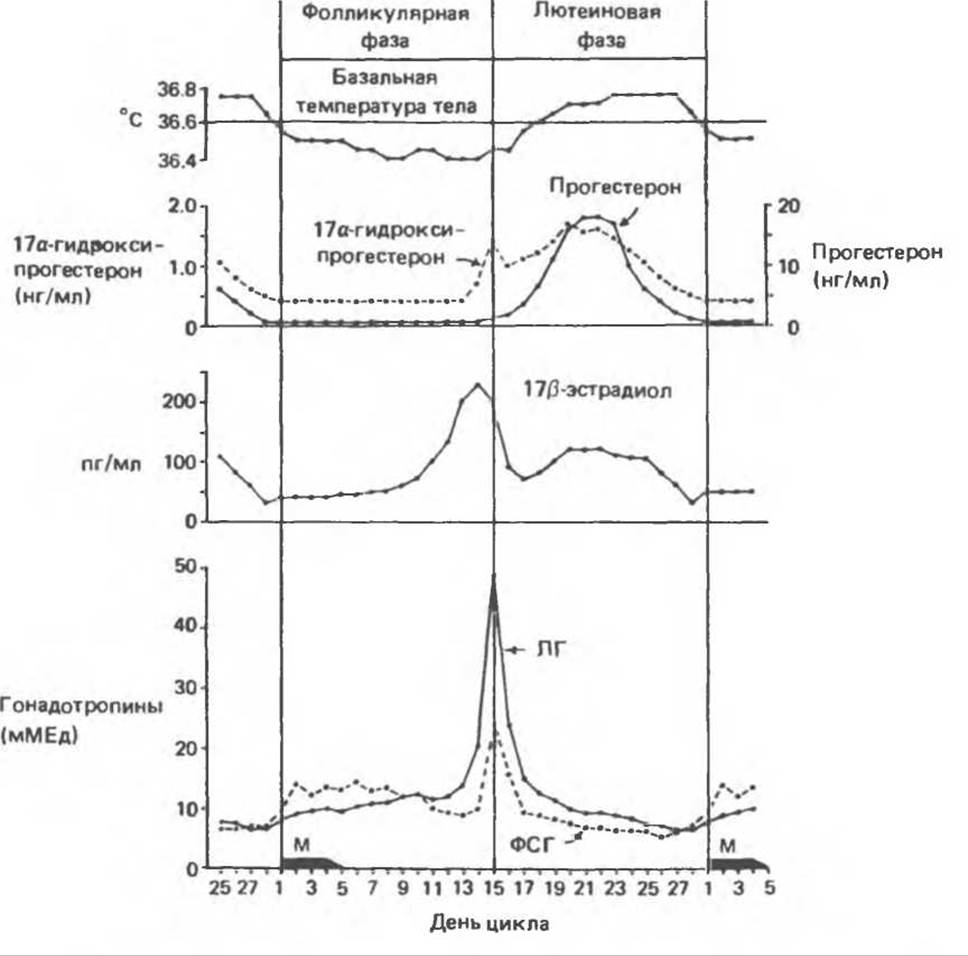
Fig. 50.8. Hormonal and physiological changes during a typical human menstrual cycle. M — menstruation, LH — luteinizing hormone, FSH — follicle-stimulating hormone. (Reproduced, with permission, from Midgley A. R. In: Human Reproduction, Hafez ESE, Evans TN [editors]. Harper and Row, 1973.)
The LH surge is triggered either by this high level of E2 via a positive feedback mechanism or by a sharp drop in its level. Prolonged administration of high doses of estrogens (such as in oral contraceptives) suppresses both LH and FSH secretion as well as the action of GnRH on the pituitary. Progesterone levels during the follicular phase are very low.
B. Luteal Phase. Following ovulation, the granulosa Cells of the ruptured follicle luteinize and form the corpus luteum—a Structure that soon begins to secrete progesterone and a certain amount of estradiol. Estradiol levels peak around the middle of the luteal phase and then decline sharply. The primary hormone of the cycle's luteal phase is progesterone. It is essential for The formation of a secretory endometrium, which provides the proper conditions for the development of the implanted blastocyst. Initially, the Maintenance of the corpus luteum requires the presence of LH, and the pituitary secretes this hormone for about ten days. If implantation occurs (days 22–24 of the cycle), the function of LH is taken over by human chorionic gonadotropin (hCG)—a placental hormone very similar to LH, produced by the cytotrophoblast cells of the early-stage implanted embryo (see Chapter 45). hCG sustains progesterone synthesis by the corpus luteum until the Placenta begins producing large amounts of this steroid. In the absence of implantation (and hCG), the corpus luteum degenerates and menstruation ensues. Following endometrial shedding, a new cycle begins. The luteal phase invariably lasts 14 + 2 days. Variations in cycle length are almost universally attributable to differences in the duration of the follicular phase.
Pregnancy and Placental Hormones
The implanted blastocyst forms the trophoblast, which subsequently organizes into the placenta. The placenta serves as the vital link between the fetal and maternal circulatory systems and secretes a variety of hormones.
A. Human Chorionic Gonadotropin (hCG).
The primary function of this glycoprotein hormone is to maintain the corpus luteum until the placenta begins producing progesterone in quantities sufficient to sustain a normal pregnancy. hCG is detectable within just a few days after implantation, making it useful for the early Diagnosis of pregnancy. The concentration of this hormone peaks around the middle of the first trimester and gradually declines throughout the remainder of gestation. The levels of hCG and other pregnancy-related hormones are illustrated in Fig. 50.9.
B. Progestins. During the first 6–8 weeks of pregnancy, the corpus luteum serves as the primary source of progesterone; thereafter, the placenta assumes this function. Although the corpus luteum continues to function, in later stages of pregnancy the placenta produces 30 to 40 times more progesterone than the corpus luteum does. Because the placenta is incapable of synthesizing Cholesterol de novo, it must acquire it from the maternal Circulation.
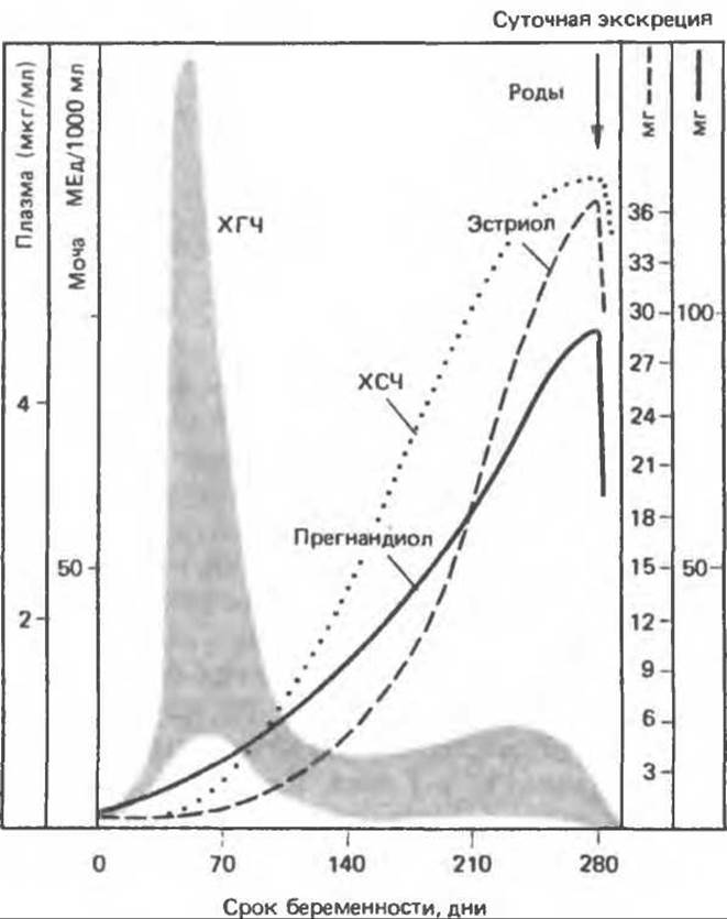
Fig. 50.9. Hormone concentrations during normal pregnancy. hCG — human chorionic gonadotropin, hCS — human chorionic somatomammotropin (data compiled from various authors). (Reproduced, with permission, from Ganong W. F. Review of Medical Physiology, 13th ed. Appleton and Lange, 1987.)
C. Estrogens. Throughout pregnancy, the plasma concentrations of estradiol, estrone, and estriol progressively rise. Estriol is produced in the greatest Abundance, and its synthesis reflects various fetoplacental Functions. The fetal Adrenal Glands produce dehydroepiandrosterone (DHEA) and DHEA sulfate, which are converted in the fetal Liver into 16α-hydroxy derivatives; these, in turn, are transformed into estriol within the placenta. The resulting estriol enters the maternal bloodstream, is conjugated with glucuronides in the maternal liver, and is excreted in the urine in this form (Fig. 50.10). Urinary estriol levels are used as an index to evaluate various aspects of fetal and maternal well-being.
Equally interesting is the exchange of substrates required for the synthesis of cortisol and DHEA in the fetus. Due to the absence of the 3β-hydroxysteroid dehydrogenase/Δ5,4-isomerase complex in the embryonic adrenal glands, fetal cortisol synthesis relies on placental progesterone. Pregnenolone, which is required for dehydroepiandrosterone synthesis, is also supplied by the placenta (Fig. 50.10).
D. Placental Lactogens. The placenta produces placental lactogen (PL)—a hormone also known as chorionic somatomammotropin or placental Growth Hormone, as it exhibits the biological properties of both prolactin and growth hormone. The genetic relationship between these hormones is discussed in Chapter 45. The Physiological Role of placental lactogen has not been definitively established, as women lacking this hormone experience normal pregnancies and deliver healthy infants.
Parturition
Pregnancy lasts for a strictly determined number of days, specific to each species. However, the precise trigger responsible for the termination of pregnancy remains elusive. It is hypothesized that hormones play a critical role, potentially estrogens and progestins, given their regulatory effects on uterine contractility. There is also evidence implicating catecholamines in the initiation of labor. Oxytocin stimulates uterine contractions and is used clinically to facilitate labor; however, it does not initiate labor prior to term. The concentration of oxytocin receptors in the myometrium increases dramatically toward the end of gestation, exceeding early-pregnancy levels by a factor of 100. This upregulation correlates with the late-gestational surge in estrogen levels, which enhance oxytocin receptor expression (Chapter 45). At the onset of labor, the uterine cervix stretches, triggering a reflex stimulation of oxytocin release and, consequently, further uterine contractions. Mechanical factors, such as myometrial stretch or pressure, may also play an important role in this process. During parturition, both the mother and the newborn experience a dramatic shift in their hormonal environment, and following the expulsion of the placenta, plasma levels of progesterone (measured as pregnanediol) and estriol rapidly decline (Fig. 50.9).
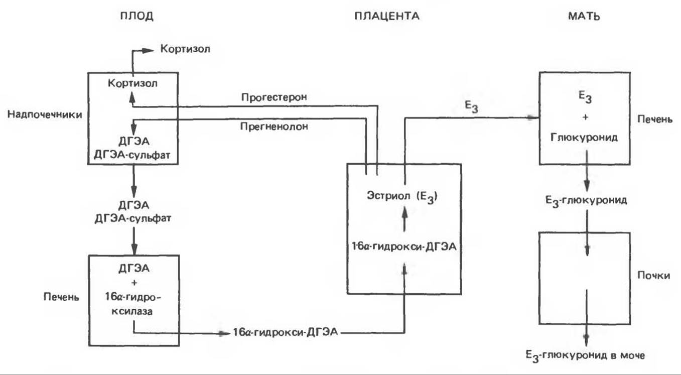
Fig. 50.10. Steroid metabolism in the maternal–fetal unit. DHEA — dehydroepiandrosterone.
Mammary Gland Development and Lactation
The differentiation and function of the mammary gland are regulated by the coordinated action of several hormones. This process is initiated by Female Sex Hormones: estrogens are responsible for ductal growth, whereas progestins stimulate alveolar proliferation. Some proliferation of glandular tissue, accompanied by fat deposition, occurs during puberty; however, the gland achieves its greatest development during pregnancy, when the glandular tissue is exposed to high concentrations of estradiol and progesterone. Studies conducted primarily on rat mammary gland explants have shown that full differentiation also requires the action of prolactin, glucocorticoids, Insulin or a growth factor, and an unidentified serum factor. Among these hormones, only the concentration of prolactin undergoes dramatic changes during pregnancy, increasing from less than 2 ng% to over 200 ng% in late gestation. The influence of hormones on the synthesis of various milk Proteins, including lactalbumin, lactoglobulin, and casein, has been studied in detail. These hormones increase the rate of synthesis of these proteins by elevating the levels of specific mRNAs, which, at least in the case of casein, results from enhanced Gene Transcription. Notably, enhanced gene transcription was observed only when cortisol, prolactin, and insulin were added simultaneously to the explant culture.
During late stages of pregnancy, milk production and secretion are suppressed by progesterone, which is required for alveolar differentiation. Lactation begins only after childbirth, when the levels of this hormone decline sharply. Postpartum prolactin levels also decrease rapidly but rise with each nursing episode (Chap. 45), thereby maintaining continuous lactation. If the infant is weaned for any reason, lactation gradually ceases. Parenteral administration of large doses of androgens prior to the onset of nursing can induce a rapid cessation of lactation.
Nursing also stimulates the secretion of oxytocin by the posterior pituitary gland. Oxytocin promotes the contraction of myoepithelial cells surrounding the alveolar ducts, thereby facilitating milk ejection from the gland. The regulatory mechanisms governing the Synthesis and Secretion of oxytocin are discussed in Chap. 45.
Menopause
In women of the Western hemisphere at approximately 53 years of age, menstrual cycles become increasingly irregular; simultaneously, ovarian follicles disappear, and their function ceases. There are no other sources of progesterone in the body, but the aromatization of the adrenal steroid androstenedione produces significant amounts of estrone, which possesses weak estrogenic activity (Fig. 50.6). The amount of estrone produced is not sufficient to suppress pituitary gonadotropins, and the postmenopausal period is characterized by a sharp increase in LH and FSH levels. Postmenopausal women are particularly prone to two Problems associated with tissue metabolism. The first is that estrone is not always able to prevent the atrophy of secondary sex tissues, particularly the epithelium of the lower urogenital tract and Vagina. The second problem faced by older women is Osteoporosis. In women with a particularly marked reduction in bone mass, estrone levels are below normal.
Synthetic Agonists and Antagonists
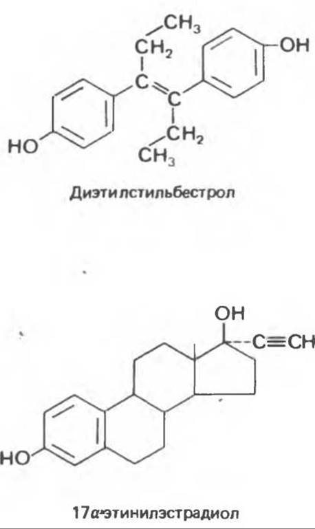
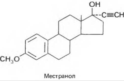
A. Estrogens. Several synthetic compounds exhibit estrogenic activity while offering pharmacological advantages. Most modifications are aimed at reducing hepatic metabolism so that the drugs can be administered orally. One of the first synthetic estrogens was diethylstilbestrol. Examples of modified steroids also include 17α-ethinylestradiol and mestranol, which are used as oral contraceptives.
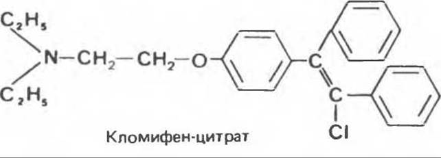
Numerous compounds with antiestrogenic activity have also been synthesized, some of which have found clinical application. Most of these antagonists act by competing with estradiol for its intracellular receptors (see below). Clomiphene citrate (Clomid) is distinguished by a particularly high affinity for estrogen receptors in the hypothalamus. Clomiphene was originally intended for use as a contraceptive, but it proved to have the opposite effect. Because clomiphene competes with estradiol for hypothalamic receptors, the release of gonadotropin-releasing hormone is no longer restricted, leading to the release of large amounts of LH and FSH. Under the influence of clomiphene, the simultaneous maturation of multiple follicles is frequently observed, which can result in multiple pregnancies. Nafoxidine (a nonsteroidal compound) and tamoxifen interact with estrogen receptors to form a very stable complex with Chromatin. Because receptors bound in this manner cannot enter a new cycle, these compounds inhibit the action of estradiol for prolonged periods. Such antagonists are used in the Treatment of estrogen-dependent breast Cancer.
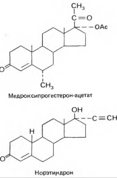
B. Progestins. Synthesizing compounds that possess progestational activity without estrogenic or androgenic side effects has proved to be a challenging task. 17α-Alkyl-substituted derivatives of 19-nortestosterone (e.g., norethindrone) exhibit minimal androgenic activity in most women and are used as oral contraceptives. Another potent progestin is medroxyprogesterone acetate. Intramuscular administration of long-acting medroxyprogesterone results in the suppression of ovulation for several months. However, this drug is much more commonly used to treat differentiated endometrial carcinoma. It is believed to block the division of normal and malignant endometrial cells by forming stable complexes with progesterone receptors (thereby preventing the action of the natural hormone).
Last update: 06/08/2026
Editorial and Educational Adaptation: This material has been compiled based on the primary/original source text. The project team performed an editorial review, corrected technical inaccuracies, structured sections, and adapted the content for an educational format.
What was processed:
- elimination of formatting defects (OCR errors, structural breaks, corrupted characters);
- editorial organization of content;
- standardization of terminology in accordance with academic sources;
- verification of factual statements against the original source text.
All mentions of the author, publication year, and origin of the primary text have been preserved in accordance with the source.